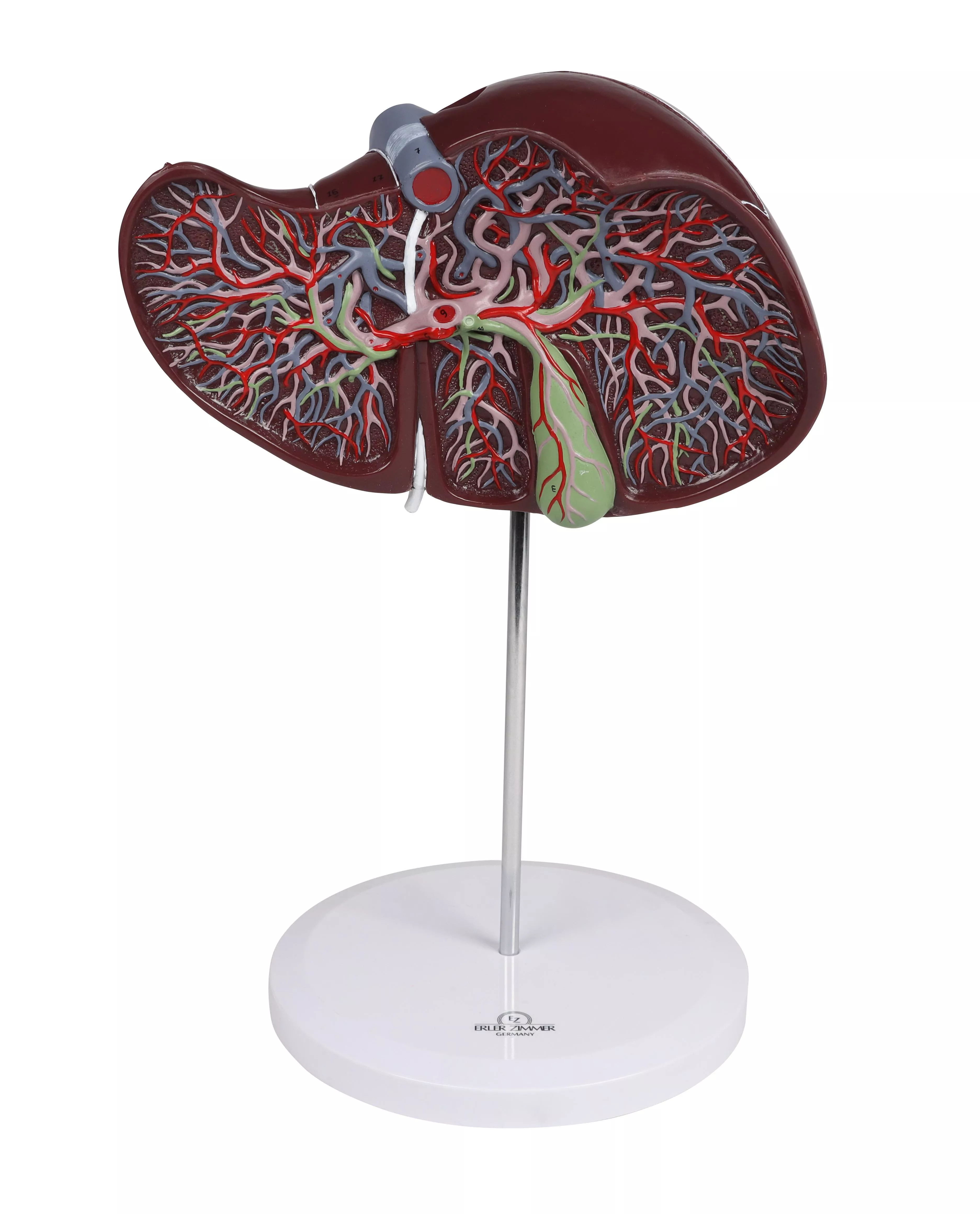
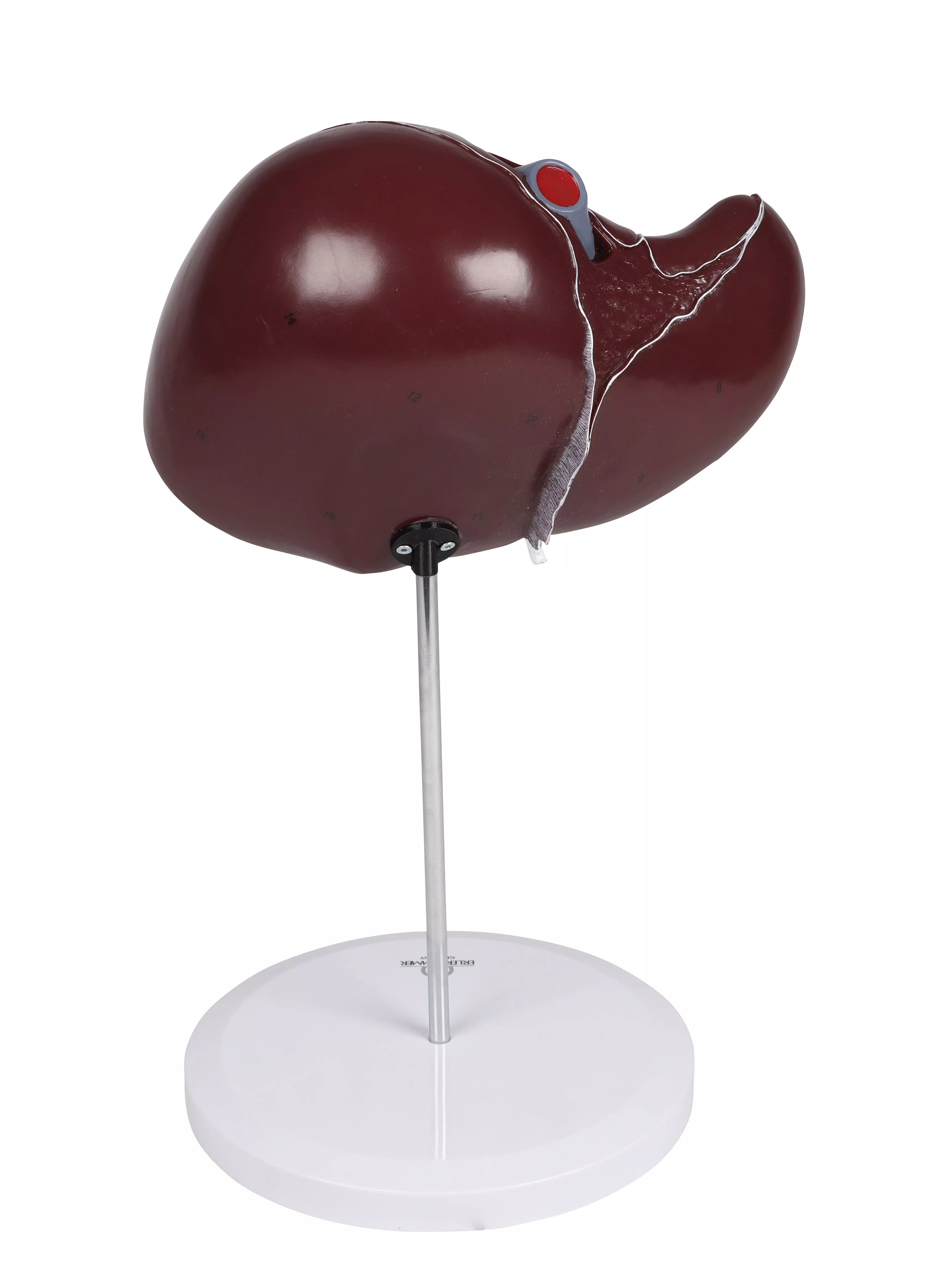
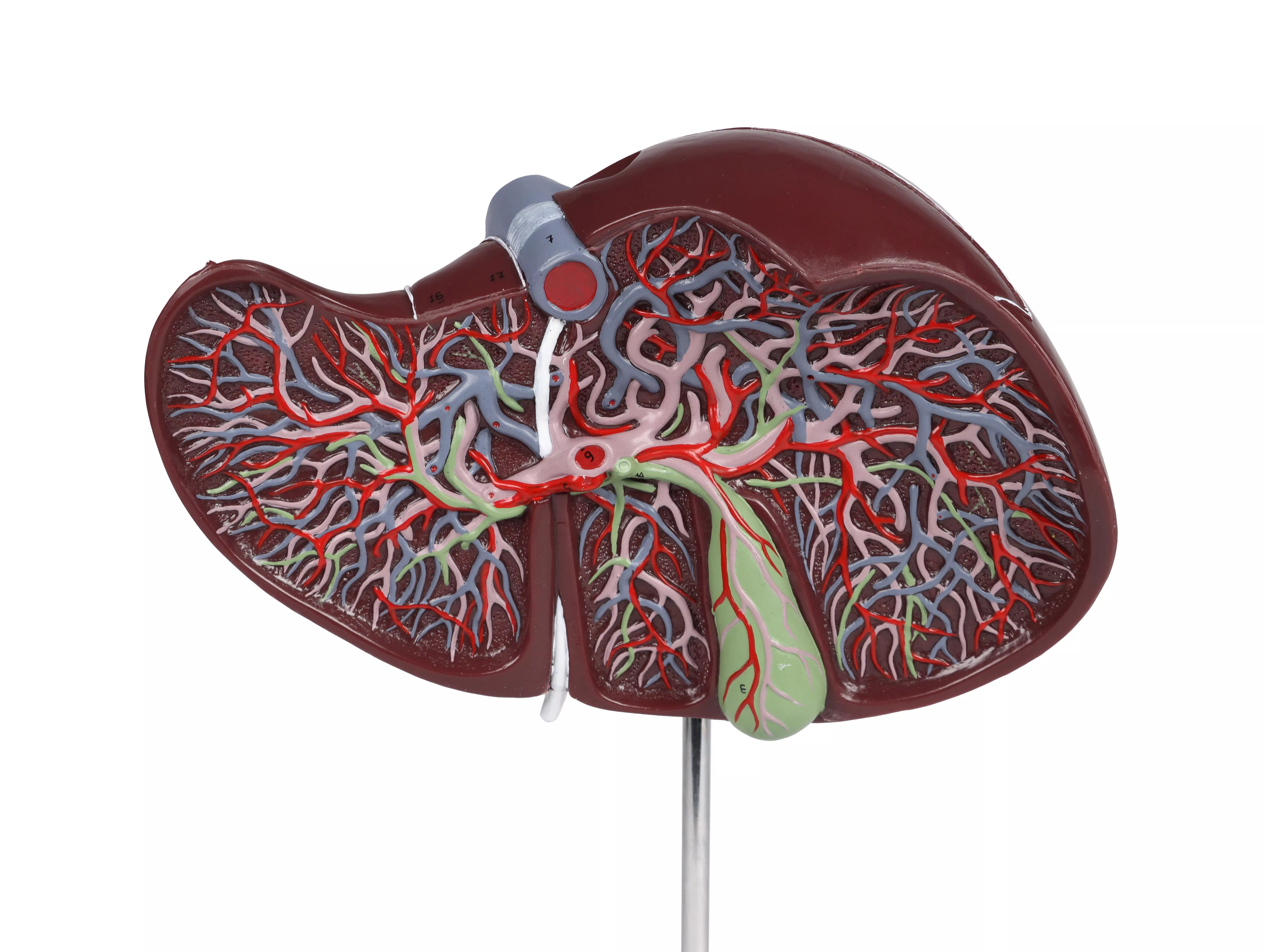
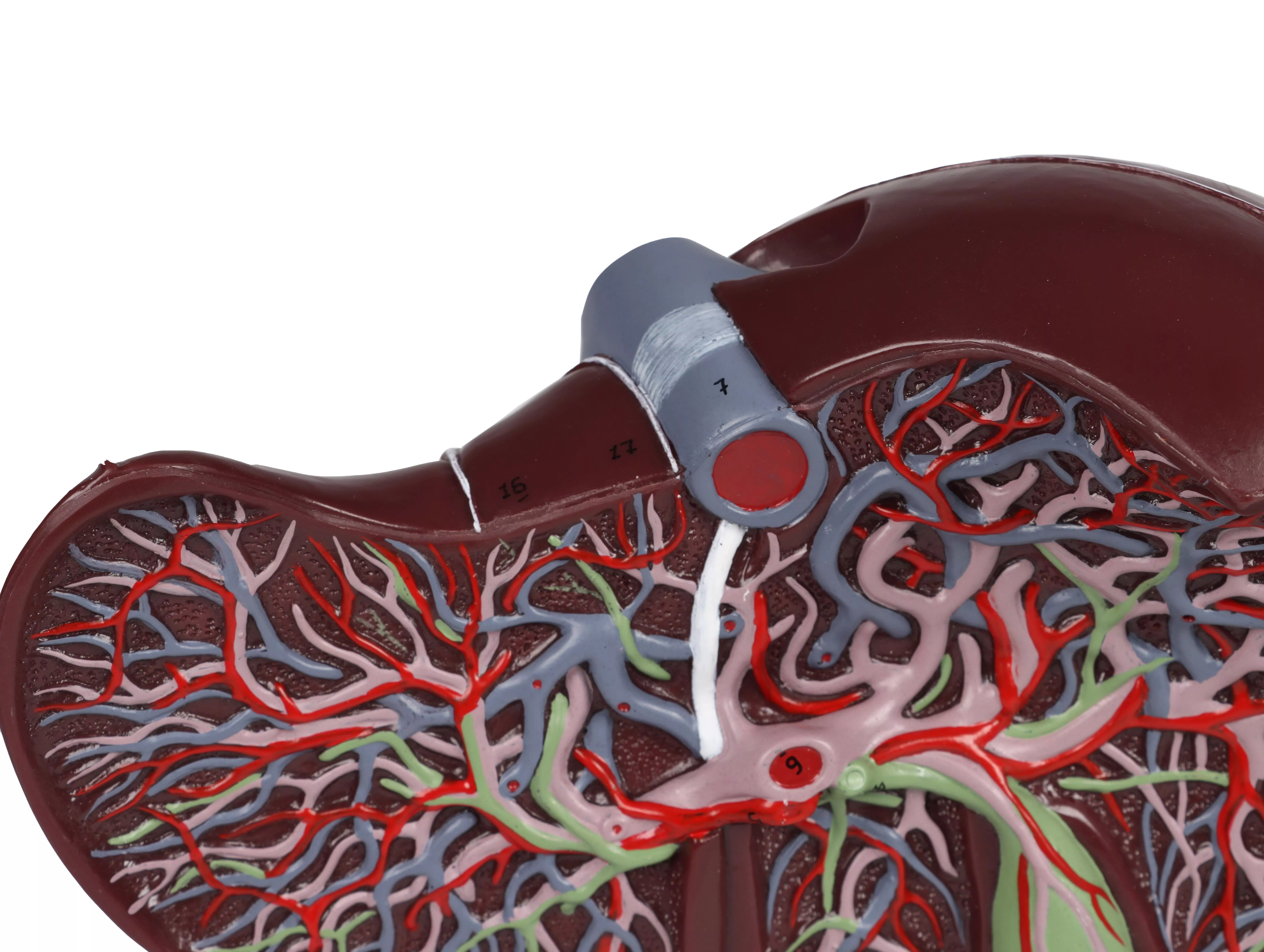
Product information "Human liver, 1.5 times life size"
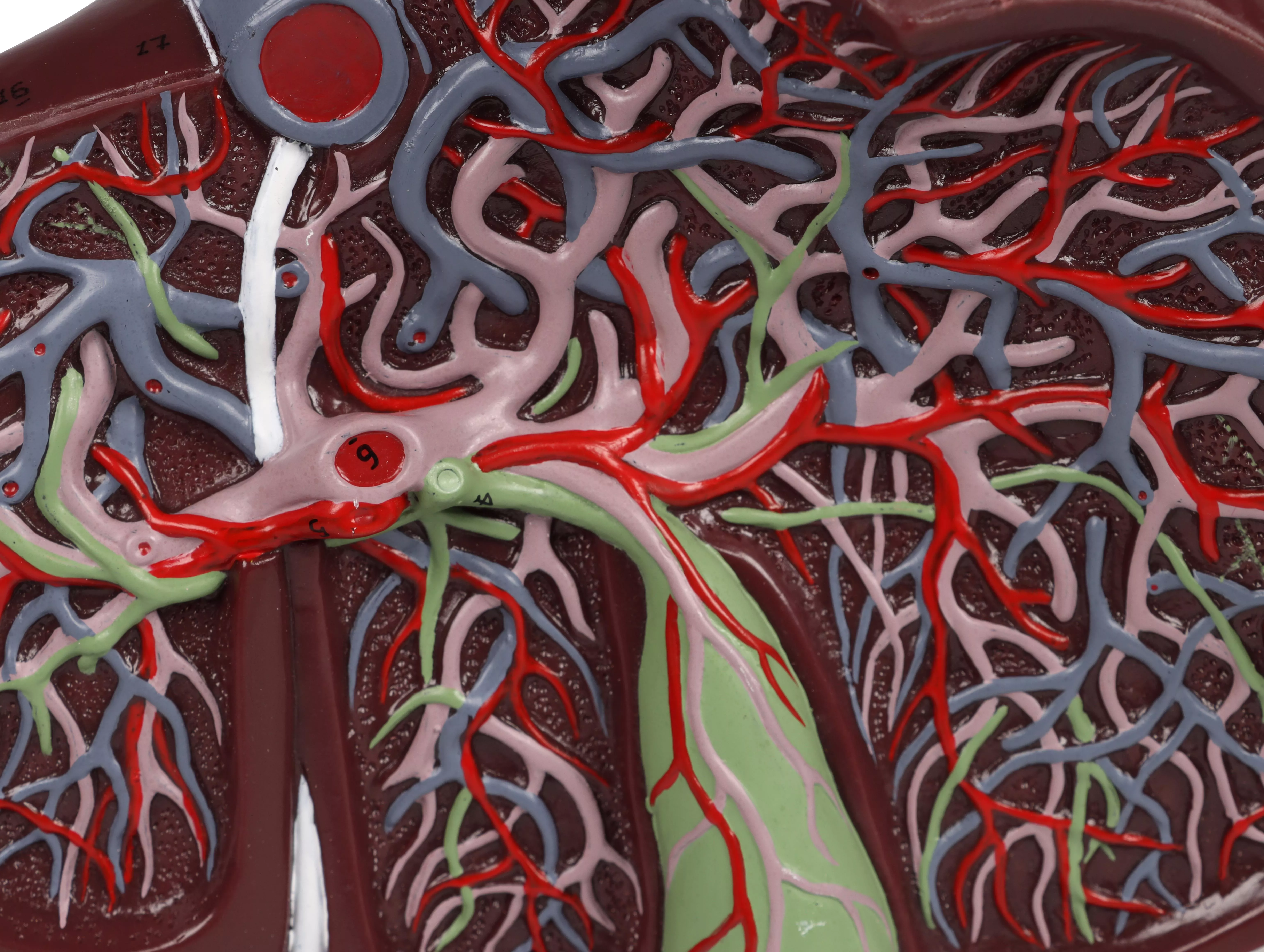
Enlarged 1.5 times, this model shows a liver that is dissected to expose the internal distribution of arteries and veins, the portal vein and the bile duct. Mounted on stand. Size: 15 x 26 x 12 cm, Weight: approx. 1 kg
0 of 0 reviews
Leave a review!
Share your experiences with other customers.
Login
No reviews found. Share your insights with others.
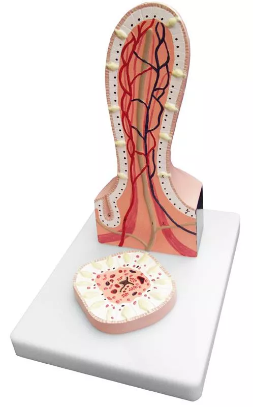
This greatly enlarged 2-part model of a villus from the small intestine shows detail from a transverse and longitudinal section. Mounted on base, with key card.Size: 17.5 x 15 cm
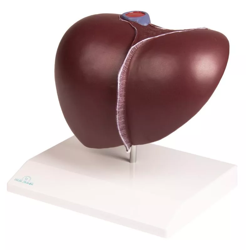
This realistic model reproduces a liver with the gall bladder. The hilus vessels are shown as well as the extrahepatic ducts and the main ligaments. Mounted on removable stand.Size: 16 x 12 x 11 cm
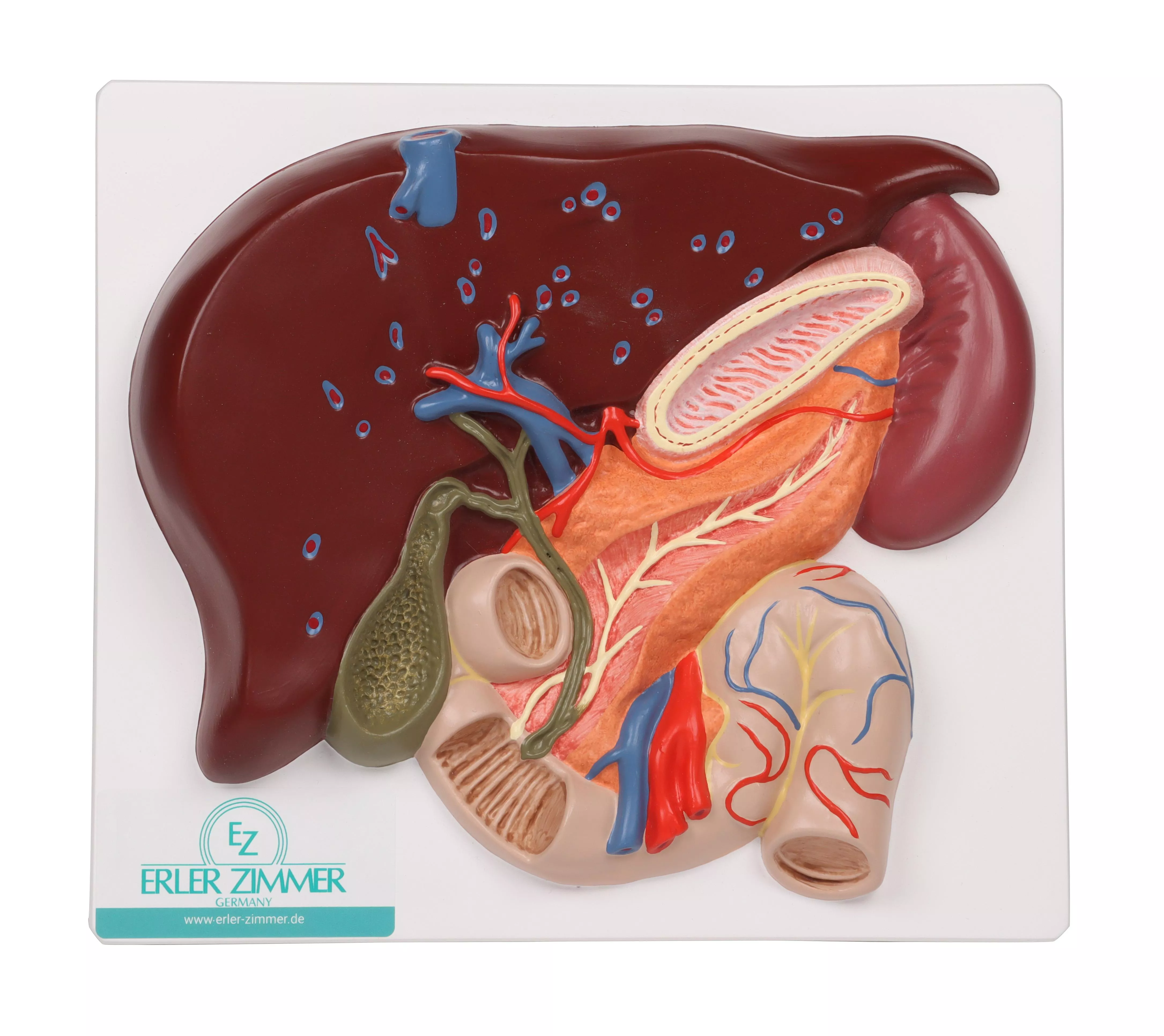
This life size model shows a section of the liver with gall bladder, pancreas and duodenum; includes hepatic and pancreatic ducts. Mounted on board.Size: 4 x 20 x 18 cm
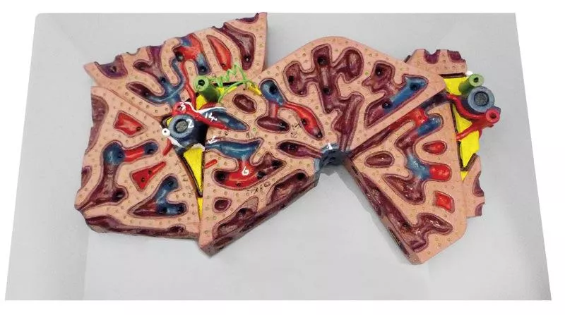
This greatly enlarged model shows the fine detail of a single liver lobule, which is sectioned and shown in relationship to portions of surrounding lobules. The fine colouring distinguishes the portal veins and vessels, venous sinusoids and central veins with sections through the bile canaliculi. With key.Size: 25 x 15 x 6 cm
This life size 5-part model represents the major components of the urinary system, plus the vena cava and abdominal aorta; the right kidney is dissected to show the cortex, medulla, pyramids, calyces, pelvis and origins of the renal artery and vein. The bladder can be opened to reveal the mucosa, trigone, urethra, seminal vesicles, ejaculatory ducts and vas deferens. Mounted on base.Size: 24.5 x 18 x 36 cm, weight: 0.7 kg
The human circulatory system as overview. The perfect opportunity to explain and understand the blood supply in the human body. The model pictures clearly the lung circulation, the heart with ist chambers, artriums and valves, the large vessels and the blood supply to the extremities. Of course liver, kidneys, spleen and a part of the intestines are shown. The front wall of the heart can be removed. To locate the vessels the model shows the skeleton, allowing to understand the three-dimensional paths of the vessels. On base board, also suitable for hanging.Size: 90 x 35 x 5 cm
Longitudinal section of an artery with constriction caused by plaque adsorption. Four cross sections on the base show step-by-step build-up of plaque at the artery wall.Size: 14 x 18 x 13 cm, weight: 0.4 kgAugmented- Anatomy- AppThe new Augmented Anatomy App in combination with this high quality anatomical model makes learning now even easier and more efficient!This app automatically recognises our anatomical models and displays the nomenclature in augmented reality. As an Erler Zimmer customer, you can use it completely free of charge and for an unlimited period of time. - High-quality Augmented Reality learning app- Free of charge and without registration- Nomenclature always and everywhere available- Further online links in the learning lexiconOur augmented anatomy app works on all common smartphones or tablets. Click here to Download the App
This improved model of a female pelvis with pelvic floor muscles depicts the pelvic floor in its layers. The following muscles are represented and magnetically detachable: - M. obturatorius internus (left and right),- M. piriformis (left and right),- M. coccygeus (left and right), - Diaphragma pelvis (M. levator ani consisting of M. puborectalis, M. pubococcygeus, and M. iliococcygeus), - Diaphragma urogenitale (consisting of M. transversus perinei profundus, M. transversus perinei superficialis, and M. ischiocavernosus), and the - sphincter muscles of the urogenital and digestive tracts (consisting of M. sphincter ani externus, M. sphincter urethrae, and M. bulbospongiosus).The model consists of a total of 12 parts, including the two halves of the pelvis and the sacrum. The model is held together by magnets, allowing individual parts to be easily removed for demonstration of the different layers.This model is highly suitable for courses in pelvic floor exercises, childbirth preparation, midwifery training, or simply for studying the pelvic floor muscles.Size: 27 x 18 x 17 cm, Weight: 960 g.
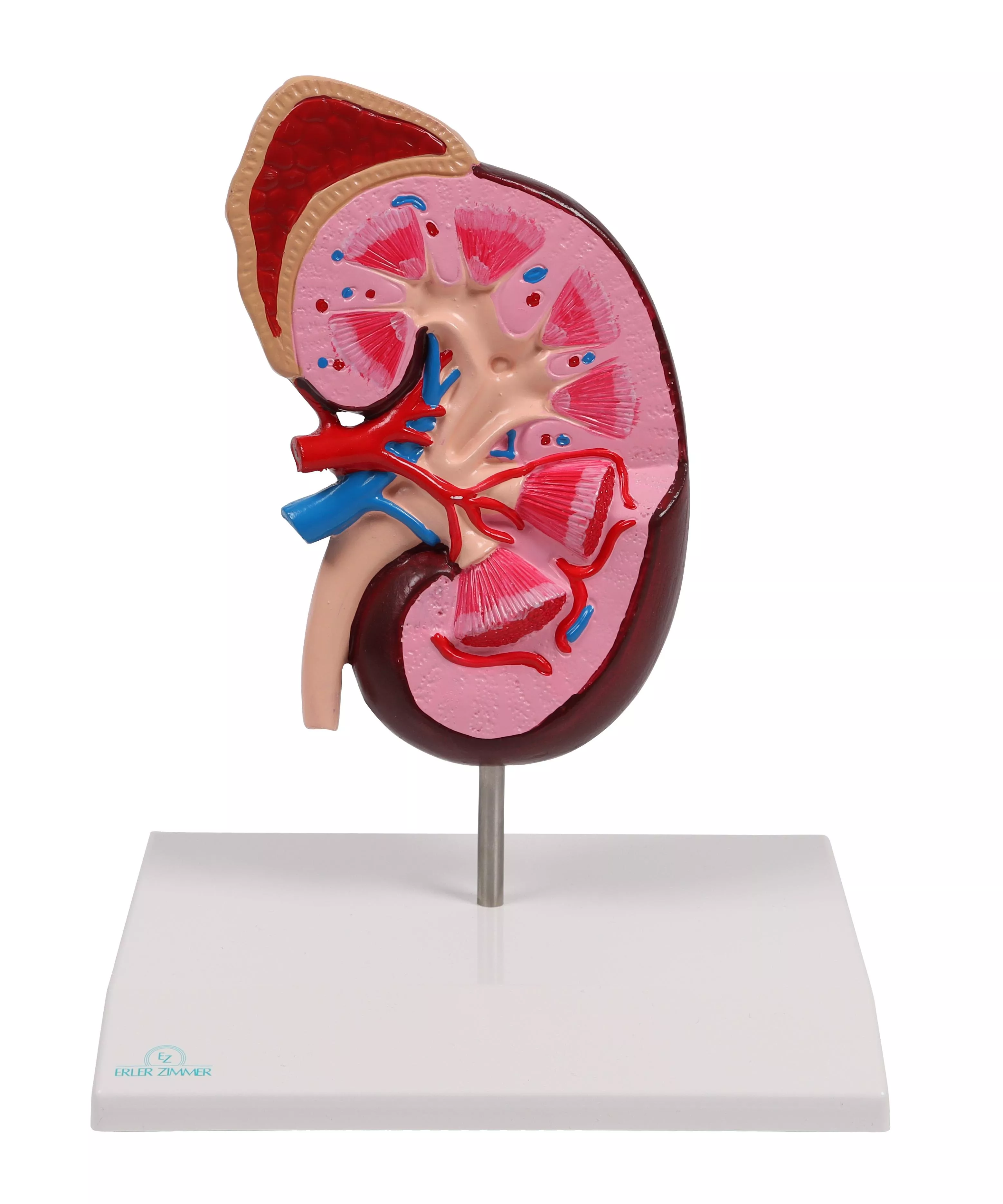
This 3 models set shows the basic structure of the kidney. We can find as a first model a frontal section of the kidney, enlarged 3 times, illustrates adrenal gland, cortex, medulla, pyramids with papillae, renal pelvis and blood vessels. The second model, representing a nephron enlarged120 times, shows renal tubules, a collecting tube system and Henle‘s loop. The third one illustrates Malpighian corpuscle with the Bowman‘s capsule, 700 times life size. All these models are a great instrument to understand the kidney anatomy in all the inner details. The three models aremounted on board.Size: 70 x 29 x 12 cm, Weight: 4.1 kg
This life size model is dissected along the medial plane and can be opened to show the internal structure of the stomach, including the cardia, the mucosa and the pylorus. The model also shows the blood vessels. Mounted on stand.Size: 15 x 15 x 6 cm, weight: 0.5 kg