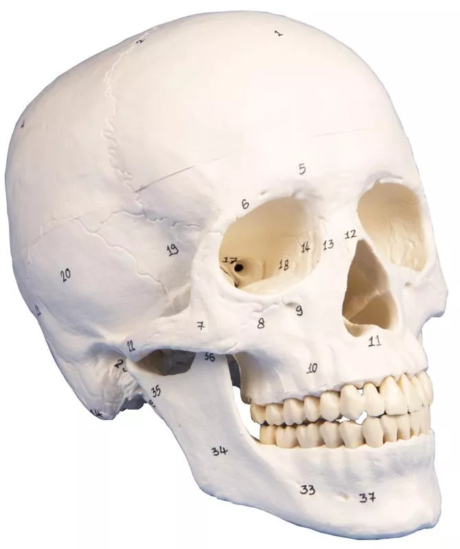
Product information "Skull model for dentistry and oral surgery, 5-part"
In addition to the detailed reproduction of the skull structures, this first-class model of a human skull focuses specifically on teeth and jaws and is therefore the perfect teaching and demonstration tool for the field of dentistry and oral surgery.
The front half of the upper and lower jaw can be removed to reveal teeth with roots, unilateral nerves and blood vessels as well as the maxillary sinuses. The skull is not only suitable as a perfect aid for learning anatomy, but also as an explanatory aid for oral surgery and implantology.

5 of 5 reviews

5 out of 5 stars


100%

0%

0%

0%

0%


Leave a review!

Share your experiences with other customers.


October 4, 2021 09:47

Good price-performance

The skull fits well in the hand and all important structures are shown as described. In addition, the delivery was super fast.

November 20, 2021 11:51

Fantastic Skull model

good teaching tool for physiotherapy education.The skull has very accurate anatomical structures and is a relief when learning.

November 16, 2021 15:06

fast delivery

very good skull model, we are using this for osteopathy students.

October 4, 2021 10:12

Die Lieferung kam prompt, das Modell entspricht genau unseren Erwartungen

Der Schädel hat wirklich eine Top Qualität - hochwertig und gut verarbeitet. Schöne Details und anatomisch korrekt. Lieferung wie gewohnt, schnell und zuverlässig.Klare Kaufempfehlung!

October 4, 2021 08:41

Zahnmedizin Studium

Ich nutze den Dental Schädel in meinem Zahnmedizin Studium, dieser ist für das erlernen der Anatomie eine echte hilfe! :)

Erler-Zimmer

Erler-Zimmer GmbH & Co.KG
Hauptstrasse 27
77886 Lauf
Germany
info@erler-zimmer.de


Achtung! Medizinisches Ausbildungsmaterial, kein Spielzeug. Nicht geeignet für Personen unter 14 Jahren.

Attention! Medical training material, not a toy. Not suitable for persons under 14 years of age.


Other customers also bought

Luxury demonstration Children skull; 14 pieces; for advanced studies
This skull model is a cast of a real human children's skull specimen and shows all anatomical structures in great detail. It was developed for students of anatomy, medicine, surgery, ENT medicine, ophthalmology and dentistry. The skull is complexly cut and joined together with metal and magnetic connections.The top of the skull is open and removable. Bony impressions of the sinus sagittalis, the sinus transversus and the sinus sigmoideus as well as the meninges are painted. The base of the skull is cut sagittally in such a way that the cut runs through a sieve plate on one side and another cut with the same plane through the other sieve plate of the ethmoid bone, which leaves the Christa galli and the perpendicular plate as well as the entire nasal septum intact. The structures of the anterior, middle and posterior fossa are easily accessible. One can see the nasal cavity, the turbinate, the septum and the bony and nasopharynx directly. The nasal septum can be removed from the surrounding bone structures. The removable temporal bone shows a complete external auditory canal.The upper jaw and the lower jaw show the structures of the teeth, the roots, the bony edge of the alveolar process as well as dental nerves and vessels. The maxillary sinus can be opened by removing a flap. The teeth, including the permanent teeth still embedded in the bone, can be made accessible by removing a bone flap.Estimated age: 3 years

€696.15*
Deluxe demonstration skull; 14-part; for advanced studies
This skull model is an actual cast of a real human specimen and shows all anatomical structures in highest detail. It is made for students in anatomy, medicine, surgery, otolaryngology, ophthalmology and dentistry. The Skull is intricately sectioned and reassembled with metal and magnet connections. The calvarium is sectioned horizontally leaving the temporal bone and its sutures intact. Bony impressions of the superior sagittal sinus, transverse sinus and sigmoid sinus as well as the meningeal vessels have been painted. The base portion of the skull has been sagittaly sectioned in the way that it passes through the one cribriform plate on one side and another section in the same plane passes through the other cribriform plate of the ethmoid leaving the crista galli perpendicular plate of the ethmoid intact as also the whole nasal septum. The structures of anterior, middle and posterior cranial fossae are easily accessible. One can directly visualize the nasal cavity, the concha, the nasal septum, the bony pharyngeal and naso-pharyngeal spaces. The nasal septum is separable from surrounding bones. The frontal sinuses have been dissected on one side to show the sinus as whole and on the other side chiselled out for full access to the sinus. The relation of this sinus to the nasal cavity is clearly shown and is especially valuable for otolaryngologists. On one side of the skull the temporal bone has been left in situ. The other temporal bone is removable from the skull. A portion of the mastoid and squama can be removed along with the tympanic antrum, baring internal ear in full view. All three semicircular canals are visible along with the course of the facial nerve coursing backwards and then downwards emerging finally through the stylo-mastoid foramen. The removable temporal bone has the external auditory meatus intact. An almost vertical section through the squama mastoid process and carried inwards along the petro-squamosal junction has been made and when apart, one sees the position of tympanic membrane. The carotid canal has been opened as also the cochlea , showing the internal canal, and the course of the facial nerve has been depicted. Oval window, the semi-circular canals, and aditus of the tympanic antrum are visible. The maxilla and mandible expose the structures of dentition, the roots, the bony margin of the alveolar process, dental vessels and nerves are visible. The maxillary sinus can by opened by removing a bone flap. Teeth of the right mandible are sectioned to show the inner tooth structure.

€696.15*
Osteopathic Skull Model, 22 part, didactical version - EZ Augmented Anatomy
This fascinating model of an average European adult skull can be disassembled into 22 single bones. During development of this model one of the main targets was to make the model easy to assemble and dismantle. Stable parts with convenient magnet connections make handling of the product a child‘s play. The detailed bones do not need any complicated pins to be stuck into holes, they almost slide into position, guided by realistic bone sutures and held by strong magnets. Due to the very good anatomy and the easy handling this skull is the ideal training aid for osteopathy.This version is perfect for visual distinction of the single bones. The bones are produced in different colours, commonly used in anatomy, geminate bones do have identical colour. The following individual bones are shown:•Parietal bone left and right •Occipital bone •Temporal bone left and right •Sphenoid bone •Frontal bone •Ethmoid bone •Vomer •Palatine bone, left and right •Inferior nasal concha left and right •Maxilla with teeth, left and right •Lacrimal bone left and right •Nasal bone left and right •Zygomatic bone left and right •Mandible with teeth Scope of delivery:•User manual in English and German •Detailed description on CD in Latin, English, French, German, Spanish, Portuguese, Italian, Polish, Russian, Arabic, Korean and Japanese. Augmented- Anatomy- AppThe new Augmented Anatomy App in combination with this high quality anatomical model makes learning now even easier and more efficient!This app automatically recognises our anatomical models and displays the nomenclature in augmented reality. As an Erler Zimmer customer, you can use it completely free of charge and for an unlimited period of time. •High-quality Augmented Reality learning app •Free of charge and without registration •Nomenclature always and everywhere available •Further online links in the learning lexicon Our augmented anatomy app works on all common smartphones or tablets. Click here to Download the App.

€332.01* €324.87 (lowest price)*
Skull model for dentistry with TMD syndrome, 8 parts
In addition to the detailed reproduction of the skull structures, this first-class model of a human skull focuses specifically on teeth and jaws and is therefore the perfect teaching and demonstration tool for the field of dentistry and oral surgery. The front half of the upper and lower jaw can be removed to reveal teeth with roots, unilateral nerves and blood vessels as well as the maxillary sinuses. The skull is not only suitable as a perfect aid for learning anatomy, but also as an explanatory aid for oral surgery and implantology.This special design serves to illustrate the TMD syndrome. In temporomandibular dysfunction, there is a dysfunction in the interaction between temporomandibular joints, masticatory muscles and teeth. The wear of the tooth enamel due to grinding, wear of the discus as well as relevant masticatory muscles M. masseter, pars superficialis, pars profundus as well as the relatively newly discovered pars coronoidea are shown. All three muscles are detachable.

€439.11* €427.21 (lowest price)*
Skull model, 3-part, numbered
Skull model like ref.no. 4500, but additionally with numbered bones and structures. With nomenclature. Size: 18 x 19 x 12 cm, weight: 0.7 kg

€127.33*

Continuous innovation

Social responsibility

Active customer orientation

Understanding quality

Sustainable actions

ISO 9001 certification

Your last viewed products