


Luxury demonstration Children‘s skull; 14 pieces; for advanced studies
Learn more€675.92*
Available, within 1-3 working days
Product number:
4810
Article number: 4810
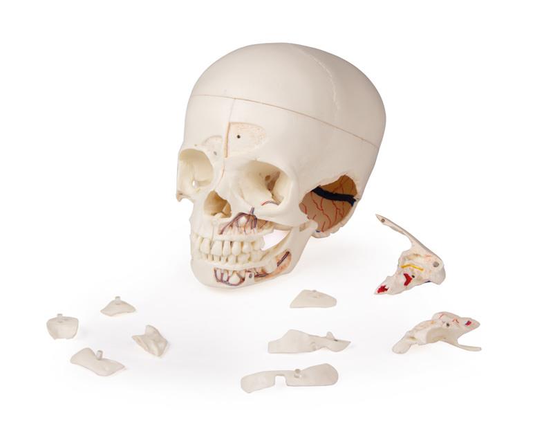
Product information "Luxury demonstration Children‘s skull; 14 pieces; for advanced studies"
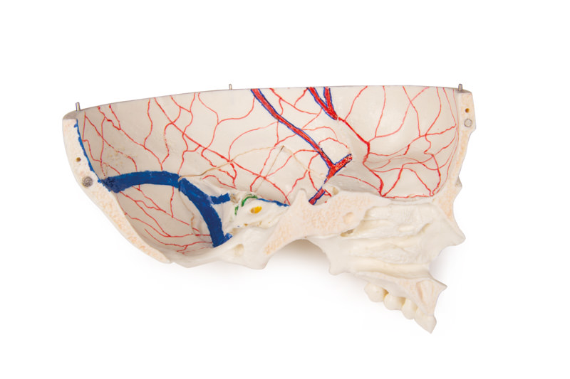
This skull model is a cast of a real human children‘s skull specimen and shows all anatomical structures in great detail. It was developed for students of anatomy, medicine, surgery, ENT medicine, ophthalmology and dentistry. The skull is complexly cut and joined together with metal and magnetic connections.
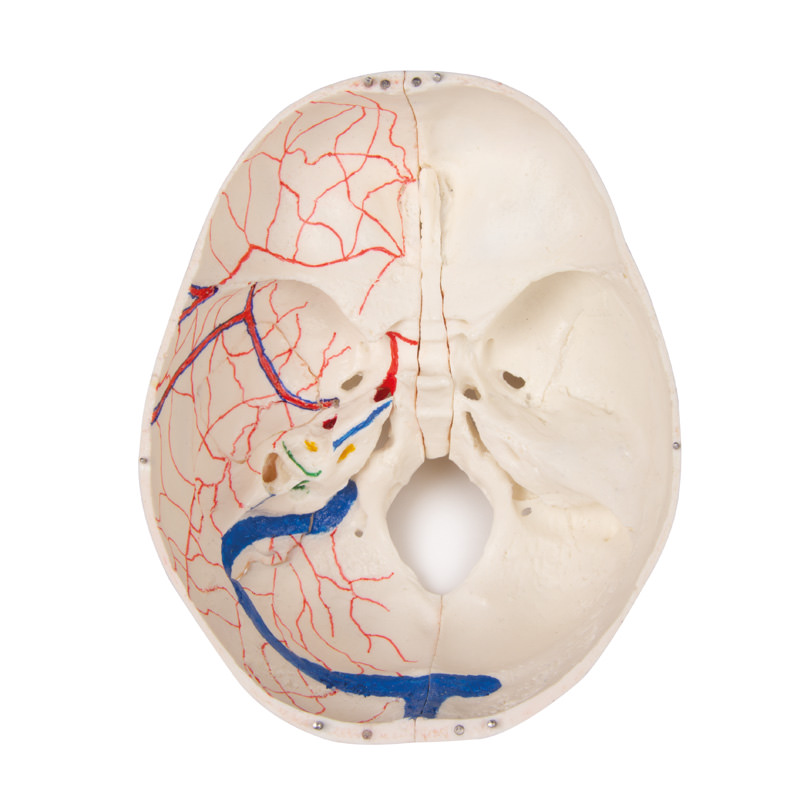
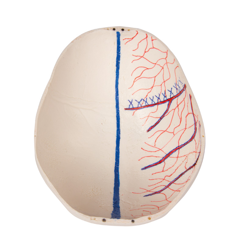
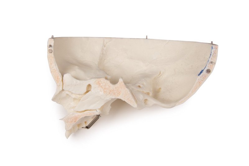
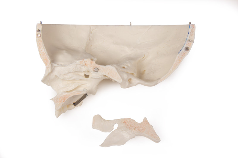
The top of the skull is open and removable. Bony impressions of the sinus sagittalis, the sinus transversus and the sinus sigmoideus as well as the meninges are painted. The base of the skull is cut sagittally in such a way that the cut runs through a sieve plate on one side and another cut with the same plane through the other sieve plate of the ethmoid bone, which leaves the Christa galli and the perpendicular plate as well as the entire nasal septum intact. The structures of the anterior, middle and posterior fossa are easily accessible. One can see the nasal cavity, the turbinate, the septum and the bony and nasopharynx directly. The nasal septum can be removed from the surrounding bone structures. The removable temporal bone shows a complete external auditory canal.
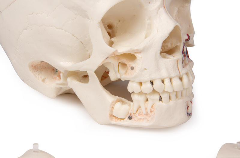
The upper jaw and the lower jaw show the structures of the teeth, the roots, the bony edge of the alveolar process as well as dental nerves and vessels. The maxillary sinus can be opened by removing a flap. The teeth, including the permanent teeth still embedded in the bone, can be made accessible by removing a bone flap.
Estimated age: 3 years
The top of the skull is open and removable. Bony impressions of the sinus sagittalis, the sinus transversus and the sinus sigmoideus as well as the meninges are painted. The base of the skull is cut sagittally in such a way that the cut runs through a sieve plate on one side and another cut with the same plane through the other sieve plate of the ethmoid bone, which leaves the Christa galli and the perpendicular plate as well as the entire nasal septum intact. The structures of the anterior, middle and posterior fossa are easily accessible. One can see the nasal cavity, the turbinate, the septum and the bony and nasopharynx directly. The nasal septum can be removed from the surrounding bone structures. The removable temporal bone shows a complete external auditory canal.
The upper jaw and the lower jaw show the structures of the teeth, the roots, the bony edge of the alveolar process as well as dental nerves and vessels. The maxillary sinus can be opened by removing a flap. The teeth, including the permanent teeth still embedded in the bone, can be made accessible by removing a bone flap.
Estimated age: 3 years
Login
Other customers also bought

Deluxe demonstration skull; 14-part; for advanced studies
€675.92*
This skull model is an actual cast of a real human specimen and shows all anatomical structures in highest detail. It is made for students in anatomy, medicine, surgery, otolaryngology, ophthalmology and dentistry. The Skull is intricately sectioned and reassembled with metal and magnet connections. The calvarium is sectioned horizontally leaving the temporal bone and its sutures intact. Bony impressions of the superior sagittal sinus, transverse sinus and sigmoid sinus as well as the meningeal vessels have been painted. The base portion of the skull has been sagittaly sectioned in the way that it passes through the one cribriform plate on one side and another section in the same plane passes through the other cribriform plate of the ethmoid leaving the crista galli perpendicular plate of the ethmoid intact as also the whole nasal septum. The structures of anterior, middle and posterior cranial fossae are easily accessible. One can directly visualize the nasal cavity, the concha, the nasal septum, the bony pharyngeal and naso-pharyngeal spaces. The nasal septum is separable from surrounding bones. The frontal sinuses have been dissected on one side to show the sinus as whole and on the other side chiselled out for full access to the sinus. The relation of this sinus to the nasal cavity is clearly shown and is especially valuable for otolaryngologists. On one side of the skull the temporal bone has been left in situ. The other temporal bone is removable from the skull. A portion of the mastoid and squama can be removed along with the tympanic antrum, baring internal ear in full view. All three semicircular canals are visible along with the course of the facial nerve coursing backwards and then downwards emerging finally through the stylo-mastoid foramen. The removable temporal bone has the external auditory meatus intact. An almost vertical section through the squama mastoid process and carried inwards along the petro-squamosal junction has been made and when apart, one sees the position of tympanic membrane. The carotid canal has been opened as also the cochlea , showing the internal canal, and the course of the facial nerve has been depicted. Oval window, the semi-circular canals, and aditus of the tympanic antrum are visible. The maxilla and mandible expose the structures of dentition, the roots, the bony margin of the alveolar process, dental vessels and nerves are visible. The maxillary sinus can by opened by removing a bone flap. Teeth of the right mandible are sectioned to show the inner tooth structure.

Continuous innovation

Social responsibility

Active customer orientation

Understanding quality

Sustainable actions

ISO 9001 certification