


Atrial septal defect
Learn more€1,148.35*
Article in production, available in about 2-3 weeks
Product number:
MP2032
Article number: MP2032
Product information "Atrial septal defect"
Clinical History
A 10-year-old girl with a known congenital heart was admitted for surgical repair because of the recent onset of cyanosis and cardiac failure. On examination, she was breathless with a blood pressure of 105/60mm/Hg and a pulse rate of 140/min. There was a loud heart murmur in the fourth left intercostal space adjacent to the sternum. The jugular venous pressure was elevated, and there were bilateral pulmonary basal crepitations but no peripheral oedema. At operation, the defect was repaired; however, death followed a sudden post-operative deterioration of unknown cause.
Pathology
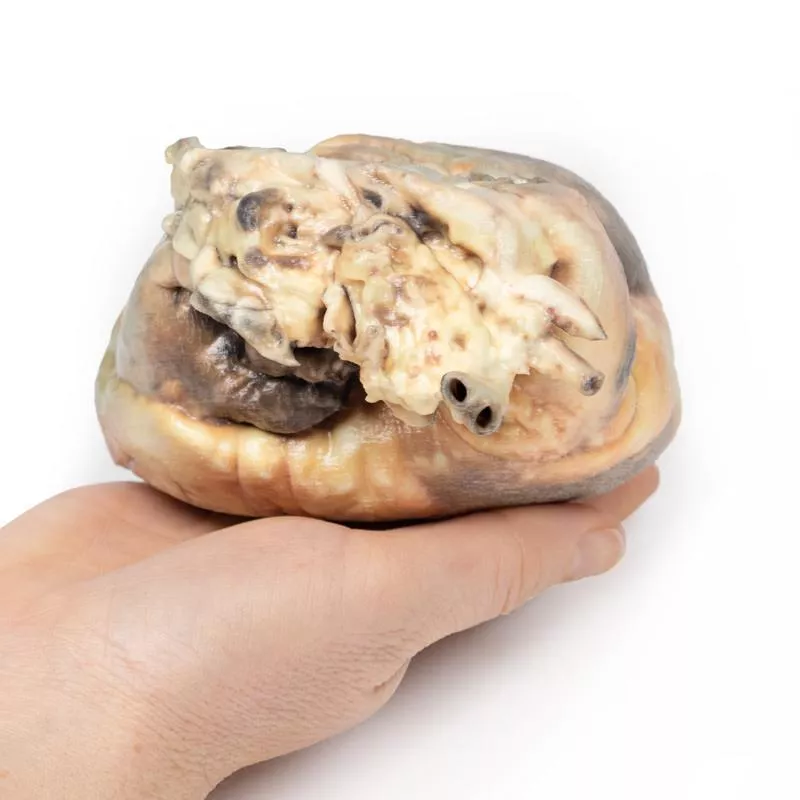
The heart is viewed from the left side. The left atrium has been opened to display a large ovoid defect 3.5 cm in greatest diameter in the inter-atrial septum. Only a small postero-inferior crescentic rim of septum remains. The left ventricle is small, and the right ventricle is hypertrophied (see posterior aspect of specimen where part of the right postero-lateral wall of the right ventricle has been cut away to demonstrate the thickened wall). The pulmonary artery, seen to the left of the atrial cavities, is greatly enlarged. The smaller vessel seen lying above the cut end of the pulmonary artery is the aortic arch. The cut edge of a lumen 8 mm in diameter immediately below the cut end of the pulmonary artery is the left auricular appendage.
Further Information
Atrial septal defect is usually asymptomatic early in life, even when large. Symptoms may not develop until adult life. The onset of symptoms is due to reversal of the initial left-to-right shunt as a result of increasing right ventricular hypertrophy and pulmonary hypertension. The ensuing right-to-left shunt is associated with cyanosis and dyspnoea, and ultimately leads to congestive cardiac failure.
There are several types of atrial septal defects, including:
Secundum - This is the most common type of ASD and occurs in the middle of the wall between the atria (atrial septum). Primum - This defect occurs in the lower part of the atrial septum and might occur with other congenital heart problems.
Sinus venosus - This rare defect usually occurs in the upper part of the atrial septum and is often associated with other congenital heart problems.
Coronary sinus - In this rare defect, part of the wall between the coronary sinus — which is part of the vein system of the heart — and the left atrium is missing.
It is not known why all atrial septal defects occur, but some congenital heart defects appear to be familial and sometimes occur with other genetic problems, such as Trisomy 21 (Down’s syndrome). Some conditions during pregnancy can increase the risk of having a baby with a heart defect, including acute infections such as Rubella infection; drug, tobacco or alcohol use, or exposure to certain substances (such as cocaine) during the first trimester of pregnancy; and underlying systemic conditions, such as diabetes or systemic lupus erythematosus.
A 10-year-old girl with a known congenital heart was admitted for surgical repair because of the recent onset of cyanosis and cardiac failure. On examination, she was breathless with a blood pressure of 105/60mm/Hg and a pulse rate of 140/min. There was a loud heart murmur in the fourth left intercostal space adjacent to the sternum. The jugular venous pressure was elevated, and there were bilateral pulmonary basal crepitations but no peripheral oedema. At operation, the defect was repaired; however, death followed a sudden post-operative deterioration of unknown cause.
Pathology
The heart is viewed from the left side. The left atrium has been opened to display a large ovoid defect 3.5 cm in greatest diameter in the inter-atrial septum. Only a small postero-inferior crescentic rim of septum remains. The left ventricle is small, and the right ventricle is hypertrophied (see posterior aspect of specimen where part of the right postero-lateral wall of the right ventricle has been cut away to demonstrate the thickened wall). The pulmonary artery, seen to the left of the atrial cavities, is greatly enlarged. The smaller vessel seen lying above the cut end of the pulmonary artery is the aortic arch. The cut edge of a lumen 8 mm in diameter immediately below the cut end of the pulmonary artery is the left auricular appendage.
Further Information
Atrial septal defect is usually asymptomatic early in life, even when large. Symptoms may not develop until adult life. The onset of symptoms is due to reversal of the initial left-to-right shunt as a result of increasing right ventricular hypertrophy and pulmonary hypertension. The ensuing right-to-left shunt is associated with cyanosis and dyspnoea, and ultimately leads to congestive cardiac failure.
There are several types of atrial septal defects, including:
Secundum - This is the most common type of ASD and occurs in the middle of the wall between the atria (atrial septum). Primum - This defect occurs in the lower part of the atrial septum and might occur with other congenital heart problems.
Sinus venosus - This rare defect usually occurs in the upper part of the atrial septum and is often associated with other congenital heart problems.
Coronary sinus - In this rare defect, part of the wall between the coronary sinus — which is part of the vein system of the heart — and the left atrium is missing.
It is not known why all atrial septal defects occur, but some congenital heart defects appear to be familial and sometimes occur with other genetic problems, such as Trisomy 21 (Down’s syndrome). Some conditions during pregnancy can increase the risk of having a baby with a heart defect, including acute infections such as Rubella infection; drug, tobacco or alcohol use, or exposure to certain substances (such as cocaine) during the first trimester of pregnancy; and underlying systemic conditions, such as diabetes or systemic lupus erythematosus.
Login
Other customers also bought

Continuous innovation

Social responsibility

Active customer orientation

Understanding quality

Sustainable actions

ISO 9001 certification