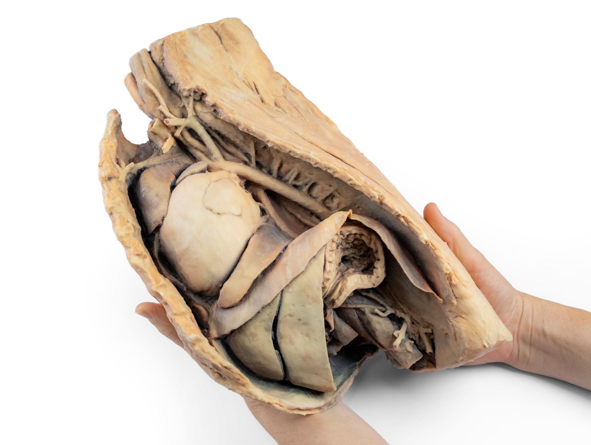
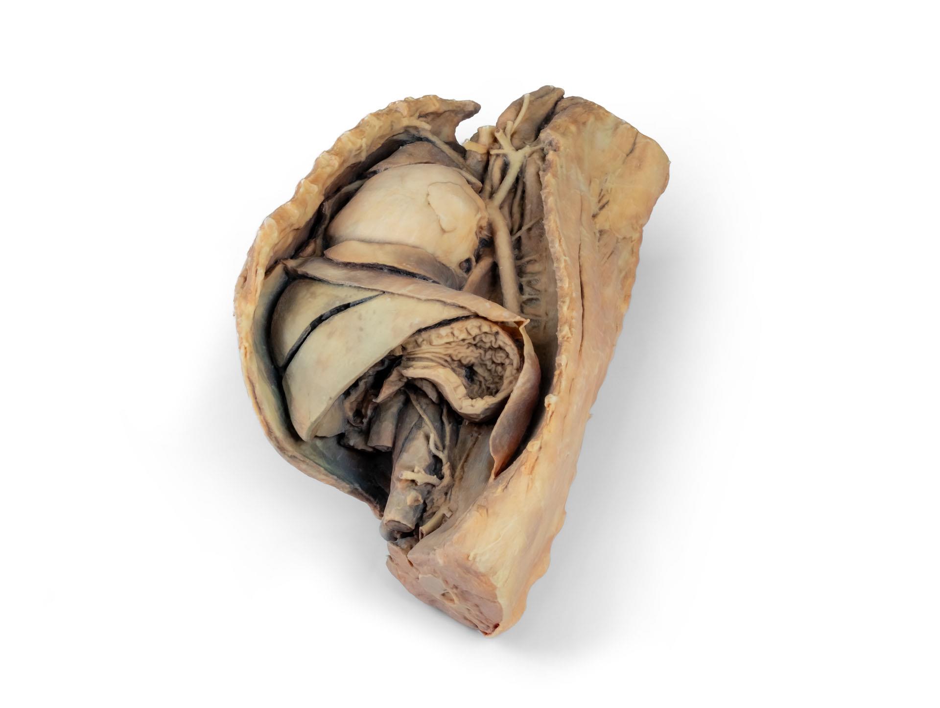
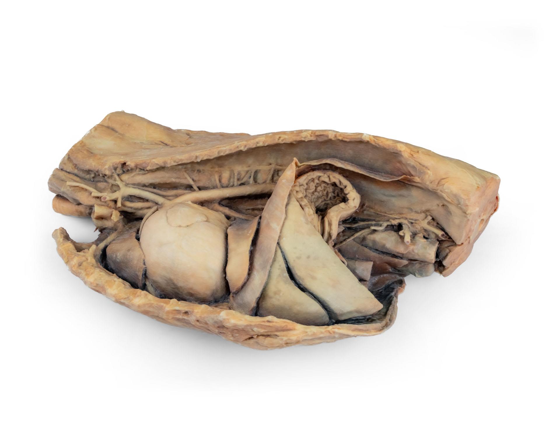
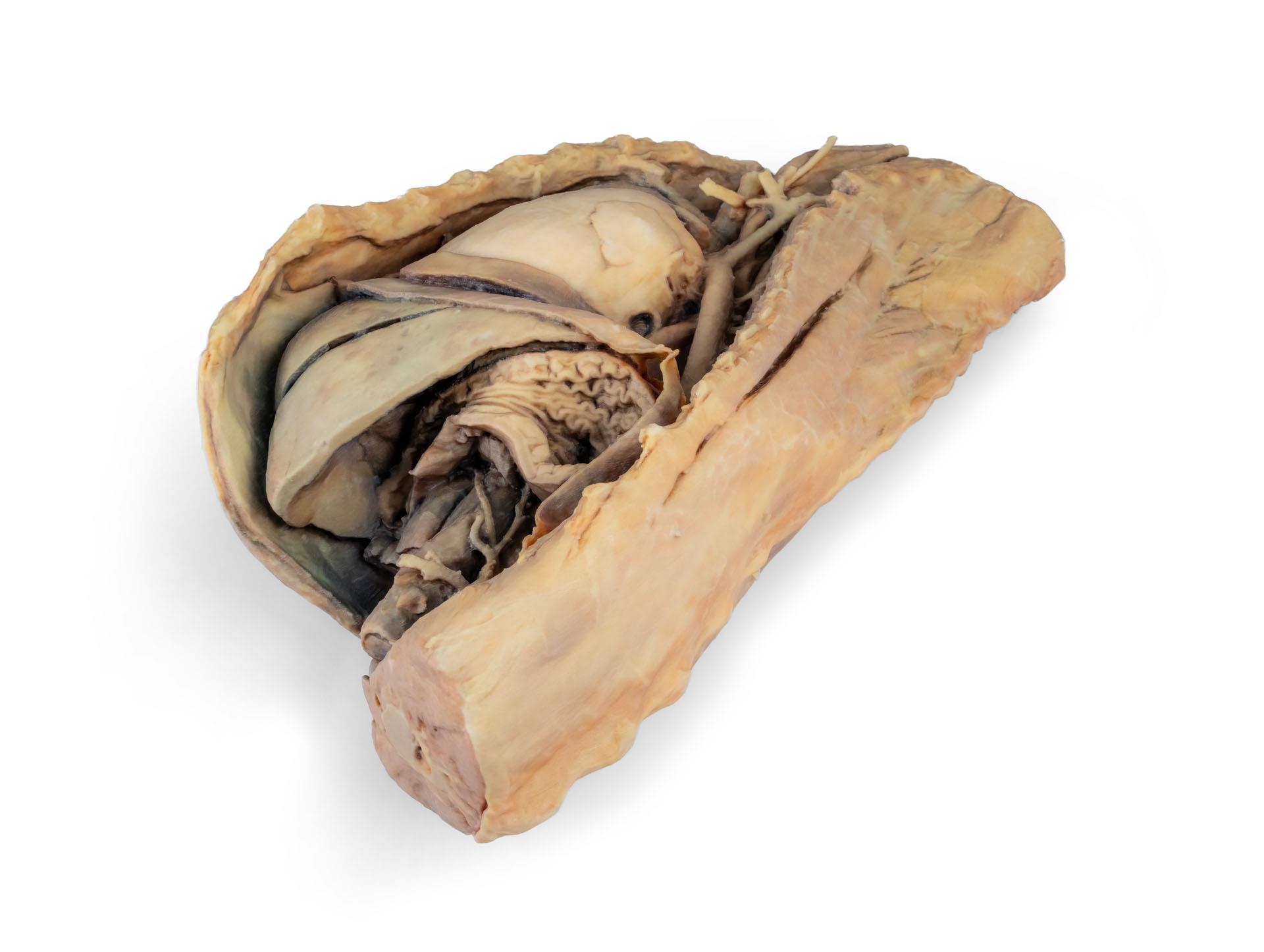
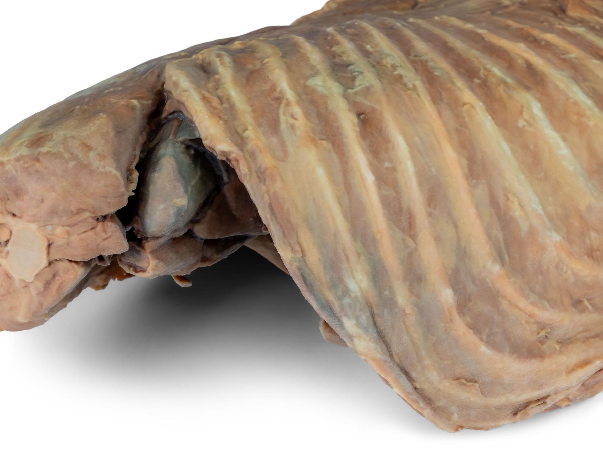
Informations sur le produit "Cavité thoracique canine côté gauche"
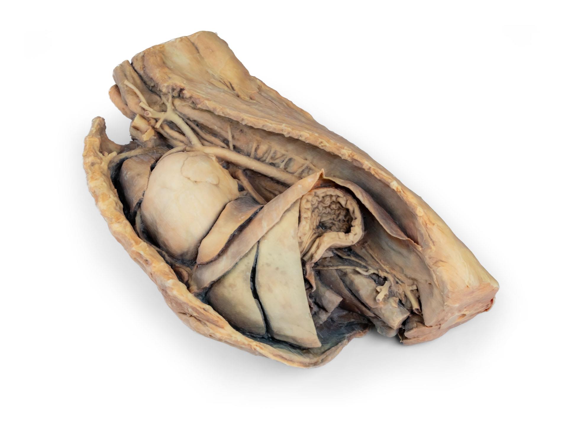
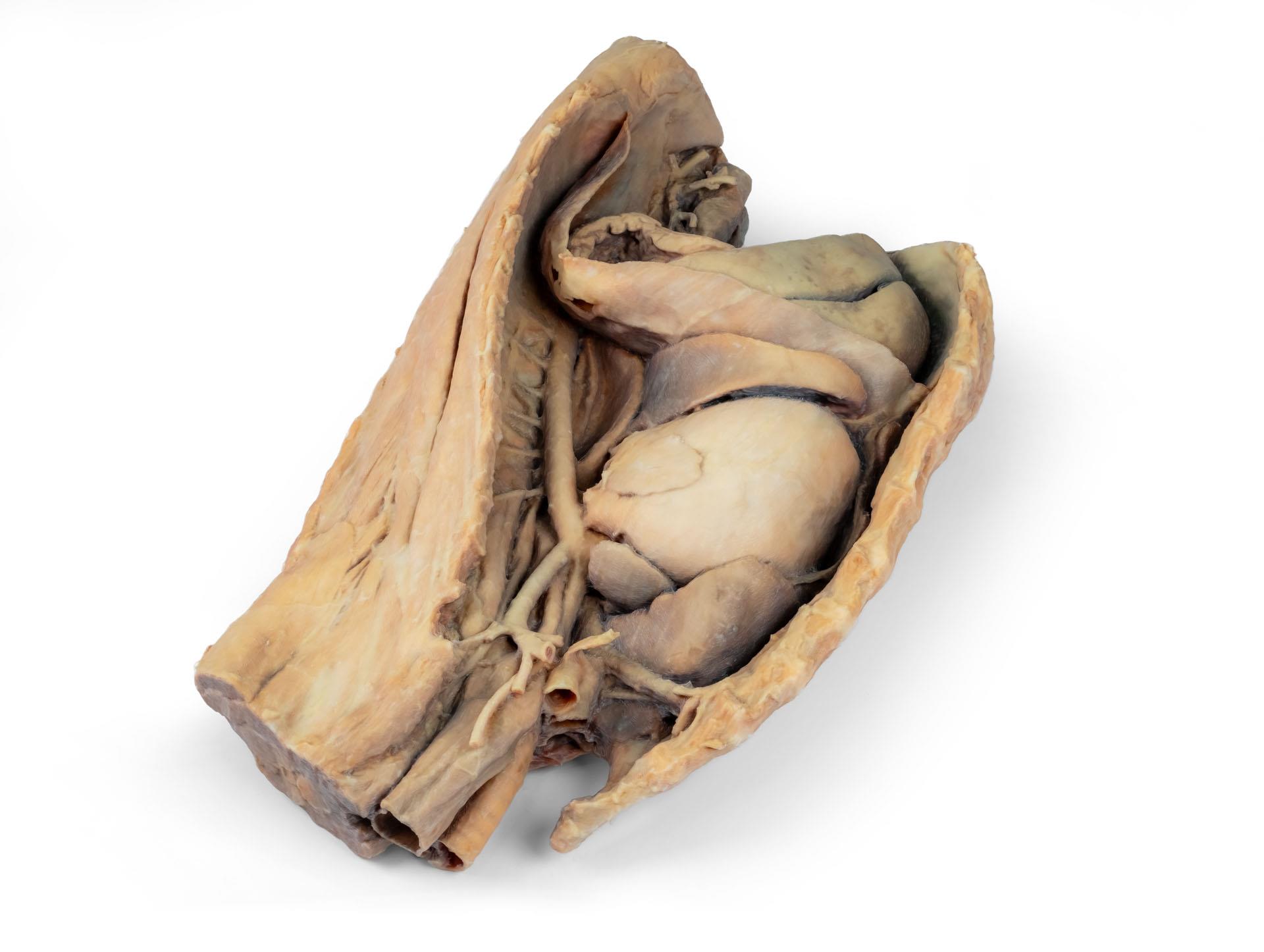
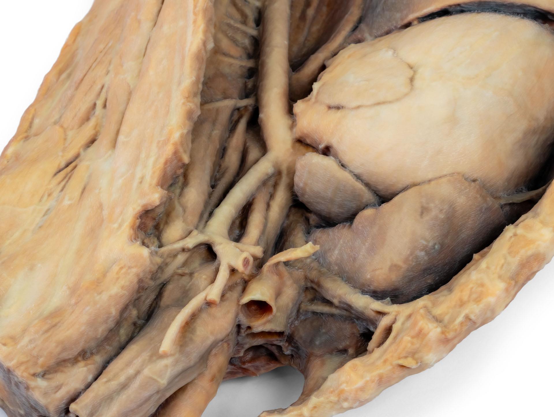
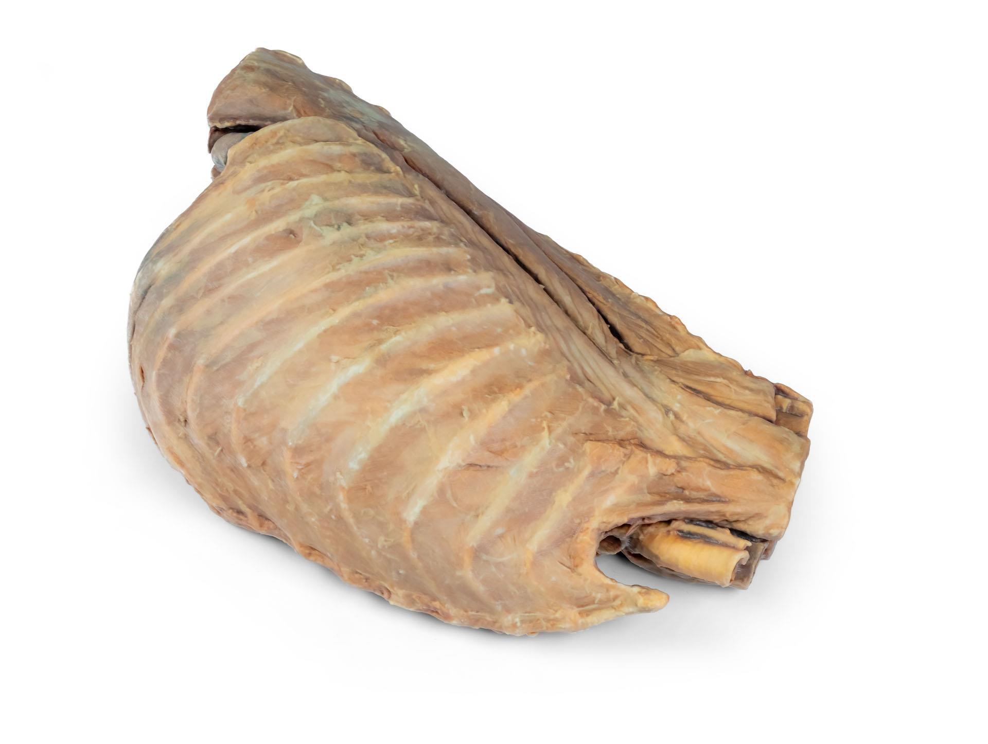
Ce spécimen représente la topographie du cœur après l'ablation du poumon gauche lors d'une dissection de la cavité thoracique et de l'abdomen crânien d'un chien abordé par le côté gauche. Les lobes crâniens et accessoires du poumon droit restent in situ et servent de points de référence anatomiques. Dans le médiastin crânien, le tronc brachiocéphalique et l'artère sous-clavière gauche sont identifiables en tant que branches principales issues de l'arc aortique.

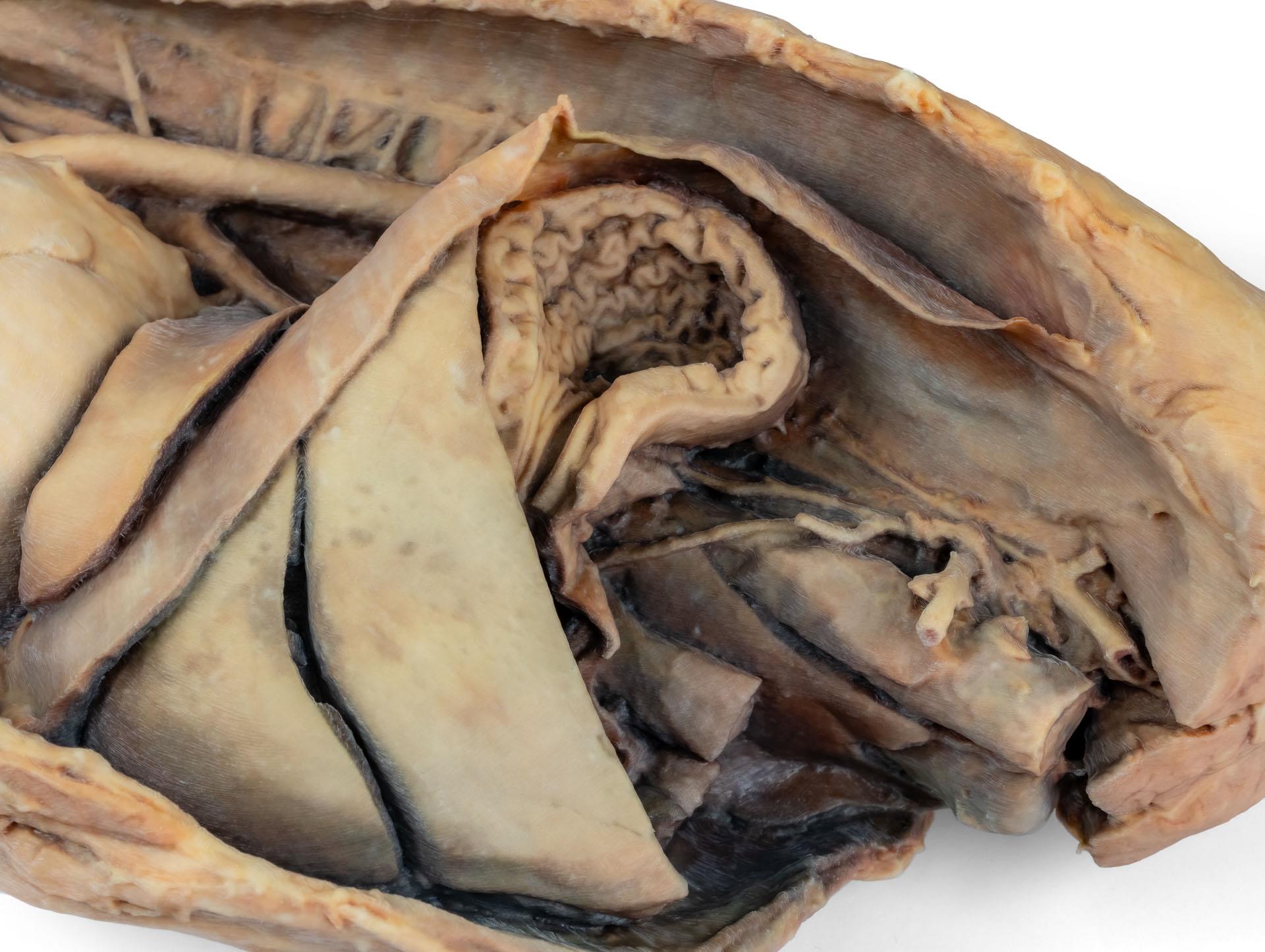
La partie thoracique de l'œsophage traverse le médiastin en direction craniocaudale. Le diaphragme a été préservé pour servir de repère aux organes rétrodiaphragmatiques tels que le foie et l'estomac.Les crêtes diaphragmatiques et leur relation avec le hiatus aortique sont conservées, de même que les branches viscérales initiales de l'aorte abdominale, à savoir le tronc cœliaque et l'artère mésentérique crâniale. Une partie de la paroi auriculaire droite a été enlevée pour exposer la valve auriculo-ventriculaire droite (tricuspide), y compris les muscles chordaetendineae et papillaires.

En outre, la trabécule septomarginale (bande modératrice) est visible dans le ventricule droit, ainsi que la trajectoire du ventricule vers le tronc pulmonaire. La paroi auriculaire gauche est également ouverte pour montrer sa lumière.
Documents
Datenblatt / Factsheet VP9050 Herunterladen

0 sur 0 évaluations

Laissez une évaluation !

Partagez avec d'autres clients votre avis sur le produit.


Erler-Zimmer

Erler-Zimmer GmbH & Co.KG
Hauptstrasse 27
77886 Lauf
Germany
info@erler-zimmer.de


Achtung! Medizinisches Ausbildungsmaterial, kein Spielzeug. Nicht geeignet für Personen unter 14 Jahren.

Attention! Medical training material, not a toy. Not suitable for persons under 14 years of age.


Documents
Datenblatt / Factsheet VP9050 Herunterladen

D'autres clients ont également acheté

Hinterbein des Hundes – Muskeln, Sehnen, Bänder, Gefäße und Nerven, distal zum Kniegelenk
This specimen preserves a superficial dissection of the lower hindlimb musculature from the mid-thigh to the foot, as well as nerves and vessels of the femoral canal and popliteal fossa.The insertions of the muscles of the thigh in the stifle joint and tibial bone are visible. The capsule of the stifle joint has been opened to demonstrate the menisci and the collateralligaments. Extensor and flexor muscles of the tarsus and fingers are exposed. The dissection preserves the topography of the lateral saphenous vein on the foot surface and caudal aspect of the leg Dorsal and palmar anatomical structures of the foot are dissected. Detailed anatomical description on request.

2 145,57 €*
Vorderbein des Hundes – Muskeln, Sehnen, Bänder, Gefäße und Nerven, distal bis zum Ellenbogen
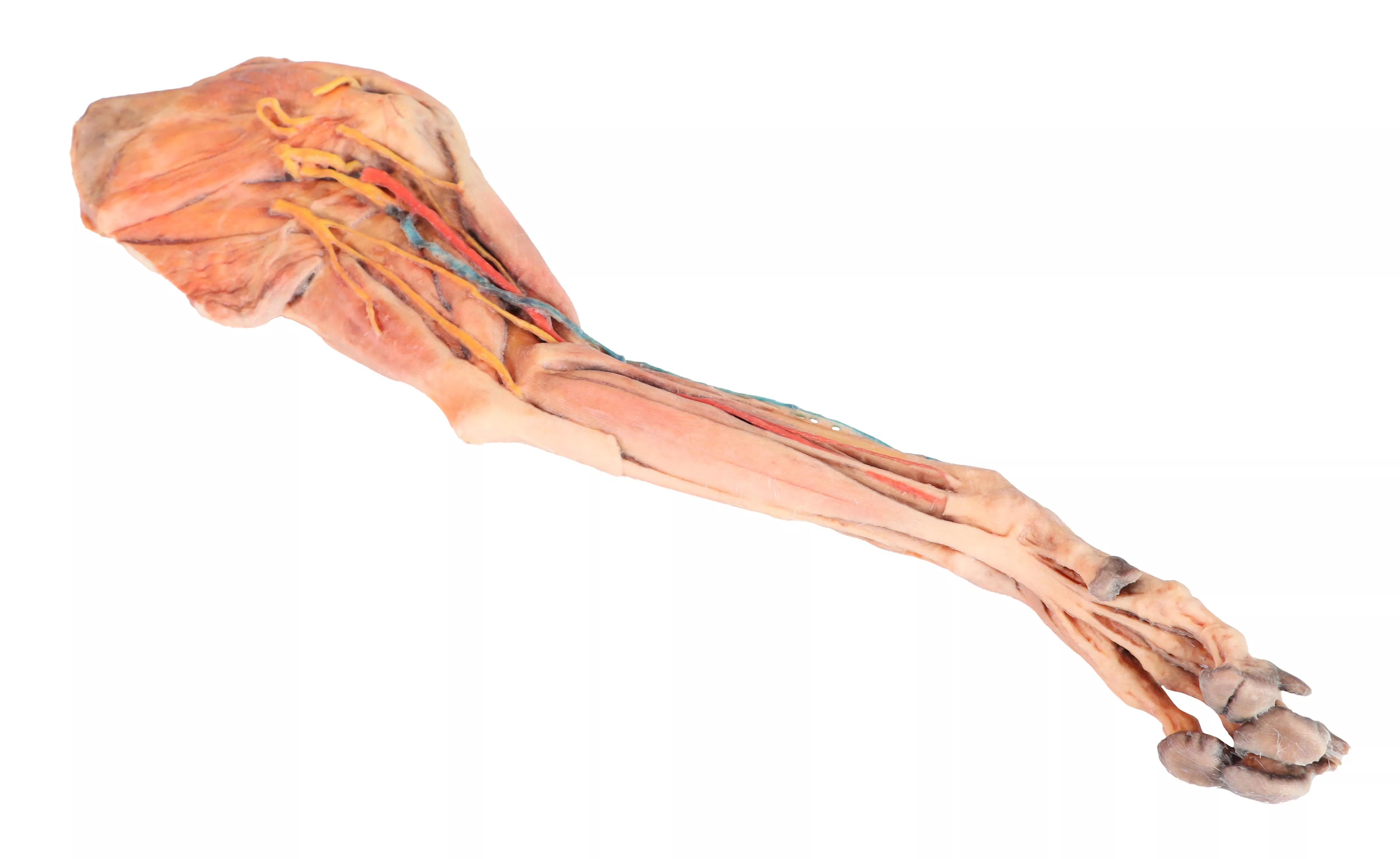
This specimen demonstrates the superficial anatomy of a dog's right thoracic limb from the scapula to the hand. The shoulder flexor and extensor muscles of the scapular region have been preserved, along with the arm flexor and extensor muscles of the elbow. In the forearm and hand are the flexor and extensor muscles of the carpus and fingers. On the medial side of the axillary region the main nerves of the brachial plexus have been preserved. Similarly, the paths of the brachial and median arteries and their respective veins are identified. In a superficial position, the cephalic vein has been preserved together with the antebrachialis lateral cutaneous nerve of the forearm. Detailed anatomical description on request.

1 949,22 €*
Tête de chien - dissections superficielles et profondes
Superficial anatomical structures:Tip of the nose (Apez nasi)Right and left wings of the nose (Alae nasi)Nonglandular skin on the tip of the nose (Planun nasale)Superficial disscetion on the left side:On the left side, the skin has been removed to identify the main anatomical structures,which are described grouped below.Muscles of the facial neuromuscular system:• M. nasolabial levator (M. levator nasolabialis)• M. canine (M. caninus)• M. buccinator (M. buccinator)• M. Zygoma9c (M. zygoma:cus)• M. parodidoauricularisMuscles of the mandibular neuromuscular system (mas9cators):• M. masseter (M. masseter)• M. Temporary (M.temporalis)Nerves:• Facial nerve: (N . facialis)• Dorsal buccal Branch (Rami buccales)• Ventral buccal Branch (Rami buccales)• Bucolabial branches (Rami buccolabiales)• N. Auriculopalpebral (N. Auriculopalpebralis)Vascular:• Facial artery (Arteria facialis)• External jugular vein: (V. Jugularis externa)o Maxillary vein (V. Maxillaris)o Linguofacial vein (V. Linguofacialis)Salivary glands:• Parotid gland and paro9d duct (Glandula parotis) (Ductus parotideus)• Mandibular gland Glandula mandibularisLymph nodes:• N. L. mandibular (Lymphonodi mandubulares)Deep dissection on the right side:On this right side mandible has been removed to see deeper anatomical structures. You can see the surfaces of the temporal bone for the temporomandibular joint (Articulatio Temporomandibularis). The medial pterygoid muscle (M. pterygoideus medialis) is identified and sectioned in its insertion area to the ramus of the mandible (Ramus mandibulae). Next to this muscle, the branches of the mandibular nerve (N. mandibularis) are identified, as well as the maxillary nerve (nevus maxillaris) and the maxillary artery (arteria maxillaris). Near the external acoustic meatus (Meatus acusticus externus), the facial nerve (N . facialis) has been maintained, with one of its branches,the auriculopalpebral nerve (N. Auriculopalpebralis), running parallel to the zygomatic arch (Arcus zygomaticus), visible atier removing the parotd salivary gland (Glandula parotis). The tongue is identified in its entire caudal extension, and the styloglossus (M. stylogossus), genioglossus (M. genioglossus) and genihyoid (M. geniohydeus) muscles reach it. Next to these muscles, the hypoglossal nerve (N. hypoglossus) is identified. In relation to the pharynx (Pharynx), the constrictor muscles of the pharynx (Mm. constrictors phyngis caudalis) are identified. In the most caudal area, in relation to the neck, the course of the common caro9d artery (arteria caarotis communis) and the vagosympathetic trunk nerve (Truncus vagosympathicus) are identified.

4 001,97 €*

Une force d'innovation constante

Responsabilité sociale

Clients fidèles - Orientation

Compréhension de la qualité

Action durable

Certification ISO 9001

Vos derniers produits consultés