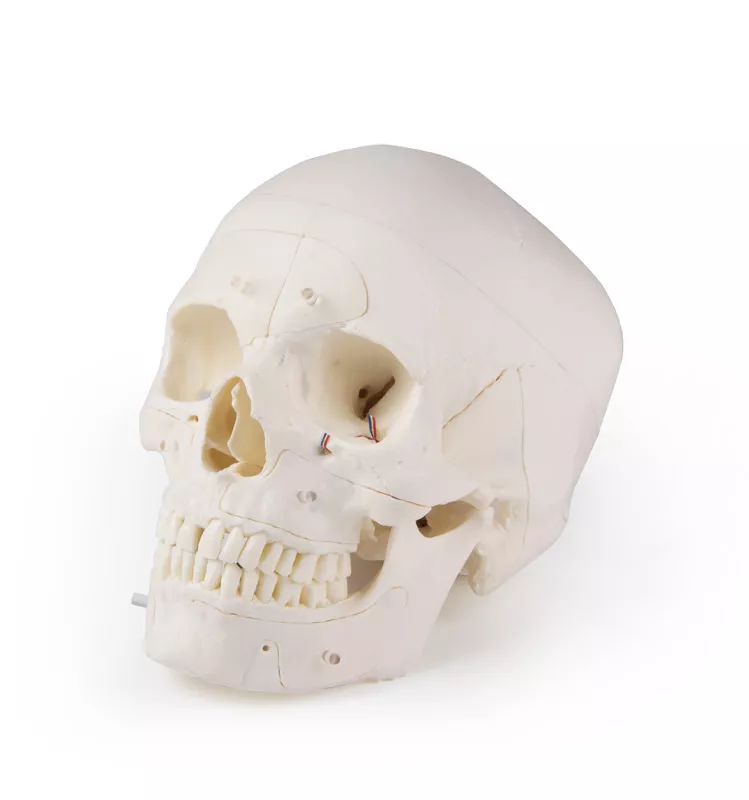
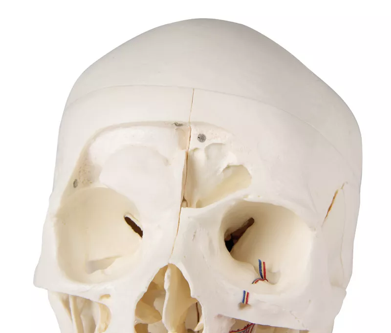
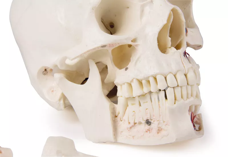
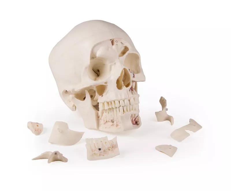
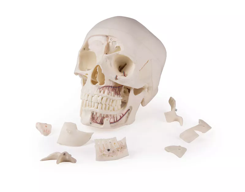
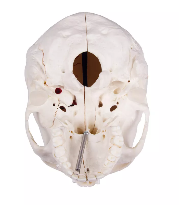
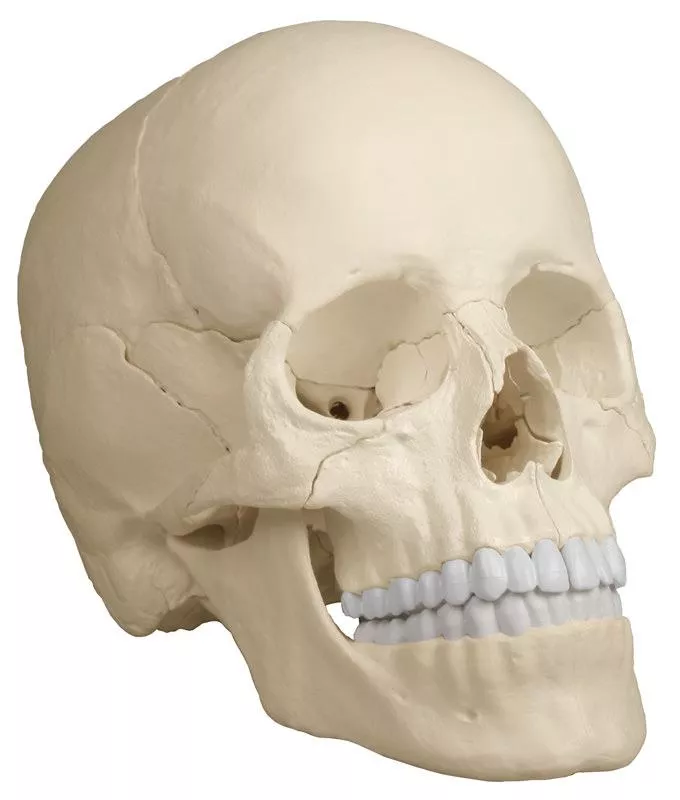
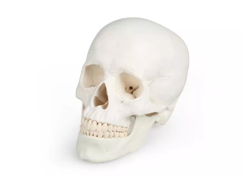
Crâne de démonstration de luxe ; 14 pièces ; pour études avancées
733,04 €*
Disponible, sous 1-3 jours ouvrables
Numéro d'article: 4800
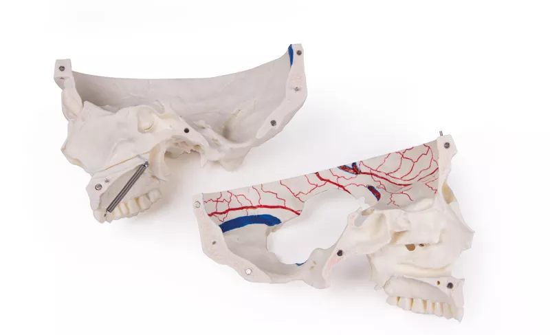
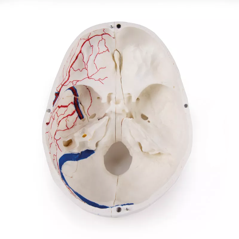
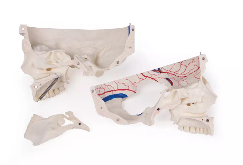
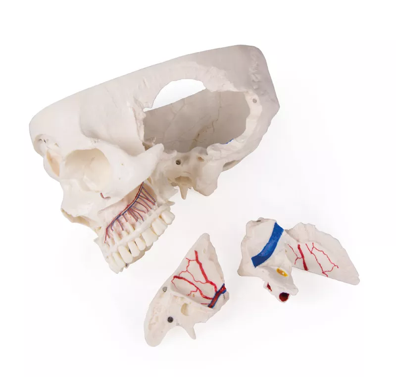
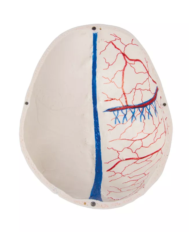
The calvarium is sectioned horizontally leaving the temporal bone and its sutures intact. Bony impressions of the superior sagittal sinus, transverse sinus and sigmoid sinus as well as the meningeal vessels have been painted. The base portion of the skull has been sagittaly sectioned in the way that it passes through the one cribriform plate on one side and another section in the same plane passes through the other cribriform plate of the ethmoid leaving the crista galli perpendicular plate of the ethmoid intact as also the whole nasal septum. The structures of anterior, middle and posterior cranial fossae are easily accessible. One can directly visualize the nasal cavity, the concha, the nasal septum, the bony pharyngeal and naso-pharyngeal spaces. The nasal septum is separable from surrounding bones. The frontal sinuses have been dissected on one side to show the sinus as whole and on the other side chiselled out for full access to the sinus. The relation of this sinus to the nasal cavity is clearly shown and is especially valuable for otolaryngologists.
On one side of the skull the temporal bone has been left in situ. The other temporal bone is removable from the skull. A portion of the mastoid and squama can be removed along with the tympanic antrum, baring internal ear in full view. All three semicircular canals are visible along with the course of the facial nerve coursing backwards and then downwards emerging finally through the stylo-mastoid foramen. The removable temporal bone has the external auditory meatus intact. An almost vertical section through the squama mastoid process and carried inwards along the petro-squamosal junction has been made and when apart, one sees the position of tympanic membrane. The carotid canal has been opened as also the cochlea , showing the internal canal, and the course of the facial nerve has been depicted. Oval window, the semi-circular canals, and aditus of the tympanic antrum are visible.
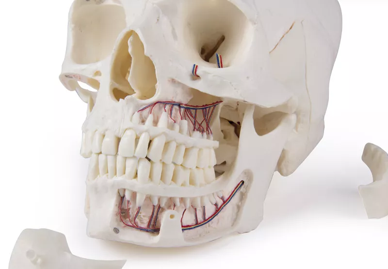
The maxilla and mandible expose the structures of dentition, the roots, the bony margin of the alveolar process, dental vessels and nerves are visible. The maxillary sinus can by opened by removing a bone flap. Teeth of the right mandible are sectioned to show the inner tooth structure.
Connexion
October 4, 2021 08:39
einfaches zusammenbauen
Ich bin positiv von der Qualität des Schädels überrascht! Das zusammenbauen ist Dank der Magnete ein Kinderspiel.
November 20, 2021 11:53
awesome details / dental skull
great teaching tool for my dental studies :)The skull has very accurate anatomical structures and is a relief when learning. Through the openings in the jaw, all the important nerve tracts can be shown.
February 5, 2021 13:50
sehr schöne Strukturen, Schädel hält gut zusammen
sehr schöne Strukturen, Schädel hält gut zusammen
October 4, 2021 09:45
great structures
very detailed structures are visible, especially in the dental area.
Erler-Zimmer GmbH & Co.KG
Hauptstrasse 27
77886 Lauf
Germany
info@erler-zimmer.de
Achtung! Medizinisches Ausbildungsmaterial, kein Spielzeug. Nicht geeignet für Personen unter 14 Jahren.
Attention! Medical training material, not a toy. Not suitable for persons under 14 years of age.


