Extrémités inférieures
22
infinitescrolling.productCount.produkte
Female right pelvis superficial and deep structures
This 3D printed female right pelvis preserves both superficial and deep structures of the true and false pelves, as well as the inguinal ligament, the obturator membrane and canal, and both the greater and lesser sciatic foramina. Somewhat unique is the removal of portions of the peritoneum (a grayish colour) to create ‘windows’ displaying extraperitoneal structures. Detailed anatomical description on request.
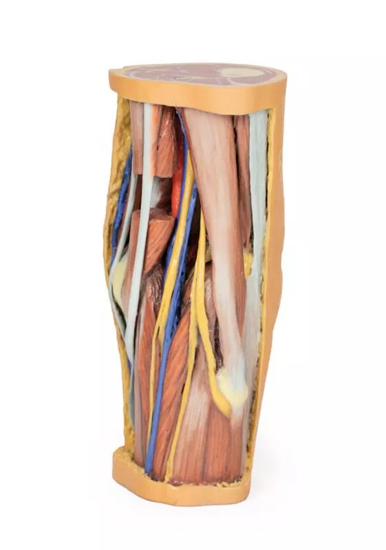
Popliteal Fossa
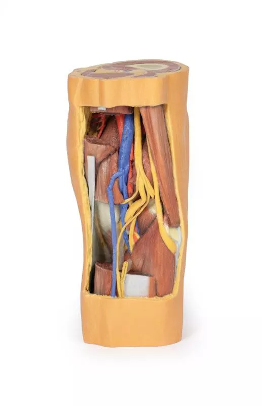
This 3D printed specimen preserves the distal thigh and proximal leg, dissected posteriorly to demonstrate the contents of the popliteal fossa and surrounding region. The proximal cross-section demonstrates the anterior, posterior and medial compartment muscles, with the femoral artery and vein visible within the adductor canal. The sciatic nerve and great saphenous vein are also visible. Detailed anatomical description on request.
Female left pelvis and proximal thigh
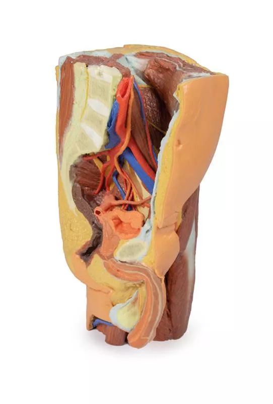
This 3D printed female left pelvis and proximal thigh preserves both superficial and deep structures of the true and false pelves, inguinal region, femoral triangle, and gluteal region. The specimen has been sectioned transversely through the fourth lumbar vertebra, displaying thecross-section of the musculature (epaxial musculature, psoas and quadratus lumborum muscles) and cauda equina within the vertebral canal. The ventral and dorsal roots of the cauda equina are also visible exiting the intervertebral and sacral foramina in the sagittal section. Detailed anatomical description on request.
Female right pelvis
This 3D printed specimen represents a female right pelvis, sectioned along the midsagittal plane and transversely across the level of the L4 vertebrae and the proximal thigh. The specimen has been dissected to demonstrate the deep structures of the true and false pelves, the inferior anterior abdominal wall and inguinal region, femoral triangle and gluteal region. Detailed anatomical description on request.
Foot - Plantar surface & superficial dissection on the dorsum
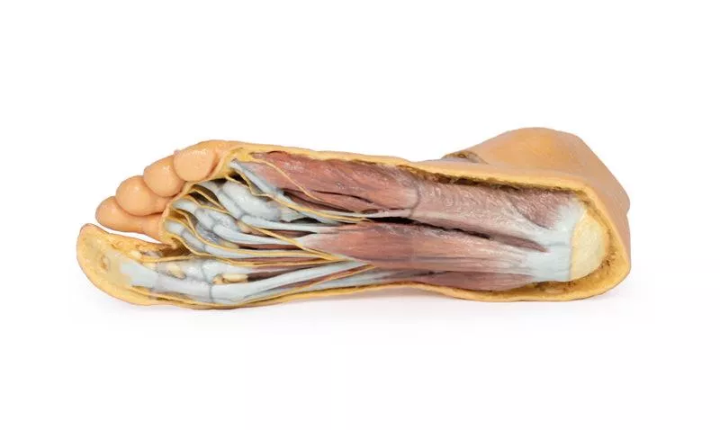
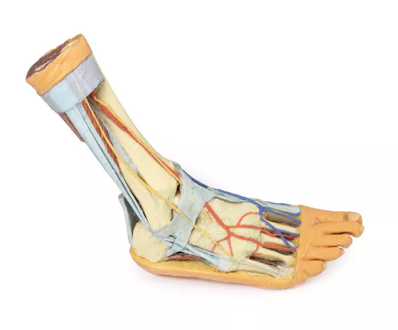
This 3D printed specimen is a left foot with superficial structures exposed on the dorsum, and the superficial layer of muscles and nerves on the plantar surface. The anterior portion of the plantar aponeurosis has largely been removed to expose the first layer of muscles. Detailed anatomical description on request.
Male left pelvis and proximal thigh
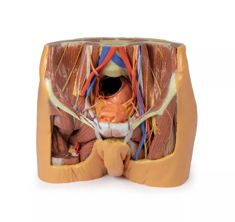
This 3D printed male left pelvis and proximal thigh (sectioned through the midsagittal plane in the midline and transversely through the L3/4 intervertebral disc) shows superficial and deep structures of the true and false pelves, inguinal and femoral region. In the transverse section, the epaxial musculature, abdominal wall musculature (rectus abdominis, external and internal abdominal obliques, transversus abdominis), psoas major and quadratus lumborum are visible and separated from each other and the superficial fat by fascial layers such as the rectus sheath and the thoracolumbar fascia. The psoas major muscle lies lateral to the external iliac artery, with the left testicular artery and vein lying on its superficial surface. More laterally (and moving inferiorly), the ilioinguinal nerve, the lateral cutaneous nerve of the thigh and the femoral nerve are positioned over the superficial surface of the iliacus muscle. Detailed anatomical description on request.
Foot - Structures of the plantar surface
This 3D print records the anatomy of a right distal leg and the deep structures of the plantar surface of the foot. Proximally, the tibia, fibula, interosseous membrane, and leg muscles are discernable in cross-section. Medially, at the level of the ankle joint, the long tendons of the dorsi- and plantar-flexors are visible superficial to the capsular and extra capsular ligaments. Detailed anatomical description on request.
Foot - Superficial and deep dissection of distal leg and foot
This 3D printed specimens preserves a mixed superficial and deep dissection of a left distal leg and foot. Posteriorly, the compartment muscles and neurovascular structures have been removed to isolate the tendocalcaneous and expose the body of the calcaneus. Detailed anatomical description on request.
Foot - Deep plantar structures
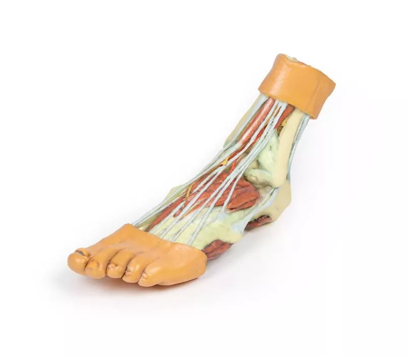
This 3D printed specimen provides a view of deep plantar structures of a right foot. Medially, the cut edge of the great saphenous vein is visible within the superficial fascia, just anterior to the cut edges of the medial and lateral plantar arteries and nerves overlying the insertion of the tibialis posterior muscle. The superficial fascias, the plantar aponeurosis, and superficial musculature have been removed to expose the deep (third layer) of musculature. Detailed anatomical description on request.
Lower limb – superficial dissection with male left pelvis
This 3D printed specimen combines the Lower limb – superficial dissection (Ref.no. MP1816) with the male left pelvis (Ref.no. MP1765). Detailed anatomical description on request.
Popliteal Fossa distal thigh and proximal leg
This 3D printed specimen preserves the distal thigh and proximal leg, dissected posteriorly to demonstrate the contents of the popliteal fossa and surrounding region. Detailed anatomical description on request.
Male Pelvis
This multipart 3D printed specimen represents the inferior portions of our larger posterior abdominal wall print (MP1300) that displays the inferior posterior abdominal wall, the pelvic cavity and the proximal thigh (including the gluteal regions and femoral triangles).Lower posterior abdominal wall and false pelvis: The specimen is transected at approximately the level of the L2/L3 intervertebral disc. The common iliac veins unite to form the inferior vena cava. The common iliac arteries are close to uniting at the top of the print. The iliacus and psoas musclesare easy to identify, the latter has a prominent psoas minor tendon. They can be seen to unite as they pass under the inguinal ligament. The nerves of the iliac fossa (from superior to inferior: ilioinguinal nerve, lateral cutaneous nerve of thigh, femoral nerve ) and their course is clearly visible, as is the genitofemoral nerves on the surface of psoas muscle. The ureters also descend on the superficial surface of the psoas and cross from its lateral to its medial border. They enter the pelvis at the bifurcation of the common iliac arteries into external and internal arteries. The external iliac arteries and veins running along the pelvic brim are clearly visible, as is the vas deferens crossing the brim from the deep inguinal ring to enter the pelvis. Detailed anatomical description on request.
Lower Limb - deep dissection of a left pelvis and thigh
This 3D printed specimen presents a deep dissection of a left pelvis and thigh to show the course of the femoral artery and sciatic nerve from their proximal origins to the midshaft of the femur. Proximally, the pelvis has been sectioned along the mid-sagittal plane and the pelvic viscera are removed. In the pelvis the coccygeus muscle spans between the sacrum and iliac spine and the obturator artery and nerve entering the obturator canal superior to the obturator membrane. Detailed anatomical description on request.
Lower limb – superficial dissection
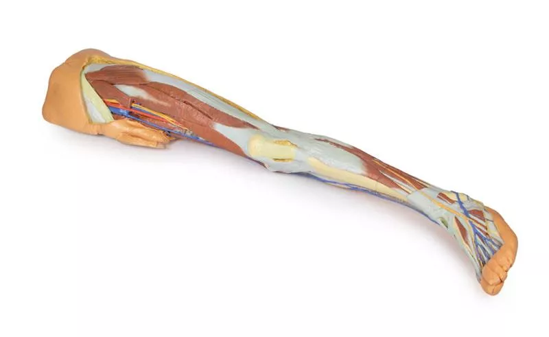
This 3D printed specimen represents the remainder of the lower limb portions of our male abdominopelvic and proximal thigh specimen (MP1765), sectioned proximally near midthigh and continuous to the partially dissected foot. The transverse section through the thigh exposes the neurovascular structures of the anterior, medial and posterior compartments. Detailed anatomical description on request.
Knee Joint, flexed
This 3D printed specimen demonstrates the ligaments of the knee joint with the leg in flexion. In the anterior view, with the patella and part of the patellar ligament removed, the medial and lateral menisci and anterior and posterior cruciate ligaments are visible. Detailed anatomical description on request.
Flexed knee joint deep dissection
This 3D printed specimen displays a deep dissection of a left knee joint with the internal joint capsule structures relative to superficial tissues in a flexed position. Detailed anatomical description on request.
Lower Limb - deep dissection
This 3D printed specimen consists of a right partial lower limb sectioned just proximal to the knee joint and complete through a partially dissectedfoot exposing the structures on the dorsum. Detailed anatomical description on request.
Lower Limb Musculature
This 3D printed specimen preserves a superficial dissection of the lower limb musculature from the mid-thigh to mid-leg, as well as nerves and vessels of the popliteal fossa. The insertions of the muscles of the anterior, middle and posterior compartments of the thigh are visible, including the pes anserinus medially and the iliotibial tract laterally. The capsule of the knee joint has been opened anteriorly to demonstrate the menisci and the tibial and fibular collateral ligaments. Detailed anatomical description on request. Detailed anatomical description on request.
Foot - Parasagittal cross-section
This 3D printed specimen provides a parasagittal cross-section through the medial aspect of the right distal tibia and foot, displaying the skeletal structures of the medial longitudinal arch of the foot and surrounding soft-tissue structures. Detailed anatomical description on request.
Knee Joint, extended
This 3D printed specimen demonstrates the ligaments of the knee joint with the leg in extension; it represents the same specimen as MP1800 knee joint printed in a flexed position. Both tibial and fibular collateral ligaments are intact. Detailed anatomical description on request.
Lower Limb superficial veins
This 3D printed specimen presents a superficial dissection of a left lower limb, from just proximal to the knee joint to a complete foot. The skin and superficial fascia have been removed to display the superficial venous structures of the leg including the dorsal venous plexus, great saphenous vein (including numerous tributaries), and the small saphenous vein (including numerous tributaries) on the crural fascia. Detailed anatomical description on request.
Foot - Superficial and deep structures of the distal leg and foot
This 3D printed specimen presents both superficial and deep structures of a right distal leg and foot. Proximally, the posterior compartment of the leg has been dissected to remove the triceps surae muscles and tendocalcaneous to demonstrate the deep muscles of the compartment (tibialis posterior, flexor digitorum longus, flexor hallucis longus). Detailed anatomical description on request.
Des répliques de corps humains pour améliorer l'enseignement !
La série révolutionnaire d'anatomie d'Erler- Zimmer comprend une collection unique et inégalée de répliques de corps humains colorées, spécialement conçues pour améliorer l'enseignement et l'apprentissage. Cette collection haut de gamme d'anatomie humaine extrêmement précise a été créée directement à partir de données radiologiques ou de préparations réelles à l'aide des dernières techniques d'imagerie. La série d'anatomie humaine 3D offre un moyen rentable de répondre à vos besoins spécifiques en matière d'enseignement et de démonstration dans l'ensemble du programme d'études de médecine, de sciences de la santé et de biologie. Une description détaillée de l'anatomie représentée dans chaque préparation imprimée en 3D est fournie. Quels sont les avantages de la série Anatomie 3D de Monash par rapport aux modèles en plastique ou aux véritables plastinats humains ?
Chaque réplique de corps a été soigneusement développée à partir de données radiologiques de patients ou de corps humains préparés de la plus haute qualité, sélectionnés par une équipe d'anatomistes hautement qualifiés au Centre d'enseignement de l'anatomie humaine de l'Université Monash, afin de représenter des zones cliniquement importantes de l'anatomie avec une qualité et un niveau de détail impossibles à obtenir avec des modèles conventionnels - il s'agit d'une véritable anatomie, pas d'une stylisation. Chaque réplique corporelle a été rigoureusement contrôlée par l'équipe d'anatomistes hautement qualifiés du Centre d'enseignement de l'anatomie humaine de l'Université de Monash, afin de garantir l'exactitude anatomique du produit final. Les répliques de corps ne sont pas de véritables tissus humains et ne sont donc soumises à aucune restriction en matière de transport, d'importation ou d'utilisation dans les établissements d'enseignement qui ne sont pas autorisés à utiliser des cadavres. Le site
série exclusive Anatomie 3D évite ces problèmes et d'autres problèmes éthiques qui surviennent lorsqu'on manipule des restes humains plastinés.


