


")


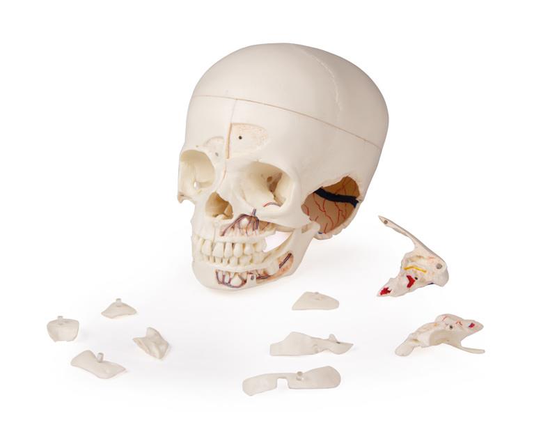

Démonstration de luxe Crâne d‘enfants; 14 pièces; pour les études avancées
produktDetailSeite.product.mehr-erfahren675,92 €*
Disponible, sous 1-3 jours ouvrables
Réf. produit :
4810
produktDetailSeite.product.artikelnummer 4810
Informations sur le produit "Démonstration de luxe Crâne d‘enfants; 14 pièces; pour les études avancées"
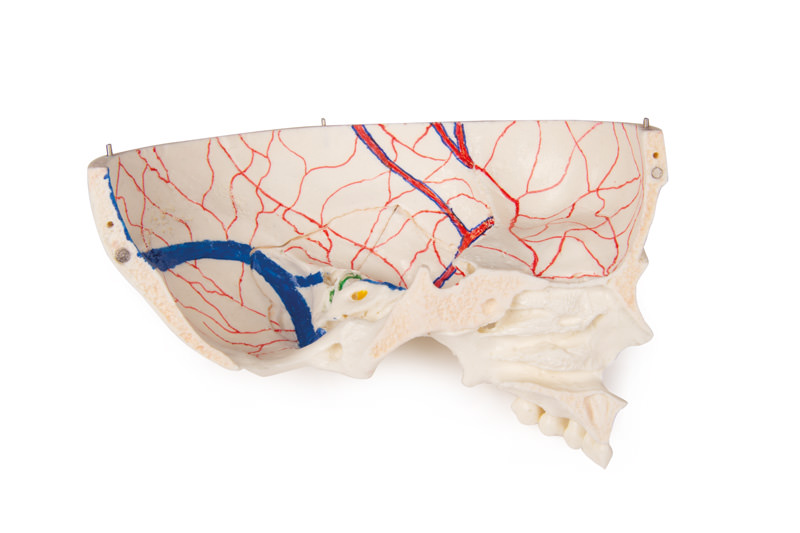
Ce modèle de crâne est un moulage d‘un véritable spécimen de crâne d‘enfant humain et montre toutes les structures anatomiques en détail. Il a été développé pour les étudiants en anatomie, médecine, chirurgie, médecine ORL, ophtalmologie et dentisterie. Le crâne est découpé et assemblé de manière complexe avec des connexions métalliques et magnétiques.
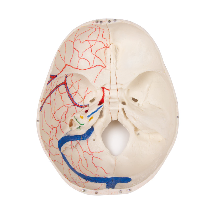
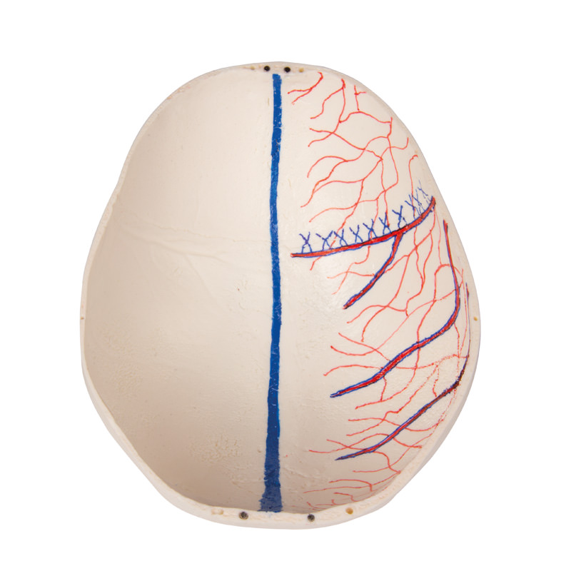
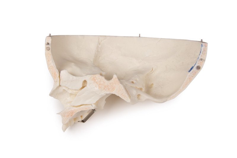
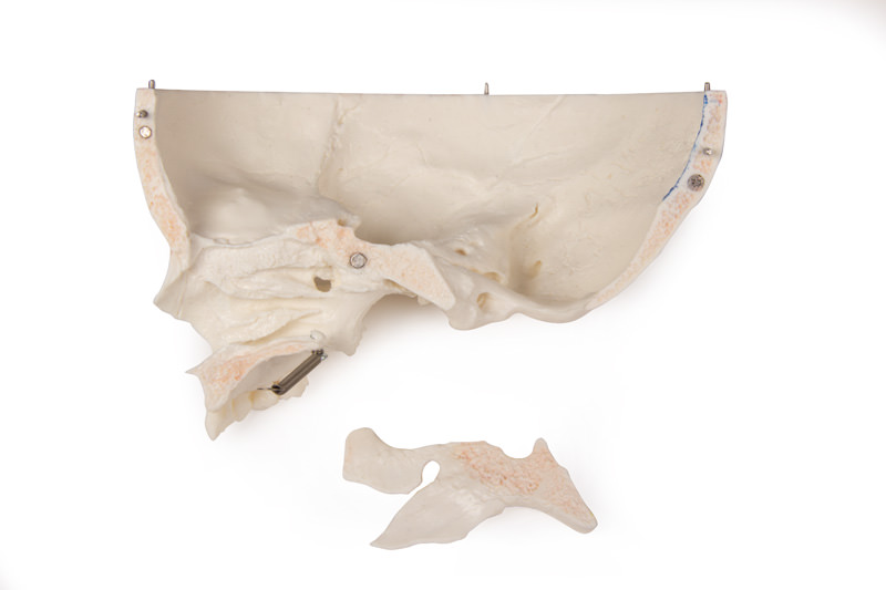
Le haut du crâne est ouvert et amovible. Les empreintes osseuses du sinus sagittal, du sinus transversus et du sinus sigmoïde ainsi que des méninges sont peintes. La base du crâne est coupée sagittalement de telle manière que la coupe traverse une plaque de tamis d‘un côté et une autre coupée avec le même plan à travers l‘autre plaque de tamis de l‘os ethmoïde, qui laisse le Christa galli et la plaque perpendiculaire comme ainsi que le septum nasal entier intact. Les structures de la fosse antérieure, moyenne et postérieure sont facilement accessibles. On peut voir directement la cavité nasale, le cornet, le septum et l‘os et le nasopharynx. Le septum nasal peut être retiré des structures osseuses environnantes. L‘os temporal amovible présente un conduit auditif externe complet.
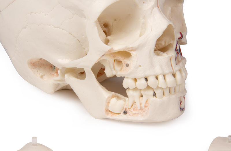
La mâchoire supérieure et la mâchoire inférieure montrent les structures des dents, des racines, du bord osseux du processus alvéolaire ainsi que des nerfs et des vaisseaux dentaires. Le sinus maxillaire peut être ouvert en retirant un lambeau. Les dents, y compris les dents permanentes encore incrustées dans l‘os, peuvent être rendues accessibles en retirant un lambeau osseux.
âge estimé 3 ans.
Le haut du crâne est ouvert et amovible. Les empreintes osseuses du sinus sagittal, du sinus transversus et du sinus sigmoïde ainsi que des méninges sont peintes. La base du crâne est coupée sagittalement de telle manière que la coupe traverse une plaque de tamis d‘un côté et une autre coupée avec le même plan à travers l‘autre plaque de tamis de l‘os ethmoïde, qui laisse le Christa galli et la plaque perpendiculaire comme ainsi que le septum nasal entier intact. Les structures de la fosse antérieure, moyenne et postérieure sont facilement accessibles. On peut voir directement la cavité nasale, le cornet, le septum et l‘os et le nasopharynx. Le septum nasal peut être retiré des structures osseuses environnantes. L‘os temporal amovible présente un conduit auditif externe complet.
La mâchoire supérieure et la mâchoire inférieure montrent les structures des dents, des racines, du bord osseux du processus alvéolaire ainsi que des nerfs et des vaisseaux dentaires. Le sinus maxillaire peut être ouvert en retirant un lambeau. Les dents, y compris les dents permanentes encore incrustées dans l‘os, peuvent être rendues accessibles en retirant un lambeau osseux.
âge estimé 3 ans.
Connexion
D'autres clients ont également acheté

Crâne de démonstration de luxe ; 14 pièces ; pour études avancées
675,92 €*
This skull model is an actual cast of a real human specimen and shows all anatomical structures in highest detail. It is made for students in anatomy, medicine, surgery, otolaryngology, ophthalmology and dentistry. The Skull is intricately sectioned and reassembled with metal and magnet connections. The calvarium is sectioned horizontally leaving the temporal bone and its sutures intact. Bony impressions of the superior sagittal sinus, transverse sinus and sigmoid sinus as well as the meningeal vessels have been painted. The base portion of the skull has been sagittaly sectioned in the way that it passes through the one cribriform plate on one side and another section in the same plane passes through the other cribriform plate of the ethmoid leaving the crista galli perpendicular plate of the ethmoid intact as also the whole nasal septum. The structures of anterior, middle and posterior cranial fossae are easily accessible. One can directly visualize the nasal cavity, the concha, the nasal septum, the bony pharyngeal and naso-pharyngeal spaces. The nasal septum is separable from surrounding bones. The frontal sinuses have been dissected on one side to show the sinus as whole and on the other side chiselled out for full access to the sinus. The relation of this sinus to the nasal cavity is clearly shown and is especially valuable for otolaryngologists. On one side of the skull the temporal bone has been left in situ. The other temporal bone is removable from the skull. A portion of the mastoid and squama can be removed along with the tympanic antrum, baring internal ear in full view. All three semicircular canals are visible along with the course of the facial nerve coursing backwards and then downwards emerging finally through the stylo-mastoid foramen. The removable temporal bone has the external auditory meatus intact. An almost vertical section through the squama mastoid process and carried inwards along the petro-squamosal junction has been made and when apart, one sees the position of tympanic membrane. The carotid canal has been opened as also the cochlea , showing the internal canal, and the course of the facial nerve has been depicted. Oval window, the semi-circular canals, and aditus of the tympanic antrum are visible. The maxilla and mandible expose the structures of dentition, the roots, the bony margin of the alveolar process, dental vessels and nerves are visible. The maxillary sinus can by opened by removing a bone flap. Teeth of the right mandible are sectioned to show the inner tooth structure.

Une force d'innovation constante

Responsabilité sociale

Clients fidèles - Orientation

Compréhension de la qualité

Action durable

Certification ISO 9001