


")


Informations sur le produit "Intracerebral Haemorrhage"
Histoire clinique
Un homme de 80 ans a perdu connaissance soudainement. L’examen a révélé une parésie du regard droit, une hémiplégie gauche et une hémiparésie droite.
Pathologie
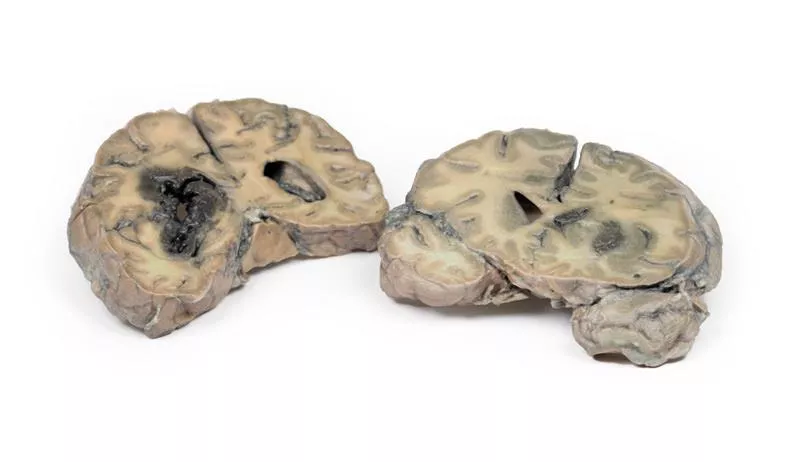
Les coupes coronales au niveau des corps mamillaires, incluant le tronc cérébral et les lobes temporaux antérieurs, illustrent un énorme caillot remplaçant le tissu des ganglions de la base gauche et de la capsule interne. L’hémorragie a rompu dans le ventricule latéral gauche et sa corne temporale, détruisant ses parois et infiltrant le tissu cérébral adjacent. Le ventricule latéral droit est aussi rempli de sang mais conserve des parois intactes. Le caillot agit comme lésion expansive, élargissant l’hémisphère gauche et décalant les structures médianes vers la droite. Une hernie sous-falcine du gyrus cingulaire gauche sous la faux du cerveau est observée.
Informations complémentaires
Les hémorragies intracérébrales de ce type sont généralement causées par hypertension artérielle systémique. L’hémorragie est due à la rupture d’un micro-anévrisme d’une branche des artères striées alimentant les ganglions de la base.
Un homme de 80 ans a perdu connaissance soudainement. L’examen a révélé une parésie du regard droit, une hémiplégie gauche et une hémiparésie droite.
Pathologie
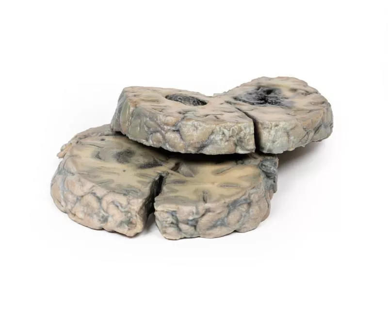
Les coupes coronales au niveau des corps mamillaires, incluant le tronc cérébral et les lobes temporaux antérieurs, illustrent un énorme caillot remplaçant le tissu des ganglions de la base gauche et de la capsule interne. L’hémorragie a rompu dans le ventricule latéral gauche et sa corne temporale, détruisant ses parois et infiltrant le tissu cérébral adjacent. Le ventricule latéral droit est aussi rempli de sang mais conserve des parois intactes. Le caillot agit comme lésion expansive, élargissant l’hémisphère gauche et décalant les structures médianes vers la droite. Une hernie sous-falcine du gyrus cingulaire gauche sous la faux du cerveau est observée.
Informations complémentaires
Les hémorragies intracérébrales de ce type sont généralement causées par hypertension artérielle systémique. L’hémorragie est due à la rupture d’un micro-anévrisme d’une branche des artères striées alimentant les ganglions de la base.
Connexion
Erler-Zimmer
Erler-Zimmer Medical GmbH
Hauptstrasse 27
77886 Lauf
Germany
info@erler-zimmer.de
Achtung! Medizinisches Ausbildungsmaterial, kein Spielzeug. Nicht geeignet für Personen unter 14 Jahren.
Attention! Medical training material, not a toy. Not suitable for persons under 14 years of age.
D'autres clients ont également acheté

Metastatic carcinoma in the brain
Histoire cliniqueCette femme de 51 ans avait subi une intervention pour un carcinome mammaire deux ans avant de se présenter avec une ataxie gauche évoluant depuis deux semaines. Elle avait auparavant eu un épisode de syncope suivi d’une faiblesse du côté gauche. L’examen a révélé une parésie spastique gauche. L’apparition rapide des symptômes orientait vers une lésion vasculaire. Elle a été renvoyée chez elle, mais a été réadmise six semaines plus tard avec des crises convulsives du côté gauche. Les examens (ponction lombaire, réévaluation clinique) étaient non concluants. L’EEG a montré une anomalie temporale antérieure droite, et une angiographie a révélé une lésion expansive dans le cerveau droit. Son état s’est détérioré progressivement jusqu’au décès.PathologieLa coupe horizontale du cerveau montre trois tumeurs kystiques principalement situées dans la région pariétale droite. La plus grande mesure 5 cm de diamètre. Une autre est visible à l’arrière, et une troisième plus petite dans la région pariétale gauche. Les tumeurs affectent surtout la substance blanche avec des parois grises, irrégulières et friables. La plus grande avait ulcéré dans le ventricule latéral droit. Une herniation sous-falciforme avec déplacement des noyaux gris centraux et de la capsule interne a été observée. L’analyse histologique a révélé un carcinome métastatique. Des métastases supplémentaires ont été trouvées dans le foie et les os, compatibles avec un cancer du sein primitif.

Ruptured Berry Aneurysm
Histoire clinique Cinq jours avant son hospitalisation, une femme de 38 ans a ressenti une douleur soudaine derrière l’œil droit, suivie d’une faiblesse progressive de la jambe gauche. L’examen a révélé un état confus et hypertendu, une hémiparésie gauche, une hémianopsie homonyme droite et une atteinte du VIe nerf crânien droit. Des clonus au genou et à la cheville gauches étaient présents, ainsi qu’un réflexe plantaire en extension. La ponction lombaire a montré une pression élevée avec du liquide céphalo-rachidien teinté de sang. L’angiographie a révélé un anévrisme cérébral qui a été clipé chirurgicalement. La patiente est décédée subitement le lendemain de l’intervention.Pathologie La surface basale du cerveau montre un anévrisme sacculaire de 5 mm à la jonction de l’artère carotide interne droite et de l’artère communicante postérieure, rompu. Du sang est visible dans la cisterna magna et sous le lobe frontal droit. Un anévrisme similaire non rompu est observé du côté gauche. Le lobe frontal droit paraît ramolli et friable. Informations complémentaires Les anévrismes de l’artère communicante postérieure sont les troisièmes plus fréquents du cercle de Willis et peuvent comprimer des nerfs crâniens voisins comme le VIe nerf. La douleur rétro-orbitaire pourrait être liée à une irritation du nerf trijumeau. Le déficit visuel résulte probablement d’une compression du tractus optique droit. Les signes neurologiques dépendent du territoire cérébral affecté par le trouble vasculaire causé par la rupture de l’anévrisme.

Craniopharyngioma
Histoire cliniqueUne femme de 62 ans s’est présentée avec une désorientation dans le temps, le lieu et la personne. L’examen clinique ne révélait aucun signe neurologique localisé. L’imagerie a montré une lésion occupant l’espace au niveau du plancher du 3ème ventricule. Un prélèvement tissulaire a été réalisé lors de la chirurgie, mais l’exérèse complète n’a pas été possible. L’histologie a confirmé un craniopharyngiome. En postopératoire, la patiente a développé des dérèglements métaboliques complexes, probablement d’origine hypothalamique. Son état s’est progressivement aggravé et elle est décédée dix semaines après son admission, suite à une aspiration gastrique.PathologieLe cerveau sectionné sagittalement montre une tumeur ovoïde rose-gris de 2,5 x 1,5 cm, centrée dans la région de l’hypothalamus. La tumeur est encapsulée sauf au pôle ventral où un tissu a été retiré lors d’une intervention précédente. La coupe montre un aspect microkystique ou spongieux. La tumeur déforme le 3ème ventricule et obstrue le foramen de Monro. Le chiasma optique est déplacé vers le bas. Un shunt ventriculo-atrial antérieur a empêché la dilatation des ventricules latéraux malgré cette obstruction. Informations complémentairesLes craniopharyngiomes représentent 1-3 % de tous les tumeurs cérébrales et 5-10 % chez l’enfant, avec une distribution bimodale d’âge entre 5-14 ans et 50-75 ans. Leur incidence est plus élevée au Japon et en Afrique. Ce sont des tumeurs épithéliales, généralement issues de la tige pituitaire, mais peuvent aussi provenir de la selle turcique, du système optique ou du 3ème ventricule. Elles comportent souvent des composantes solides et kystiques, ces dernières contenant des cristaux de cholestérol. Deux types principaux existent : adamantinome et papillaire, différant par leur histologie et leur génétique, bien que l’impact pronostique soit incertain. Le traitement associe résection chirurgicale et radiothérapie pour traiter la maladie résiduelle. Le pronostic dépend du contrôle tumoral et des complications locales ou endocriniennes.

Glioblastoma multiforme
Histoire cliniqueUn homme de 56 ans s’est présenté avec une crise généralisée, restant inconscient par la suite et décédant plus tard. L’anamnèse a révélé 6 mois de confusion progressive, perte de mémoire à court terme et changements de personnalité.PathologieLes coupes coronales post-mortem du cerveau montrent une tumeur nécrotique et hémorragique de 4 cm envahissant le lobe frontal inférieur jusqu’au ventricule latéral. Une dissémination méningée est visible à la face postérieure. Informations complémentairesLes gliomes sont les deuxièmes cancers du système nerveux central après les méningiomes. Ils proviennent de cellules gliales telles que les astrocytes, oligodendrocytes ou cellules épendymaires. Le glioblastome multiforme (GBM), un astrocytome de grade IV, dérive des astrocytes et peut apparaître de novo ou évoluer à partir de tumeurs de bas grade. Les GBM se caractérisent par une nécrose entourée de cellules anaplasiques et des vaisseaux sanguins hyperplasiques. Plus fréquent chez l’homme, il touche surtout les personnes dans la sixième décennie. Les facteurs de risque incluent neurofibromatose de type 1, syndrome de Li-Fraumeni et radiothérapie cérébrale antérieure. Les symptômes varient selon la localisation : maux de tête persistants, troubles visuels, vomissements, perte d’appétit, modifications de l’humeur et de la personnalité, troubles cognitifs, crises épileptiques et difficultés d’élocution. Le diagnostic repose sur le scanner (CT) et l’IRM. Environ 50 % des GBM occupent plus d’un hémisphère, envahissent souvent les ventricules ou les méninges, la dissémination médullaire étant rare. Les métastases extra-CNS sont peu fréquentes. La croissance tumorale entraîne un œdème cérébral et une augmentation de la pression intracrânienne. Ces tumeurs agressives ont une survie médiane d’environ 3 mois sans traitement. Le traitement principal associe chirurgie, radiothérapie et chimiothérapie.

Glioblastoma multiforme
Histoire clinique Sur une période de 3 ans, une femme de 57 ans a souffert de céphalées frontales intermittentes et de troubles de la mémoire, évoluant vers des troubles psychiatriques, vomissements et signes méningés. Les signes neurologiques localisés sont apparus tardivement.Pathologie Une coupe coronale de l’hémisphère cérébral montre une tumeur ronde, hémorragique et hétérogène dans le lobe temporal gauche. Le tissu tumoral s’étend au-delà de la ligne médiane en remplaçant le corps calleux, avec une quasi-oblitération du système ventriculaire. D’autres coupes ont confirmé qu’il s’agissait d’une seule masse tumorale étendue. Informations complémentaires Les gliomes sont le deuxième cancer le plus fréquent du SNC après les méningiomes. Ils ressemblent aux cellules macrogliales normales — astrocytes, oligodendrocytes et cellules épendymaires — et proviennent de cellules progénitrices. Le glioblastome multiforme (GBM), un astrocytome de grade IV, peut apparaître de novo ou à partir de gliomes de bas grade. Histologiquement, le GBM présente une nécrose entourée de cellules anaplasiques et des vaisseaux hyperplasiques. Plus fréquent chez l’homme, il est souvent diagnostiqué vers la sixième décennie. Les facteurs de risque incluent neurofibromatose de type 1, syndrome de Li-Fraumeni et radiothérapie cérébrale antérieure. Les symptômes varient selon la localisation et incluent céphalées persistantes, troubles visuels, vomissements, perte d’appétit, modifications de l’humeur et de la personnalité, troubles cognitifs, crises épileptiques récentes et difficultés d’élocution. Le diagnostic repose sur le CT et l’IRM. Environ 50 % des GBM touchent plusieurs hémisphères, envahissent souvent les parois ventriculaires ou les méninges, atteignant parfois le LCR. Les métastases hors SNC sont rares. La croissance tumorale provoque un œdème cérébral et une pression intracrânienne élevée. Ces tumeurs agressives ont une survie médiane d’environ 3 mois sans traitement. Le traitement principal associe chirurgie, radiothérapie et chimiothérapie.

Cerebral Arterio-Venous Malformation
Histoire cliniqueCe patient de 58 ans est décédé à la suite de complications post-opératoires après une résection transurétrale de la prostate. À 28 et 35 ans, il avait déjà présenté des épisodes neurologiques transitoires. À 50 ans, il a développé une hémiparésie permanente de la jambe gauche, principalement au niveau de la cheville.PathologieCette coupe coronale du cerveau à travers les lobes pariétaux montre une lésion de 4 cm dans l'hémisphère droit médial, s'étendant de la surface corticale au toit du ventricule latéral. Le tissu anormal est composé de canaux vasculaires tortueux. L’examen histologique a confirmé une malformation artérioveineuse (MAV) avec du tissu glial autour de vaisseaux artériels et veineux dilatés. Informations complémentairesLes MAV peuvent provoquer des céphalées, crises d’épilepsie ou des troubles des nerfs crâniens, mais restent parfois asymptomatiques. En fonction de leur emplacement, elles peuvent entraîner faiblesse, troubles sensoriels ou altérations visuelles. En cas de rupture, une hémorragie cérébrale peut survenir, causant des symptômes comme perte de conscience, maux de tête soudains, nausées, crises ou hémiparésie. Les MAV rompues sont associées à un risque élevé de mortalité et de morbidité.

Intracranial space-occupying lesion
Histoire clinique Une femme de 56 ans a été admise à l'hôpital dans un état comateux après une crise tonico-clonique généralisée. Elle souffrait de céphalées intermittentes et de vomissements depuis 6 mois, sans jamais reprendre connaissance après la crise.Pathologie La coupe coronale du cerveau montre une compression latérale et vers le bas causée par une importante masse intracrânienne du côté droit, probablement un méningiome (la tumeur n’est pas présente sur cette coupe). La face antérieure révèle un déplacement de la ligne médiane avec hernie sous-falcine du gyrus cingulaire. La face postérieure montre des hémorragies d’âges variés dans le lobe temporal et le pont, typiques d'une lésion supratentorielle. Une asymétrie ventriculaire est également présente. Informations complémentairesUn méningiome occupant de l’espace peut comprimer le tissu cérébral, causant une atrophie et un déplacement, avec des répercussions sur les fonctions nerveuses, la circulation sanguine et les fonctions cérébrales normales. Les symptômes peuvent inclure : - Crises épileptiques – telles que les crises myocloniques (brèves secousses musculaires) ou tonico-cloniques (perte de conscience, rigidité musculaire, convulsions, perte de contrôle des fonctions corporelles). - Altérations sensorielles – troubles de la vision, de l’odorat ou de l’ouïe sans perte de conscience.Les symptômes varient en fonction de la localisation de la tumeur.

Astrocytoma
Histoire clinique Une femme de 73 ans a été admise avec une hémiplégie gauche récemment développée. Elle évoquait une histoire de trois mois de céphalées, nausées et instabilité progressive. Un scanner a révélé une tumeur cérébrale inopérable. Elle est décédée une semaine après son admission.Pathologie Une coupe coronale du cerveau montre une tumeur mal délimitée dans le lobe temporal droit, avec une augmentation de la taille de l’hémisphère et un aplatissement des circonvolutions. La vue postérieure montre une hernie sous-falcienne et le tissu tumoral présente des foyers de nécrose et d’hémorragie. L’histologie confirme un astrocytome de grade III/IV. Informations complémentairesLes astrocytomes appartiennent aux gliomes, deuxième groupe de cancers du SNC après les méningiomes. Issus des astrocytes, ils se classent en diffus (II), anaplasique (III) et glioblastome (IV). L’histologie caractéristique présente des gemistocytes à cytoplasme éosinophile sur fond fibrillaire. On les rencontre surtout entre 40 et 60 ans dans les hémisphères, avec des symptômes tels que convulsions, céphalées, nausées ou déficit focaux. Sans traitement, la survie médiane du grade III est d’environ 18 mois. Le traitement combine chirurgie, radiothérapie, chimiothérapie.

Metastatic melanoma
Histoire cliniqueDans les années 1970, une femme de 31 ans s’est présentée avec de violents maux de tête et une diplopie, huit mois après l’exérèse d’un mélanome cutané invasif du cou. L’examen initial n’a révélé aucune anomalie. Après son congé, elle a été réadmise pour vomissements persistants. Son état s’est rapidement détérioré, et elle est décédée peu après.PathologieLe spécimen cérébral montre de nombreuses métastases mélanocytaires intracérébrales. La face inférieure présente plusieurs nodules sombres atteignant 1,5 cm de diamètre. Des lésions similaires sont visibles sur la face supérieure coupée et sont confinées à la substance grise. Les nodules tumoraux ne sont pas encapsulés et envahissent le cortex, avec des zones de nécrose et d’hémorragie. Informations complémentairesEnviron 10 % des métastases cérébrales proviennent de mélanomes cutanés. Les facteurs de risque incluent l’âge avancé, le sexe masculin, la durée et le stade de la maladie, ainsi que certaines mutations (BRAF, NRAS). La majorité des métastases sont supratentorielles et peuvent provoquer des céphalées, troubles neurologiques ou crises d’épilepsie. Elles présentent également un risque de saignement spontané. Le diagnostic repose sur l’imagerie et, si nécessaire, une biopsie stéréotaxique. Le traitement comprend la radiochirurgie stéréotaxique, la radiothérapie et des traitements systémiques (immunothérapie ou thérapies ciblées), permettant une survie médiane allant jusqu’à 11 mois.

Une force d'innovation constante

Responsabilité sociale

Clients fidèles - Orientation

Compréhension de la qualité

Action durable

Certification ISO 9001