


")


Informations sur le produit "Lung Slab, Hilum removed"
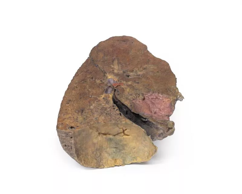
Ce modèle anatomique en 3D présente un poumon gauche disséqué dans un plan parasagittal, offrant une vue interne unique des structures pulmonaires et des repères anatomiques. La surface médiastinale a été retirée, permettant une observation détaillée de l'anatomie interne du poumon et de sa relation avec le cœur et le diaphragme.
Caractéristiques principales :
Arbre bronchique (vue interne) :
- Les bronches primaires ne sont pas visibles en raison d'une ramification antérieure.
- Les bronches subdivisées sont conservées, mais la profondeur de la dissection ne permet pas de déterminer s'il s'agit de bronches secondaires (lobaires) ou tertiaires (segmentaires).
Structures vasculaires :
- Les artères et veines pulmonaires sont généralement visibles au niveau du hile, mais les niveaux précis de subdivision ne sont pas déterminés dans cette section.
Empreinte cardiaque :
- Une empreinte concave nette reste visible sur la surface médiale du poumon, formée par le ventricule gauche du cœur appuyant contre le poumon.
- Malgré la dissection, ce repère reste clairement visible.
Surface diaphragmatique :
- La base du poumon est concave et repose sur le diaphragme.
- Bien que la plèvre ne soit pas préservée, le modèle indique l'emplacement où se formerait le renfoncement diaphragmatique, entre la plèvre costale et la plèvre diaphragmatique.
Connexion
Erler-Zimmer
Erler-Zimmer Medical GmbH
Hauptstrasse 27
77886 Lauf
Germany
info@erler-zimmer.de
Achtung! Medizinisches Ausbildungsmaterial, kein Spielzeug. Nicht geeignet für Personen unter 14 Jahren.
Attention! Medical training material, not a toy. Not suitable for persons under 14 years of age.
D'autres clients ont également acheté

Right lung, hilum removed
Ce modèle 3D offre une vue en coupe détaillée du poumon droit. Il complète le modèle TW 63 « Hilum du poumon droit » et contraste avec le modèle TW 61 « Coupe du poumon gauche ». Il met en évidence la macrostructure du poumon, de l'apex à la base, offrant ainsi une perspective précieuse pour l'étude anatomique et la comparaison entre les deux poumons.Caractéristiques principales :Organisation lobaire :- Des fissures obliques et horizontales clairement définies segmentent le poumon en lobes supérieur, moyen et inférieur.- La profondeur de ces fissures est visible, montrant leur extension dans la structure interne du poumon. Empreintes superficielles :- Des empreintes costales proéminentes s'étendent longitudinalement de l'apex à la base sur la face latérale, indiquant un contact étroit avec la cage thoracique.Surface diaphragmatique :- La base profondément concave reflète la forme bombée du diaphragme droit, qui est surélevé chez le sujet vivant en raison de la position du foie sous-jacent.

Cœur
Ce modèle 3D représente un cœur adulte grandeur nature avec une légère dissection de l'épicarde, offrant une vue claire des artères coronaires, des veines cardiaques et des gros vaisseaux à la base du cœur.Caractéristiques anatomiques clés :Gros vaisseaux :- La veine cave supérieure et la veine azygos se jettent dans l'oreillette droite.- L'arc aortique est conservé, avec deux branches principales : - Un tronc brachio-céphalique combiné (donnant naissance aux artères carotides communes droite et gauche et à l'artère sous-clavière droite).- L'artère sous-clavière gauche naît indépendamment.- Le tronc pulmonaire et les artères pulmonaires sont intacts.- Le ligament artériel est visible entre l'arc aortique et l'artère pulmonaire gauche. Artères coronaires :- L'artère coronaire droite (ACD) descend de l'aorte ascendante et s'enroule autour du sillon interventriculaire postérieur.- Branches de l'artère coronaire gauche (ACG) : - L'artère interventriculaire antérieure (AIA) s'étend vers l'apex.- Les branches diagonales plongent dans le myocarde.- L'artère circonflexe s'étend vers l'arrière, près de la grande veine cardiaque.Drainage veineux :Le sinus coronaire est clairement préservé sur la surface postérieure du cœur et se draine dans l'oreillette droite près de la veine cave inférieure.

Pericardial space
Ce modèle anatomique 3D détaillé présente la cavité péricardique et ses reflets après ablation du cœur, permettant ainsi une visualisation claire des structures clés du médiastin moyen.Caractéristiques principales :Péricarde : Montre l'étendue complète du péricarde pariétal, en continuité avec la couche viscérale (épicarde), et coloré de manière artificielle pour indiquer les positions des oreillettes, des ventricules et des gros vaisseaux. Repères médiastinaux : Met en évidence la base, l'apex, les surfaces diaphragmatique et pulmonaire du cœur par leurs empreintes dans la cavité péricardique.Grands vaisseaux :- Aorte (ascendante, crosse et descendante)- Veines caves supérieure et inférieure- Tronc pulmonaire et artères pulmonaires- Veines pulmonaires (4 au total) - Tous représentés dans leur position naturelle par rapport au péricarde.Sinus péricardiques :- Sinus transverse : situé entre les artères et les veines ; pertinent pour l'accès chirurgical.- Sinus oblique : renfoncement postérieur entre les veines pulmonaires.Utilisation pédagogique :- Idéal pour l'enseignement de l'anatomie thoracique et cardiaque, y compris les reflets péricardiques, les relations médiastinales et les repères chirurgicaux.- Utile pour la formation médicale, la planification chirurgicale et l'orientation radiologique.

Hilum of the left lung
Ce modèle 3D haute résolution offre une vue détaillée du hile pulmonaire gauche et des structures associées, sectionné sagittalement à travers l'échancrure cardiaque. Il montre clairement les relations entre les bronches, les vaisseaux pulmonaires, la plèvre et les structures de soutien, ce qui le rend idéal pour l'enseignement anatomique avancé.Caractéristiques principales :Anatomie du hile :- Point d'entrée/sortie de l'artère pulmonaire, des veines pulmonaires supérieure et inférieure, de la bronche principale, des vaisseaux lymphatiques et des nerfs.- Montre le point de rencontre de la plèvre viscérale et pariétale, formant le ligament pulmonaire, seule connexion anatomique du poumon au corps. Circulation pulmonaire :- L'artère pulmonaire (en position supérieure) transporte le sang désoxygéné depuis le cœur.- Les veines pulmonaires (antérieure et inférieure) ramènent le sang oxygéné vers le cœur.Structure bronchique :La bronche principale gauche et ses branches lobaires sont visibles, situées en position postérieure dans le hile.Vues supplémentaires :- Fissure oblique le long de la surface latérale du poumon.- Surface diaphragmatique à la base ; surface viscérale costale en position postérieure.- Ganglions lymphatiques pulmonaires entourant le hile, à la fois en position médiale et latérale.

Abdomen with bilateral Hernias
Ce modèle 3D est l'un des plus grands et des plus complexes de la série. Il représente une partie du torse, du diaphragme à la partie proximale de la cuisse, avec une cavité abdominale complète présentant différents niveaux de dissection. Ce modèle 3D reproduit également la rare occurrence simultanée d'une hernie inguinale indirecte et d'une hernie inguinale directe, ce qui permet d'étudier les fondements anatomiques de ces deux pathologies. Compte tenu de l'ampleur de la dissection, la description de ce modèle 3D est divisée en parties distinctes en fonction des vues et des régions.Le diaphragmeLe diaphragme est conservé sur la partie supérieure du modèle, les deux dômes et les renfoncements costodiaphragmatiques étant visibles malgré une certaine distorsion due au retrait des côtes. Le péricarde fibreux repose sur le tendon central, la veine cave inférieure terminale étant visible dans le foramen cave. Latéralement à cela se trouvent l'œsophage dans le hiatus œsophagien et l'aorte thoracique descendante qui s'approche du hiatus aortique près des vertèbres. Les régions épigastrique et hypochondrialeDans l'abdomen, l'ablation de la paroi antérieure, du grand omentum et d'une grande partie du tractus gastro-intestinal révèle les structures rétropéritonéales. L'œsophage terminal pénètre juste à gauche du foie. Une fois l'estomac retiré, le pancréas est entièrement exposé de la tête à la queue, atteignant la rate dans l'hypochondre gauche. Au-dessus, les artères splénique et hépatique commune enjambent l'espace étroit entre le pancréas, le diaphragme et le foie. L'artère splénique tortueuse se divise près de la veine splénique ; l'artère hépatique commune donne naissance aux artères gastroduodénale et gastrique droite, superficielles à la veine porte. Les vaisseaux mésentériques supérieurs passent près de la tête du pancréas, et l'artère iléo-colique mène au cæcum. La veine mésentérique inférieure prend naissance dans la veine rectale supérieure et traverse l'aorte descendante. Sous le foie, la vésicule biliaire se trouve entre les lobes. À gauche, les vaisseaux rénaux passent en profondeur du pancréas, les uretères descendant à travers les muscles psoas. Les régions ombilicale et lombaireLa plupart des organes abdominaux des régions ombilicale et lombaire ont été retirés afin de révéler la paroi abdominale postérieure. Au centre, l'aorte descendante et la veine cave inférieure sont bien visibles, et les vaisseaux testiculaires sont visibles jusqu'à la région inguinale. Deux artères lombaires droites partent de l'aorte, et l'artère mésentérique inférieure donne naissance aux artères coliques gauches, sigmoïdes et rectales supérieures. À droite, les nerfs sous-costaux, ilio-hypogastriques et ilio-inguinaux sont visibles, ainsi que l'artère iliaque circonflexe.Les régions hypogastrique et iliaqueL'aorte abdominale se divise en artères iliaques communes, internes et externes, avec les veines iliaques correspondantes qui se rejoignent dans la veine cave inférieure. L'artère obturatrice, les uretères et les vaisseaux testiculaires sont visibles. Dans le bassin véritable, le péritoine recouvre la vessie, tandis que le rectum reste masqué. La fosse iliaque droite contient l'iléon terminal, le cæcum et l'appendice, avec les vaisseaux et les nerfs voisins. À gauche, le côlon sigmoïde traverse la fosse iliaque, où un appendice épiploïque s'étend en une hernie indirecte près de l'artère épigastrique inférieure. La région inguinale et le périnéeCe modèle préserve de manière unique les hernies inguinales directes (à droite) et indirectes (à gauche), avec les vaisseaux épigastriques inférieurs conservés pour l'orientation anatomique. La hernie droite se trouve en position médiale par rapport à ces vaisseaux ; le sac herniaire gauche s'étend latéralement dans le cordon spermatique, contenant un appendice épiploïque. Le périnée révèle le pénis, les testicules et les cordons spermatiques. À droite, le cordon reste intact ; à gauche, il est ouvert, montrant une veine testiculaire variqueuse liée à la hernie indirecte. La cuisseLe triangle fémoral a été disséqué sur les deux cuisses. À droite, la gaine fémorale a été retirée pour révéler l'artère fémorale, la veine, les ganglions lymphatiques inguinaux profonds et le nerf fémoral. À gauche, une vue plus large expose les muscles antérieurs et médians de la cuisse, avec l'artère fémorale, l'artère profonde de la cuisse et l'artère circonflexe iliaque visibles. Le modèle se termine au milieu de la cuisse, montrant l'anatomie en coupe transversale, y compris le fémur, les vaisseaux et les muscles du canal sous-artériel.

Thorax with heart and vessels
Ce modèle 3D très détaillé représente l'anatomie clé de l'ouverture thoracique supérieure, du médiastin et des structures adjacentes du cou et du thorax, les deux clavicules et certains éléments musculaires et veineux ayant été retirés afin d'améliorer la visibilité et l'impact pédagogique.Points anatomiques importants :Ouverture thoracique supérieure :- Trachée visible en haut, avec un anneau cartilagineux robuste.- Côte 1 exposée de latéral à médial, y compris l'insertion du muscle scalène antérieur.- La suppression des clavicules permet une vue dégagée sur le corridor thoracique supérieur. Anatomie vasculaire :- L'artère sous-clavière droite, située au-dessus de la côte 1, donne naissance au tronc thyro-cervical.- L'artère sous-clavière gauche, également au-dessus de la côte 1, se ramifie en artère suprascapulaire.- Les deux artères carotides communes sont visibles ; la gaine carotidienne gauche comprend le nerf vague gauche.Système nerveux :- Le nerf vague gauche suit l'artère carotide gauche dans la gaine carotidienne ; le nerf laryngé récurrent gauche passe sous l'aorte.- Le nerf vague droit et le nerf phrénique droit sont rétractés pendant la dissection ; le nerf phrénique gauche reste antérieur au cœur, remontant jusqu'au diaphragme.- Les éléments du plexus brachial gauche sont visibles, des racines aux troncs, y compris le nerf dorsal de l'omoplate.Orientation médiastinale et cardiaque :- L'arc aortique, avec le tronc brachio-céphalique, la carotide commune gauche et l'artère sous-clavière gauche, se trouve juste au-dessus du cœur.- Le tronc pulmonaire émerge immédiatement au-dessus du cœur.- L'artère interventriculaire antérieure gauche (IVA) longe la surface antérieure du cœur.- La veine cave supérieure se trouve à droite et en arrière de l'aorte ascendante.- Le nerf phrénique droit est situé en arrière du cœur ; le nerf phrénique gauche passe dans son tissu conjonctif en avant.Thorax inférieur et diaphragme :- Les côtes 8 à 12 et les muscles intercostaux externes associés sont visibles ; les fibres musculaires s'étendent vers le bas et vers l'intérieur dans les couches fasciales.- Le demi-diaphragme droit est situé plus haut que le gauche, reflétant la présence du foie en dessous. Applications et avantagesPertinence clinique : idéal pour visualiser l'anatomie du cou et du thorax pertinente pour les procédures cardiovasculaires, respiratoires et nerveuses, y compris la chirurgie du larynx, du thorax et du plexus brachial.Utilité pédagogique : offre une vue claire et accessible des compartiments médiastinaux, des voies vasculaires et des trajectoires nerveuses sans obstruction par les os ou les structures superficielles. Apprentissage amélioré : la clarté du modèle facilite l'enseignement de l'interprétation radiographique, des approches chirurgicales thoraciques et des examens anatomiques.

Hilum of the right lung
Ce modèle 3D de haute qualité présente une coupe sagittale du poumon droit, centrée sur le hile, où les principales structures anatomiques entrent et sortent du poumon. Il constitue un outil essentiel pour comprendre l'anatomie vasculaire et bronchique pulmonaire, avec une orientation claire de l'apex à la base et des surfaces médiales aux surfaces latérales.Caractéristiques principales :Structure du hile :- Le hile marque la transition entre la plèvre viscérale et la plèvre pariétale et constitue le seul lien anatomique entre le poumon et le corps via le ligament pulmonaire.- Les principales structures entrant dans le poumon à cet endroit sont les suivantes :- Artère pulmonaire (supérieure dans le hile) – transportant le sang désoxygéné depuis le cœur.- Veines pulmonaires supérieure et inférieure (antérieure et inférieure) – ramenant le sang oxygéné vers le cœur.- La bronche principale droite et ses branches lobaires – situées à l'arrière du hile.- Les nerfs et les vaisseaux lymphatiques associés. Repères anatomiques visibles :- L'empreinte cardiaque (formée par l'oreillette droite) est visible juste en avant du hile.- Le sillon œsophagien est conservé le long de la surface postérieure, retraçant le trajet de l'œsophage descendant.- Les fissures obliques et horizontales sont bien définies sur la surface latérale, délimitant les trois lobes du poumon.- Les ganglions lymphatiques hilaires sont observés autour du hile médial.Surfaces pulmonaires :- Surface diaphragmatique (inférieure), montrant l'interface concave avec le diaphragme.- Surface viscérale costale (postérieure), où le poumon est en contact avec la paroi thoracique.

Thoracic cross section at T6
Ce modèle 3D détaillé présente une coupe transversale du thorax au niveau de la vertèbre T6, offrant une vue claire de l'anatomie thoracique en relation avec les structures squelettiques, vasculaires, respiratoires et cardiaques.Caractéristiques principales :Structures postérieures- Commence en position médiane avec la moelle épinière dans le canal vertébral- Les articulations costo-vertébrales des 6e côtes sont visibles, ainsi que les côtes environnantes formant la paroi thoracique. Paroi thoracique antérieure- Les articulations costo-sternales montrent la connexion entre les côtes et le sternum.Principaux organes thoraciques- Œsophage situé à l'avant du corps vertébral.- Aorte descendante située latéralement au corps vertébral.Poumons et cavités pleurales- Dans les espaces pleuraux (bordés par la plèvre pariétale) :- Poumon droit : lobes moyen et inférieur- Poumon gauche : lobe inférieurCœur et médiastin- Dans le médiastin moyen, le cœur est représenté à l'intérieur du péricarde, sectionné pour montrer son anatomie interne :- Oreillette gauche (postérieure)- Valve aortique, ventricule droit et oreillette droite dans le sens des aiguilles d'une montre

Une force d'innovation constante

Responsabilité sociale

Clients fidèles - Orientation

Compréhension de la qualité

Action durable

Certification ISO 9001