


")


Informations sur le produit "Uterus Bicornuate Unicollis"
Histoire clinique
Une femme de 36 ans a présenté une hémorragie post-partum sévère après l’accouchement en siège de son quatrième enfant. Ses trois précédents accouchements étaient également en siège, sans antécédent de fausse couche. Elle souffrait de douleurs abdominales légères intermittentes. L’équipe obstétricale, incapable d’arrêter l’hémorragie, a procédé à une hystérectomie radicale en urgence avec salpingo-ovariectomie bilatérale. La mère et l’enfant se sont complètement rétablis.
Pathologie
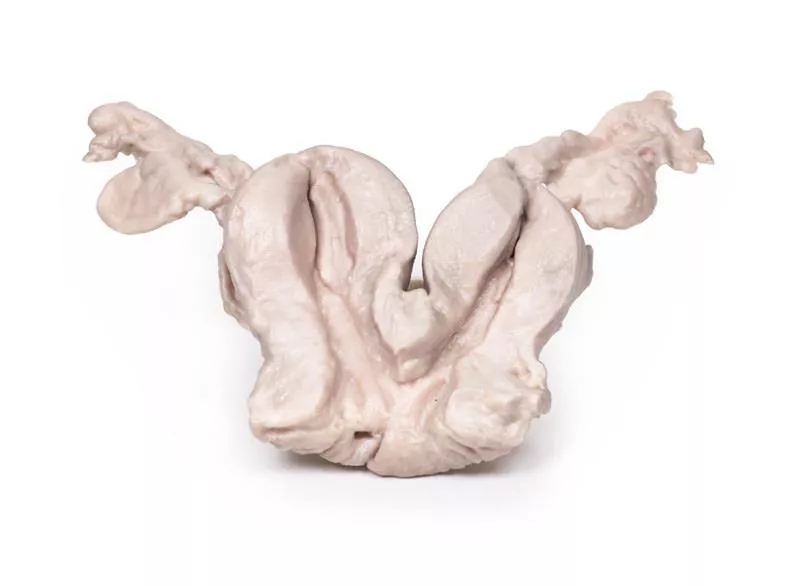
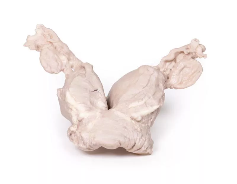
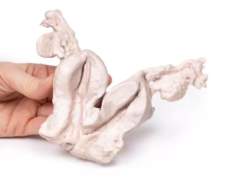
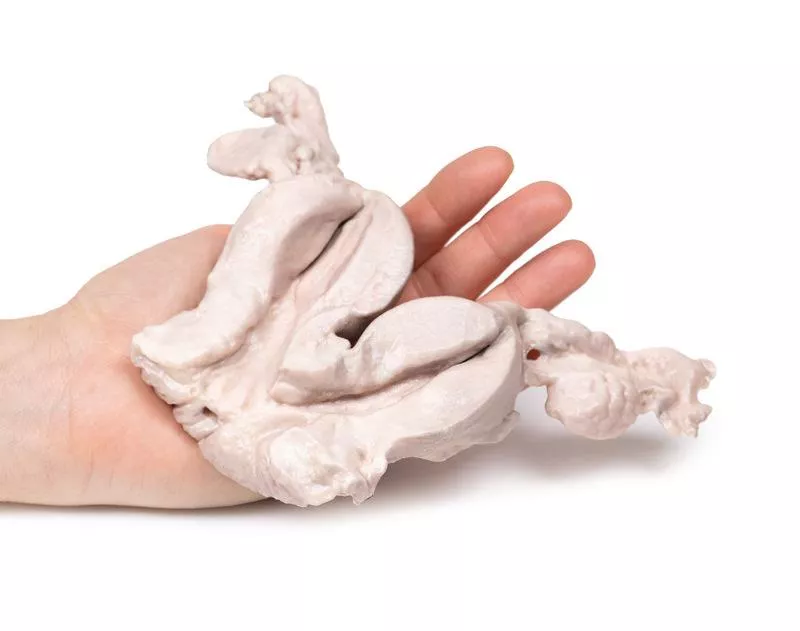
Le prélèvement comprend un utérus bicorne, les trompes de Fallope et les ovaires, coupés dans le plan coronal pour montrer les faces internes et externes. Les deux corps utérins sont de taille égale et partagent un canal cervical commun. Quelques petits kystes sont présents au niveau du col.
Informations complémentaires
Un utérus bicorne est une malformation congénitale caractérisée par une échancrure de plus de 1 cm au niveau du fond utérin. Le col de l’utérus et le vagin sont généralement normaux, mais on retrouve deux cavités endométriales partiellement séparées, en raison d'une fusion incomplète des canaux de Müller durant l’embryogenèse.
Cette anomalie touche environ 0,5?% des femmes, bien que le nombre réel soit probablement plus élevé, car de nombreuses femmes sont asymptomatiques. Les symptômes possibles incluent des douleurs pelviennes (cycliques ou non), des saignements utérins anormaux, des pertes vaginales ou des infections urinaires.
Pendant la grossesse, un utérus bicorne peut entraîner un risque accru de fausse couche, accouchement prématuré, présentation fœtale anormale, retard de croissance intra-utérin et placenta prævia. Une mauvaise présentation du fœtus augmente la probabilité de césarienne. Après l’accouchement, le risque de rétention placentaire et d’hémorragie post-partum est plus élevé.
Le diagnostic repose généralement sur une échographie pelvienne. L’IRM est rarement utilisée pour confirmation. Aucun traitement n’est nécessaire dans la plupart des cas.
Une femme de 36 ans a présenté une hémorragie post-partum sévère après l’accouchement en siège de son quatrième enfant. Ses trois précédents accouchements étaient également en siège, sans antécédent de fausse couche. Elle souffrait de douleurs abdominales légères intermittentes. L’équipe obstétricale, incapable d’arrêter l’hémorragie, a procédé à une hystérectomie radicale en urgence avec salpingo-ovariectomie bilatérale. La mère et l’enfant se sont complètement rétablis.
Pathologie
Le prélèvement comprend un utérus bicorne, les trompes de Fallope et les ovaires, coupés dans le plan coronal pour montrer les faces internes et externes. Les deux corps utérins sont de taille égale et partagent un canal cervical commun. Quelques petits kystes sont présents au niveau du col.
Informations complémentaires
Un utérus bicorne est une malformation congénitale caractérisée par une échancrure de plus de 1 cm au niveau du fond utérin. Le col de l’utérus et le vagin sont généralement normaux, mais on retrouve deux cavités endométriales partiellement séparées, en raison d'une fusion incomplète des canaux de Müller durant l’embryogenèse.
Cette anomalie touche environ 0,5?% des femmes, bien que le nombre réel soit probablement plus élevé, car de nombreuses femmes sont asymptomatiques. Les symptômes possibles incluent des douleurs pelviennes (cycliques ou non), des saignements utérins anormaux, des pertes vaginales ou des infections urinaires.
Pendant la grossesse, un utérus bicorne peut entraîner un risque accru de fausse couche, accouchement prématuré, présentation fœtale anormale, retard de croissance intra-utérin et placenta prævia. Une mauvaise présentation du fœtus augmente la probabilité de césarienne. Après l’accouchement, le risque de rétention placentaire et d’hémorragie post-partum est plus élevé.
Le diagnostic repose généralement sur une échographie pelvienne. L’IRM est rarement utilisée pour confirmation. Aucun traitement n’est nécessaire dans la plupart des cas.
Connexion
Erler-Zimmer
Erler-Zimmer Medical GmbH
Hauptstrasse 27
77886 Lauf
Germany
info@erler-zimmer.de
Achtung! Medizinisches Ausbildungsmaterial, kein Spielzeug. Nicht geeignet für Personen unter 14 Jahren.
Attention! Medical training material, not a toy. Not suitable for persons under 14 years of age.
D'autres clients ont également acheté

Retrosternal Goiter
Histoire cliniqueUne femme de 60 ans s’est présentée avec une masse anormale au cou, une toux persistante et des difficultés à avaler. Elle avait pris du poids au cours des dernières années. Elle est décédée d’une maladie cardiovasculaire sans lien avec ces symptômes, et le prélèvement a été effectué lors de l’autopsie.Pathologie Le prélèvement post-mortem comprend le larynx, la trachée et une glande thyroïde volumineuse et multilobée. Le lobe droit est particulièrement agrandi, avec deux lobes majeurs s’étendant de 7 à 8 mm vers le haut et le bas, bien au-delà des limites normales vues de face. L’œsophage a été ouvert à l’arrière pour exposer la paroi postérieure de la trachée. Le lobe droit semble encore plus grand depuis cette perspective, avec une croissance anormale centrée sur son pôle inférieur. Il n’y a pas de changements pigmentaires majeurs, mais des veines proéminentes sont visibles à la surface du lobe droit. Informations complémentairesLe goitre se manifeste souvent par une masse palpable au cou. Selon sa taille et son emplacement, il peut entraîner des symptômes de compression : gêne respiratoire, dysphagie, toux ou enrouement. Dans de rares cas, un goitre en expansion peut paralyser le nerf laryngé récurrent. Une obstruction trachéale peut provoquer stridor et essoufflement. Une douleur soudaine ou une croissance rapide peuvent indiquer une expansion kystique ou une hémorragie intra-nodulaire.Les causes fréquentes de goitre incluent les maladies auto-immunes (thyroïdite de Hashimoto, maladie de Basedow), les nodules thyroïdiens et la carence en iode. Le goitre apparaît généralement lorsqu’il y a une réduction de la synthèse des hormones thyroïdiennes due à un défaut de biosynthèse ou à une carence iodée. Cela entraîne une augmentation de la TSH (hormone stimulant la thyroïde), stimulant la croissance glandulaire de manière compensatoire. Dans la thyroïdite de Hashimoto, l’élévation de la TSH et la fibrose auto-immune contribuent à l’augmentation du volume thyroïdien. Dans la maladie de Basedow, le goitre est principalement dû aux anticorps dirigés contre le récepteur de la TSH.Référence : Hughes et al. (2012). Goitre: Causes, investigation and management. Aust Family Physician, 41, 572–576.

Carcinoma of Breast
Histoire cliniqueUne femme de 76 ans a été admise aux urgences après une perte de connaissance soudaine avec signes d’un AVC à gauche. Intubée et traitée en soins intensifs, une masse fixe dans le sein gauche ainsi qu’une adénopathie axillaire ont été détectées. Elle est décédée d’une pneumonie liée à l’intubation.PathologieLe prélèvement montre un gros nodule mammaire (11?cm) sous la peau, adhérent au muscle. La coupe révèle une structure hétérogène avec zones nécrotiques, hémorragiques et kystiques. Le diagnostic est un adénocarcinome mammaire avec envahissement ganglionnaire régional. Informations complémentairesLe cancer du sein est le deuxième cancer le plus fréquent chez la femme. Il est rare avant 30 ans, avec un pic entre 70 et 80 ans. Facteurs de risque : œstrogènes, antécédents familiaux, absence de grossesse ou allaitement, obésité et mutations génétiques (BRCA1/2…).La majorité sont des adénocarcinomes d’origine canalaire ou lobulaire, souvent sous forme de DCIS. Ils sont classés selon leur statut hormonal (ER/HER2), ce qui oriente la prise en charge. Les métastases touchent surtout os, foie, poumon et cerveau.Dans les pays développés, le diagnostic repose souvent sur une mammographie anormale. Les symptômes incluent une masse dure, irrégulière, fixe, modifications cutanées (peau d’orange), rétraction du mamelon ou adénopathie axillaire.Le traitement dépend du stade et du profil tumoral?: chirurgie (mastectomie ou tumorectomie), radiothérapie, thérapies ciblées (trastuzumab pour HER2+), hormonothérapie (tamoxifène pour ER+) et chimiothérapie.

Multinodular goitre
Histoire clinique Une femme de 53 ans s’est présentée avec une masse anormale au cou et une toux persistante. Elle signalait également une fatigue et une prise de poids sur plusieurs années. Pendant les examens, elle est décédée plusieurs mois plus tard d’une maladie cardiovasculaire non liée.Pathologie Le spécimen post-mortem comprend la base de la langue, le larynx et la trachée. La glande thyroïde est très volumineuse, surtout le lobe droit, dépassant ses limites normales. Les coupes montrent de multiples nodules hyper- et hypopigmentés ainsi que des zones kystiques dans les deux lobes. La base de la langue, le larynx et la trachée paraissent normaux. Informations complémentairesLe goitre nodulaire se manifeste généralement par une tuméfaction au cou. Selon sa taille et sa localisation, il peut provoquer des symptômes de compression comme des difficultés respiratoires, une dysphagie, une toux ou une dysphonie. La paralysie du nerf récurrent est rare. Une croissance soudaine ou une douleur peut résulter d’une expansion kystique ou d’un saignement dans les nodules.Les causes incluent les maladies auto-immunes (thyroïdite de Hashimoto, maladie de Basedow), les nodules thyroïdiens et la carence en iode. Le goitre se développe lorsque la synthèse d’hormones thyroïdiennes est réduite — par des défauts biosynthétiques ou un manque d’iode — entraînant une augmentation de la stimulation par la TSH et une croissance thyroïdienne. Dans Hashimoto, la TSH élevée combinée à la fibrose agrandit la thyroïde; dans la maladie de Basedow, les anticorps au récepteur de la TSH stimulent le goitre.

Nodular hyperplasia of the Prostate
Histoire clinique Un homme de 63 ans s’est présenté avec des douleurs abdominales aiguës et une rétention urinaire de 5 jours. Depuis deux ans, il signalait fréquence urinaire, hésitation, double miction, nycturie et jet faible. La vessie était distendue, la prostate palpablement élargie. L’échographie a montré >1?L d’urine résiduelle. Les analyses ont révélé une insuffisance rénale aiguë. Après plusieurs échecs de sondage, une prostatectomie totale a été réalisée avec bonne récupération.Pathologie La prostate sectionnée transversalement montre de nombreux nodules (2–10?mm), typiques d’une hyperplasie bénigne de la prostate (HBP). Informations complémentaires L’HBP est fréquente chez les hommes âgés, causée par une croissance nodulaire des cellules stromales et glandulaires péri-urétrales, stimulée par la dihydrotestostérone. Le lobe médian peut obstruer l’urètre.Prévalence : 20?% à 40 ans, 70?% à 60 ans, 90?% à 80 ans. Facteurs de risque : antécédents familiaux, obésité, stéroïdes anabolisants.Symptômes : fréquence urinaire, nycturie, hésitation, goutte-à-goutte, jet faible. La rétention urinaire aiguë peut provoquer des infections urinaires et une atteinte rénale.Diagnostic : histoire clinique, examen rectal, PSA, échographie ou scanner. Traitements : alpha-bloquants, inhibiteurs de la 5-alpha-réductase, ou en cas grave, résection transurétrale de la prostate (RTUP). La prostatectomie totale est rarement utilisée.

Lymphoma of the thyroid
Histoire clinique Une femme de 68 ans s’est présentée avec une petite masse dure au niveau de la thyroïde. En six semaines, la masse a rapidement augmenté, causant un stridor laryngé et une obstruction œsophagienne. Aucun ganglion lymphatique augmenté ni splénomégalie n’a été observé.Pathologie Le spécimen comprend le larynx, la thyroïde, la partie supérieure de la trachée et l’œsophage. Le lobe gauche de la thyroïde est augmenté, remplacé par un tissu tumoral pâle homogène. La tumeur comprime le larynx et l’œsophage. L’histologie a confirmé un lymphome lymphoblastique de la thyroïde. En raison de sa rareté, un carcinome anaplasique ou une dissémination lymphomateuse secondaire doivent être exclus. Informations complémentairesLe lymphome primitif de la thyroïde est rare mais doit être envisagé devant toute masse thyroïdienne. La plupart sont des lymphomes non hodgkiniens; le lymphome lymphoblastique est agressif et survient habituellement chez les enfants. Le facteur de risque principal connu est la thyroïdite auto-immune chronique (Hashimoto), présente dans environ 50 % des cas.Plus de 90 % des patients présentent un goitre en croissance rapide causant une compression de la trachée, de l’œsophage et des vaisseaux du cou, avec stridor, enrouement, dysphagie et douleurs cervicales. Les symptômes systémiques (« B-symptômes ») incluent sueurs nocturnes, fièvre et perte de poids.Le diagnostic repose sur une échographie suivie d’une ponction à l’aiguille fine ou d’une biopsie. La cytologie et l’immunohistochimie sont essentielles pour différencier le lymphome de la thyroïdite de Hashimoto ou des carcinomes.

Aorta & para-aortic lymph nodes
Histoire cliniqueCe cas concerne une femme de 75 ans présentant des symptômes de récidive cinq ans après le traitement initial d’un adénocarcinome séreux ovarien de stade IIIc. Des métastases ganglionnaires rétro-péritonéales chimiorésistantes ont été diagnostiquées, affectant les ganglions para-aortiques et pelviens, comme révélé par la TEP/TDM. La patiente est décédée de complications hépatiques avant qu’un traitement chirurgical, tel qu’une lymphadénectomie radicale, ne puisse être envisagé.Pathologie Le prélèvement comprend l’aorte abdominale et les artères iliaques communes, entourées de nombreux ganglions para-aortiques et iliaques hypertrophiés. L’examen histopathologique a confirmé la présence d’un adénocarcinome de haut grade métastatique dans plusieurs ganglions. Informations complémentairesDans certains cas de récidive du cancer de l’ovaire, les métastases ganglionnaires peuvent constituer la seule manifestation. Ici, les ganglions identifiés par TEP/TDM correspondaient exactement à ceux touchés par la maladie. L’échographie aurait aussi pu détecter ces ganglions hypertrophiés, mais ni la TEP/TDM ni l’échographie ne permettent de détecter les atteintes microscopiques, ce qui complique le suivi après chirurgie.Chez des femmes plus jeunes sans autre localisation métastatique, une dissection systématique des ganglions aortiques et pelviens peut soulager les symptômes et permettre l’accès à des thérapies innovantes, bien que cette intervention ne soit généralement pas curative.Le plus souvent, le prélèvement ganglionnaire pelvien et aortique fait partie du bilan chirurgical initial d’un cancer épithélial de l’ovaire. En cas de maladie avancée, l’ablation des ganglions rétro-péritonéaux hypertrophiés est envisagée si elle permet une réduction tumorale complète.

Endometrial Carcinoma
Histoire cliniqueUne femme de 63 ans s’est présentée avec des douleurs abdominales basses persistantes depuis deux mois et un saignement vaginal abondant depuis une semaine, 13 ans après la ménopause. Après confirmation d’un adénocarcinome de l’endomètre par biopsie, une hystérectomie abdominale radicale avec salpingo-ovariectomie bilatérale a été réalisée.PathologieLe prélèvement comprend l’utérus, les trompes et les ovaires. La muqueuse endométriale est anormale, en particulier à droite où une tumeur polypoïde brune infiltre le myomètre et s’étend dans le canal cervical. L’analyse histologique révèle un adénocarcinome bien différencié. L’ovaire gauche est volumineux avec plusieurs kystes folliculaires. Informations complémentairesLe carcinome de l’endomètre est le cancer gynécologique le plus fréquent dans les pays développés. Il en existe deux types principaux : le carcinome endométrioïde (˜80?%), souvent associé à une hyperplasie atypique et au pronostic favorable, et le carcinome séreux, plus agressif.Les mutations courantes sont PTEN, PIK3Ca, ARID1A (endométrioïde) et TP53 (séreux). Les facteurs de risque comprennent l’obésité, l’intolérance au glucose, l’infertilité et l’exposition prolongée aux œstrogènes. Le type séreux touche les femmes âgées avec utérus atrophique et IMC bas. Le syndrome de Lynch augmente considérablement le risque.Le symptôme principal est le saignement post-ménopausique. L’échographie pelvienne montre un endomètre épaissi. Le diagnostic repose sur biopsie, curetage ou hystérectomie. Le traitement dépend du stade : chirurgie, radiothérapie et chimiothérapie.

Uterine Leiomyoma
Histoire clinique Une femme de 30 ans consulte pour infertilité, douleurs pelviennes, règles abondantes et dysménorrhée. L’échographie révèle une masse hypoe´chogène dans le myomètre. Une hystérectomie en urgence a été réalisée après échec de la myomectomie. Rétablissement complet.Pathologie L’utérus est coupé sagittalement. Une masse ovale de 4?×?2?cm issue de la paroi postérieure fait saillie dans la cavité utérine jusqu’au canal cervical. Informations complémentaires Les léiomyomes utérins sont des tumeurs bénignes fréquentes chez les femmes en âge de procréer. Symptômes : saignements anormaux, douleurs pelviennes, infertilité. Diagnostic par échographie. Traitement : thérapie hormonale, myomectomie, hystérectomie, ou embolisation des artères utérines.

Hydrocoele
Histoire clinique Un patient avec diabète et infarctus du myocarde antérieurs présentait un épanchement pleural bilatéral, œdème périphérique et un scrotum gonflé à transillumination. La radiographie thoracique montrait une insuffisance cardiaque congestive. Malgré le traitement, le patient est décédé durant l’hospitalisation.Pathologie Le testicule et ses enveloppes montrent une cavité distendue entre les feuillets viscéral et pariétal de la tunique vaginale due à une accumulation de liquide, formant une hydrocèle secondaire à un œdème généralisé lié à l’insuffisance cardiaque. Informations complémentairesL’hydrocèle correspond à un liquide séreux entre les couches de la tunique vaginale. Elle peut être communicante (avec la cavité péritonéale) ou non communicante. Les hydrocèles communicantes résultent d’un défaut de fermeture du processus vaginal et peuvent être congénitales ou apparaître plus tard, souvent suite à une pression abdominale accrue, comme en cas d’insuffisance cardiaque. Les hydrocèles non communicantes sont dues à un déséquilibre de la production et résorption du liquide, infections, traumatisme, tumeur ou troubles lymphatiques.Les patients notent une masse scrotale, unilatérale ou bilatérale. Les hydrocèles communicantes peuvent varier avec la pression abdominale, les autres sont stables. Elles sont en général indolores sauf en cas de complications. Les grandes hydrocèles peuvent entraîner des problèmes cutanés.Le diagnostic repose sur l’examen clinique, la transillumination et l’échographie. Les marqueurs tumoraux AFP et B-HCG excluent un cancer. Beaucoup d’hydrocèles congénitales disparaissent avant 2 ans. Les cas persistants ou symptomatiques peuvent être opérés. Le traitement de la cause sous-jacente peut aussi les faire régresser.

Une force d'innovation constante

Responsabilité sociale

Clients fidèles - Orientation

Compréhension de la qualité

Action durable

Certification ISO 9001