


")


Hydatid Disease Affecting the Heart and Aorta
Mehr erfahren622,37 €*
Artikel in Produktion, lieferbar vorauss. in 2-3 Wochen
Produktnummer:
MP2035
Artikelnummer: MP2035
Produktinformationen "Hydatid Disease Affecting the Heart and Aorta"
Clinical History
This 11-year-old female had an 18-month history of hydatid disease (see below). In total, 17 cysts were removed from the child’s brain at craniotomy on three occasions, and subsequently cysts were found in the kidneys, mesentery, and abdominal aorta at its bifurcation. X-ray of the heart showed a calcified cyst, and the patient was referred to a tertiary hospital for its removal. The patient deteriorated and died following open-heart surgery during which a dead hydatid cyst was found in the left ventricle.
Pathology
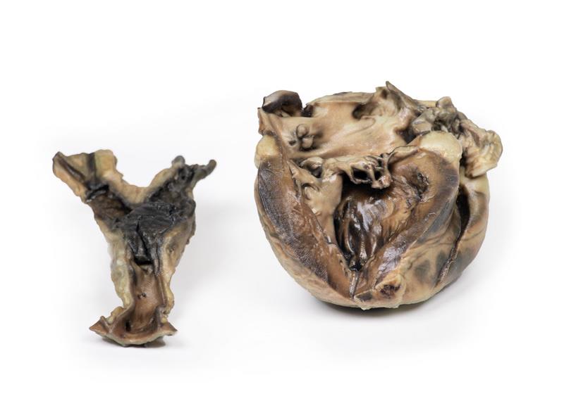
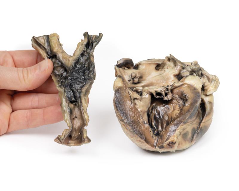
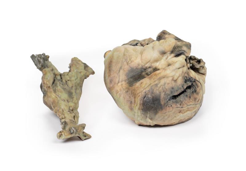
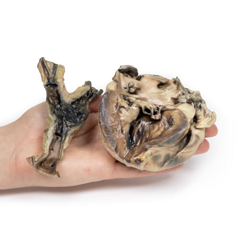
The specimens are of the heart, with the left ventricle being laid open, and of the aorta at its common iliac bifurcation. The aorta shows some atheromatous depositions in the upper portion. There is a large mass of antemortem clot at the point of iliac bifurcation with extension down both common iliac arteries.
The heart shows hypertrophy of the left ventricular wall, and an abnormal communication between the left ventricle and atrium running through the posterior cusp of the mitral valve via the papillary muscle into the left ventricular cavity. This channel is surrounded by thickened fibrous-looking tissue. The posterior cusp of the valve has been split. Sutured surgical incisions are visible on the posterior aspect of the specimen and the ventricular wall and in the left atrial appendage. Hydatid cysts occupy the abdominal aorta at its bifurcation, and the channel joining the left ventricle and left atrium.
Histology demonstrated cysts within the aorta wall comprised of 3 layers: an outermost pericyst fibrous layer; a middle ectocyst layer that was laminated, hyaline and acellular; and the inner endocyst in the germinative layer, consisting of daughter cysts and brood capsules with scolices. A focal granulomatous palisading reaction was also present within the aorta wall.
Further Information
Hydatid cyst is a human parasitic disease caused by the larval stage of the cestode tapeworm Echinococcus granulosus, which infests the gut of dogs—its definitive hosts. Human beings may serve as incidental hosts by the ingestion of ova in vegetables or water contaminated with dog faeces. Humans become infected by the ingestion of eggs passed in dog faeces. Oncospheres released from the eggs penetrate the intestinal mucosa and, via the portal system, lodge in the liver, lungs, muscle or other organs, where the hydatid cysts form.
Hydatid disease is endemic in cattle-raising areas of the world, notably in the Mediterranean countries, the Middle East, South America, Australia, and New Zealand. Although no body part can be spared from hydatid cysts, they mostly affect the liver and lungs. Cardiac involvement is much rarer, yet potentially fatal condition and comprises 0.5–2% of all hydatid cases. Cardiac complications and presentation vary depends on the location, size and integrity of the cyst(s). The myocardium of the left ventricle more frequently involved. Pericardial involvement occurs mostly in multifocal cardiac echinococcosis. Growth of the cyst leads them being pushed toward a weaker side of the cardiac wall, either the epicardium or the endocardium. LV HCs are usually located subepicardially, therefore rarely rupture into the pericardial space. However, if rupture happens, it may be silent or it may cause acute pericardial tamponade, constrictive pericarditis or secondary pericardial cysts[1].
Although E. granulosus is still found in sheep and rural dogs in Australia, the prevalence of transmission is less common than it was. The marked reduction in prevalence in rural domestic dogs, and also sheep, is the result of the highly effective cestocidal drug, praziquantel, being included in
readily available, cheap, generic, all-wormers for dogs and the development of inexpensive commercial dry dog food[2].
References
1. Oraha et al. Ann Med Surg (Lond). 2018 18–21
2. Jenkins et al. Int J Parasitol Parasites Wildl. 2019: 256–259.
This 11-year-old female had an 18-month history of hydatid disease (see below). In total, 17 cysts were removed from the child’s brain at craniotomy on three occasions, and subsequently cysts were found in the kidneys, mesentery, and abdominal aorta at its bifurcation. X-ray of the heart showed a calcified cyst, and the patient was referred to a tertiary hospital for its removal. The patient deteriorated and died following open-heart surgery during which a dead hydatid cyst was found in the left ventricle.
Pathology
The specimens are of the heart, with the left ventricle being laid open, and of the aorta at its common iliac bifurcation. The aorta shows some atheromatous depositions in the upper portion. There is a large mass of antemortem clot at the point of iliac bifurcation with extension down both common iliac arteries.
The heart shows hypertrophy of the left ventricular wall, and an abnormal communication between the left ventricle and atrium running through the posterior cusp of the mitral valve via the papillary muscle into the left ventricular cavity. This channel is surrounded by thickened fibrous-looking tissue. The posterior cusp of the valve has been split. Sutured surgical incisions are visible on the posterior aspect of the specimen and the ventricular wall and in the left atrial appendage. Hydatid cysts occupy the abdominal aorta at its bifurcation, and the channel joining the left ventricle and left atrium.
Histology demonstrated cysts within the aorta wall comprised of 3 layers: an outermost pericyst fibrous layer; a middle ectocyst layer that was laminated, hyaline and acellular; and the inner endocyst in the germinative layer, consisting of daughter cysts and brood capsules with scolices. A focal granulomatous palisading reaction was also present within the aorta wall.
Further Information
Hydatid cyst is a human parasitic disease caused by the larval stage of the cestode tapeworm Echinococcus granulosus, which infests the gut of dogs—its definitive hosts. Human beings may serve as incidental hosts by the ingestion of ova in vegetables or water contaminated with dog faeces. Humans become infected by the ingestion of eggs passed in dog faeces. Oncospheres released from the eggs penetrate the intestinal mucosa and, via the portal system, lodge in the liver, lungs, muscle or other organs, where the hydatid cysts form.
Hydatid disease is endemic in cattle-raising areas of the world, notably in the Mediterranean countries, the Middle East, South America, Australia, and New Zealand. Although no body part can be spared from hydatid cysts, they mostly affect the liver and lungs. Cardiac involvement is much rarer, yet potentially fatal condition and comprises 0.5–2% of all hydatid cases. Cardiac complications and presentation vary depends on the location, size and integrity of the cyst(s). The myocardium of the left ventricle more frequently involved. Pericardial involvement occurs mostly in multifocal cardiac echinococcosis. Growth of the cyst leads them being pushed toward a weaker side of the cardiac wall, either the epicardium or the endocardium. LV HCs are usually located subepicardially, therefore rarely rupture into the pericardial space. However, if rupture happens, it may be silent or it may cause acute pericardial tamponade, constrictive pericarditis or secondary pericardial cysts[1].
Although E. granulosus is still found in sheep and rural dogs in Australia, the prevalence of transmission is less common than it was. The marked reduction in prevalence in rural domestic dogs, and also sheep, is the result of the highly effective cestocidal drug, praziquantel, being included in
readily available, cheap, generic, all-wormers for dogs and the development of inexpensive commercial dry dog food[2].
References
1. Oraha et al. Ann Med Surg (Lond). 2018 18–21
2. Jenkins et al. Int J Parasitol Parasites Wildl. 2019: 256–259.
Anmelden
Andere Kunden kauften auch

Stetige Innovationskraft

Soziale Verantwortung

Gelebte Kundenorientierung

Verständnis für Qualität

Nachhaltiges Handeln

Zertifizierung ISO 9001