
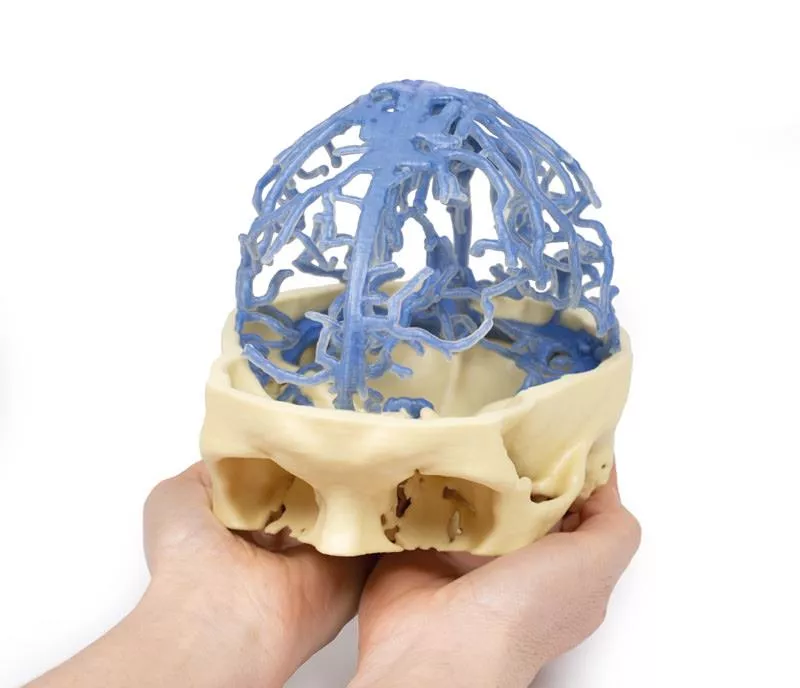


3D Printed Anatomy - Venous Circulation
Bestellen Sie für weitere 200,00 € und Sie erhalten Ihre Bestellung versandkostenfrei.
Produktinformationen "3D Printed Anatomy - Venous Circulation"
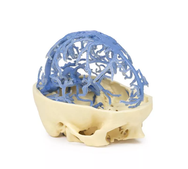
Dieser 3D-Druck basiert auf denselben Datensätzen wie unsere Modelle des Circulus Willisi und der kranialen Arterienkreislauf, die aus einer sorgfältigen Segmentierung angiographischer Daten abgeleitet wurden. Das Modell hebt das Netzwerk der duralen Venensinus hervor, das entsprechend den durch Kontrastmittelkreislauf sichtbaren Strukturen segmentiert wurde.
Venennetzwerk und Sinus
Die meisten duralen Venensinus sind dargestellt; aufgrund des begrenzten Kontrasts im vorderen Venensystem sind jedoch Strukturen wie der Sinus cavernosus und die Sinus petrosi nicht enthalten. Das Modell zeigt deutlich das ausgedehnte Netzwerk der duralen Venen und venösen Lakunen, die in der Mittellinie zum Sinus sagittalis superior zusammenlaufen.
Tiefe venöse Strukturen
Unterhalb dieses Netzwerks sind die große Hirnvene, der Sinus sagittalis inferior und der Sinus rectus sichtbar, die am Zusammenfluss der Sinus mit dem Sinus sagittalis superior zusammenlaufen.
Transversale und sigmoide Sinus
Mehrere Duralvenen münden in den linken und rechten transversalen Sinus und verlaufen anterior in Richtung des petrosalen Teils des Schläfenbeins. Die sigmoiden Sinus sind in der hinteren Schädelgrube sichtbar, bevor sie den Schädel am Foramen jugulare verlassen und die Vena jugularis interna bilden, die an der unteren Oberfläche des Schädels zu sehen ist.
Dokumente
| Datasheet MP1645 | Herunterladen |
Erler-Zimmer
Erler-Zimmer GmbH & Co.KG
Hauptstrasse 27
77886 Lauf
Germany
info@erler-zimmer.de
Achtung! Medizinisches Ausbildungsmaterial, kein Spielzeug. Nicht geeignet für Personen unter 14 Jahren.
Attention! Medical training material, not a toy. Not suitable for persons under 14 years of age.
Dokumente
| Datasheet MP1645 | Herunterladen |
Andere Kunden kauften auch
Dieser hochwertige 3D-Druck des Schädels und der Schädelhöhle bewahrt wichtige anatomische Strukturen, einschließlich der Duralfalten. Ein dünner Streifen der Schädeldecke wurde in der mittleren Sagittalebene erhalten, um die Befestigung der Falx cerebri zu erhalten.Erhaltenes Tentorium cerebelliDas Tentorium cerebelli ist in diesem Modell sorgfältig erhalten geblieben und zeigt deutlich die Tentoriumkerbe, in der normalerweise das Mittelhirn liegt. Hervorgehobene Sella turcica und innere HalsschlagadernIm Bereich der Sella turcica sind die Eintrittspunkte der inneren Halsschlagadern durch das Dach der Sinus cavernosus rot hervorgehoben, was Studenten und Fachleuten eine intuitive visuelle Orientierung bietet. Fokus auf durale VenensinusDer Hauptzweck dieses Modells besteht darin, die Anordnung der Duralfalten und die Position der duralen Venensinus zu veranschaulichen, die hellblau hervorgehoben sind. Die folgenden Sinus sind deutlich gekennzeichnet: - Sinus sagittalis superior - Sinus sagittalis inferior - Sinus rectus - Sinus transversus - Sinus petrosus superior und inferior - Sinus sphenoparietalis
Dieses einzigartige 3D-Modell basiert auf Daten der Schädeldecke (Druck Nr. 8), die mittels CT-Bildgebung und Segmentierung der inneren Räume gewonnen wurden. Teile des Schädels wurden beibehalten, während Abschnitte oder Fenster entfernt wurden, um die Nasennebenhöhlen freizulegen.StirnhöhlenDie paarigen Stirnhöhlen sind blau hervorgehoben. Die rechte Stirnhöhle ist vollständig freigelegt und zeigt das frontonasale Ostium, das als trichterförmiger Schlauch in den Infundibulum des mittleren Nasengangs mündet. Die linke Stirnhöhle bleibt teilweise vom Stirnbein umgeben, was die natürliche Anatomie verdeutlicht. Siebbeinzellen und KieferhöhlenDie Siebbeinzellen (violett) sind nur auf der linken Seite sichtbar, wobei die mediale Wand der Augenhöhle erhalten geblieben ist, die durch die Augenhöhlenplatte des Siebbeins gebildet wird. Die Kieferhöhle (grün) auf der linken Seite ist teilweise freigelegt und teilweise innerhalb des Oberkiefers erhalten geblieben, was ihre enge Beziehung zur unteren Augenhöhle verdeutlicht. Die Öffnung der Kieferhöhle in die laterale Wand der Nase ist im mittleren Nasengang subtil angedeutet. KeilbeinhöhleDie linke Keilbeinhöhle (rosa) ist in den Keilbeinknochen eingebettet, der teilweise digital entfernt wurde. Trotzdem bleibt der Abdruck der A. carotis interna deutlich erkennbar, wodurch wichtige anatomische Beziehungen hervorgehoben werden.
Dieses 3D-gedruckte Modell bietet einen detaillierten Einblick in die vordere und mittlere Schädelgrube, wobei die Schädeldecke und das Großhirn entfernt wurden, um die darunter liegenden Strukturen freizulegen.Mittelhirn und arterielle AnatomieDas Mittelhirn ist auf Höhe des Tentorium cerebelli durchtrennt, wodurch die oberen Colliculi, die Hirnstiele und die Substantia nigra sichtbar werden. Die Wirbelarterien sind deutlich sichtbar, wie sie aus der hinteren Schädelgrube aufsteigen und sich in die hinteren Hirnarterien teilen. Anterior treten die A. carotis interna aus dem Dach des Sinus cavernosus hervor, medial zu den Processus clinoideus anterior und lateral zu den Sehnerven und dem Chiasma opticum. Die N. oculomotorius durchdringen das Dach des Sinus cavernosus posterior zum Austritt der A. carotis. Olfaktorische und orbitale StrukturenIn der Mittellinie der vorderen Schädelgrube ist die Crista galli mit den Riechkolben über den Siebplatten erhalten geblieben. Auf der rechten Seite legt die Entfernung der frontalen Orbitalplatte den Nervus frontalis frei, der sich in den Nervus supraorbitalis und den Nervus supratrochlearis aufteilt und über dem Musculus levator palpebrae superioris liegt. Der Nervus trochlearis tritt medial in den Musculus obliquus superioris ein. Nach teilweiser Entfernung der Lamina papyracea sind in der medialen Orbitalwand die Siebbeinzellen sichtbar.Auf der linken Seite werden der Musculus levator palpebrae und der Musculus rectus superior zusammen mit dem Nervus frontalis geteilt, um den Nervus opticus, den Nervus nasociliaris, die Arteria ophthalmica und die Vena ophthalmica superior im intrakonalen Raum freizulegen. GesichtsanatomieDas Gesicht wurde seziert, um die Gesichtsmuskeln um die Augenhöhle auf der rechten Seite zu zeigen, während auf der linken Seite der Nervus infraorbitalis sichtbar ist. Der Nervus infratrochlearis sowie die Gesichtsvenen und -arterien sind ebenfalls dargestellt.
Dieser 3D-Druck bietet eine detaillierte Darstellung des Kopfes und der viszeralen Säule des Halses und zeigt Muskeln, Nerven, Arterien, Venen und Schädelstrukturen.GesichtsanatomieDie rechte Gesichtshälfte ist präpariert, um den Gesichtsnerv und seine Äste (Temporalis, Zygomaticus, Buccalis, Marginalis mandibularis und Cervicalis) nach Entfernung der Ohrspeicheldrüse sichtbar zu machen. Die Beziehungen zwischen Gesichtsnerv, Vena retromandibularis und Arteria carotis externa sind deutlich sichtbar. Zu den Muskeln des Kopfes gehören der Musculus temporalis, der Musculus masseter und der hintere Bauch des Musculus digastricus, während Arterien wie die Arteria facialis, die Arteria transversalis facialis und die Arteria temporalis superficialis erhalten geblieben sind. Die Vena facialis und die Vena transversalis facialis vereinigen sich zur Vena facialis communis, die sich mit der Vena retromandibularis zur Vena jugularis externa verbindet.Von vorne betrachtet sind die Gesichtsmuskeln um den Mund herum (Buccinator, Orbicularis oris, Zygomaticus major) und die Kaumuskulatur zu sehen. In der linken Fossa infratemporalis sind der Musculus pterygoideus medialis und lateralis präpariert, um den Unterkieferast des Nervus trigeminus einschließlich des Nervus lingualis und des Nervus alveolaris inferior zu zeigen. Die Äste des Nervus ophthalmicus (Nervus supraorbitalis und Nervus supratrochlearis), die die Stirn und die Kopfhaut versorgen, sind ebenfalls zu sehen. Die submandibulären Drüsen und die über den Unterkiefer verlaufenden Gesichtsgefäße sind deutlich sichtbar. Anatomie des HalsesDie muskuloskelettalen Strukturen des Halses werden entfernt, um den Pharynx posterior, den Larynx anterior und die neurovaskulären Bündel lateral freizulegen. Die suprahyoidalen und infrahyoidalen Muskeln sind sichtbar, ebenso wie die Stimmlippen, die von unterhalb der Luftröhre aus zu sehen sind. Der Ringknorpelmuskel ist ebenfalls erkennbar. Zu den neurovaskulären Strukturen gehören der Nervus hypoglossus, der Nervus vagus, die Vena jugularis interna, die Arteria carotis communis mit ihrer Gabelung in die Arteria carotis externa und die Arteria carotis interna sowie die Arteria thyroidea superior. Die Ansa cervicalis und der interne Ast des Nervus laryngeus superior sind an der Vorderseite des Halses sichtbar. Hinterer Rachenraum und vertebrobasiläre StrukturenDie oberen, mittleren und unteren Konstriktoren des Rachenraums sind zusammen mit der Speiseröhre und dem hinteren Horn des Zungenbeins zu sehen. Die Karotisscheide zeigt den Nervus vagus und seine pharyngealen Äste, und der Nervus laryngeus recurrens ist kurz sichtbar. Die Arteria occipitalis verläuft um das Mastoid herum, und die Arteria vertebralis tritt in das Foramen magnum ein. Durch die Entfernung des Kleinhirns werden der vierte Ventrikel, die durchtrennten Kleinhirnstiele und die Arteria cerebellaris posterior inferior auf der rechten Seite freigelegt. Schädelhöhle und SehbahnenDie Augenhöhlen sind geöffnet, um die Augennerven, Gefäße, Augen und Sehnerven freizulegen. Das Chiasma opticum, die Sehbahnen und die lateralen Genicula sind erhalten geblieben und zeigen die wichtigsten Sehbahnen. Der Hirnstamm ist durchtrennt, um die Ursprünge der Hirnnerven zu veranschaulichen, während auch die Riechbahnen und Riechkolben dargestellt sind.
Dieses Präparat bietet einen detaillierten Blick auf die Fossa infratemporalis und die umgebenden Strukturen des Halses.Unterkiefer und Fossa infratemporalisDer Ramus, der Processus coronoideus und der Mandibulakopf wurden entfernt, um die tiefe Fossa infratemporalis freizulegen. Die Musculi pterygoidei wurden entfernt, um die Plica pterygoidea lateralis und die hintere Oberfläche des Oberkiefers zu zeigen. Der Musculus buccinator bleibt sichtbar und entspringt aus dem Oberkiefer, der Raphe pterygomandibularis und dem Unterkiefer. Der Musculus constrictor superior entspringt aus der hinteren Raphe pterygomandibularis, und der Nervus laryngeus internus ist erhalten geblieben. Halsmuskeln und StyloidregionZu den identifizierbaren Halsmuskeln gehören der Musculus mylohyoideus, die Musculi strapii und der Musculus constrictor inferior. Die Styloidmuskeln verlaufen vom Processus styloideus zu ihren Ansatzstellen, und die Arteria carotis interna ist tief zum Styloid, nahe den Ursprüngen des Musculus stylohyoideus, Musculus styloglossus und Musculus stylopharyngeus, sichtbar. Mediale SagittalansichtDie mediale Sagittalfläche zeigt die laterale Wand der Nasenhöhle (obere, mittlere und untere Nasenmuschel; sphenoethmoidale Aussparung; oberer, mittlerer und unterer Nasengang), Nasenrachenraum, Öffnung der Eustachischen Röhre, harten und weichen Gaumen, Mundrachenraum, Kehlkopf-Rachenraum, Zungenbein und Kehlkopfknorpel. Die Zungenmuskeln sowie die Strukturen des Kehlkopfes und des Rachens sind deutlich sichtbar. HalswirbelsäuleDer mittlere Sagittalschnitt zeigt die Wirbelkörper von C2–C5, den Atlasbogen (C1) und den Dens der Achse (C2).
Dieser 3D-Druck zeigt eine erweiterte Version des Datensatzes, der für unser Modell des Circulus Willisi verwendet wurde und aus einer sorgfältigen Segmentierung angiographischer Daten abgeleitet wurde.Intrakranielle ArterienWie der ursprüngliche Druck des Circulus Willisi zeigt auch dieses Modell die A. carotis interna und die A. vertebralis, die in den Schädel eintreten und sich in die intrakraniellen Arterien verzweigen, die das Gehirn versorgen. Erweitertes ArteriennetzwerkDieses erweiterte Modell umfasst das gesamte Verzweigungsmuster der zerebralen und zerebellären Arterien und zeigt die Anastomosen der A. carotis interna und der A. vertebralis zusammen mit dem vollständigen Circulus Willisi. Detaillierte Verzweigungen der HirnarterienDas Modell hebt die Perikallosalen Arterien (aus den vorderen Hirnarterien) mit ihren benannten Verzweigungen, die oberen und unteren Abschnitte der mittleren Hirnarterie (einschließlich Sulkus-, Temporal- und Parietalarterien) sowie die Verzweigungen der hinteren Hirnarterie hervor und bietet so einen detaillierten Überblick über das arterielle Netzwerk des Gehirns.
Dieses 3D-gedruckte Präparat zeigt eine menschliche Luftröhre, die Carina sowie den kompletten rechten und linken Bronchialbaum bis hinunter zu den tertiären Lungenlappenbronchien. Jeder Satz von Lungenlappenbronchien ist farblich gekennzeichnet, um die bronchopulmonalen Segmente des rechten und linken Lungenlappens deutlich darzustellen.Anatomie der rechten LungenbronchienVom rechten Hauptbronchus verzweigt sich der Nebenbronchus zum Oberlappen in tertiäre Bronchien, die den apikalen (gelb), anterioren (braun) und posterioren (sienna) Segmenten versorgen. Der Bronchus intermedius teilt sich, um den Mittellappen zu versorgen, einschließlich des lateralen (lila) und medialen (hellbraunen) Segments. Der Bronchus des Unterlappens bildet die tertiären Bronchien des oberen (gelben) und basalen Segments, zu denen das vordere (violette), hintere (sienafarbene), laterale (braune) und mediale (hellbraune) Segment gehören. Anatomie der Bronchien der linken LungeVom linken Hauptbronchus geht der sekundäre Bronchus zum Oberlappen in tertiäre Bronchien über, die das apikale-posteriore (sienna), vordere (braune), obere linguale (hellbraune) und untere linguale (lila) Segment versorgen. Der Bronchus des unteren Lappens teilt sich in tertiäre Bronchien für die oberen (gelb) und basalen Segmente, darunter die vorderen medialen (braun), seitlichen (hellbraun) und hinteren (sienna) Segmente.
Dieses 3D-gedruckte Präparat integriert segmentierte angiographische Daten sowohl des kranialen arteriellen als auch des venösen Kreislaufs in einem einzigen Modell. Eine weitere Beschreibung der sichtbaren Strukturen finden Sie unter den Drucken „Circle of Willis“, „Cranial Arterial Circulation“ und „Cranial Venous Circulation“.
Dieser 3D-Druck zeigt eine seitliche Ansicht der Augenhöhle, wobei die seitliche Knochenwand und ein Teil der Schädeldecke entfernt wurden, um die darunter liegenden Strukturen freizulegen. Der Frontal- und der Temporallappen des Gehirns sind deutlich sichtbar, sodass die Anatomie des Schädels und der Augenhöhle detailliert beobachtet werden kann.Extraokulare Muskeln und NervenInnerhalb der Augenhöhle wurde der Musculus rectus lateralis (LR) geteilt, um den intrakonalen Raum zu zeigen. Seine Insertion wird nach vorne reflektiert, um die Insertion des Musculus obliquus inferior (IO) freizulegen, während der Teil in der Nähe seines Ursprungs am Annulus Zinn reflektiert wird, um den Nervus abducens (VI) zu zeigen, der in den bulbären Teil des Muskelbauchs eintritt. Orbitalgefäße und -strukturenWeitere Merkmale, die in diesem Modell sichtbar sind, sind die Tarsalplatte (TP), die Tränendrüse (LG), die Tränenarterie (LA), der Tränennerv (LNv) und zahlreiche andere Nerven und Gefäße, die den Sehnerv umgeben, wodurch ein umfassender Blick auf die laterale Orbita ermöglicht wird.
Dieses hochdetaillierte 3D-gedruckte Modell bietet eine präzise Darstellung der intrakraniellen Arterien, die das menschliche Gehirn versorgen, und wurde anhand segmentierter angiographischer Bilddaten erstellt. Das Modell zeigt Gefäßstrukturen, die physisch nicht präpariert und in situ betrachtet werden können, was es zu einem einzigartigen und wertvollen Lehrmittel macht.Wirbel- und BasilararterienDie paarigen Wirbelarterien werden dargestellt, wie sie durch das Foramen magnum in die Schädelhöhle eintreten, wo sie sich zur Basilararterie vereinigen. Die Arteria basilaris verläuft entlang des Hirnstamms und endet in einer Verzweigung in die hinteren Hirnarterien. Kurz vor dieser Bifurkation entspringen die Arteria cerebellaris superior, die Teile des Kleinhirns und des Mittelhirns versorgen. Arteria carotis interna und Carotis-SiphonDie Arteria carotis interna (ACI) wird von ihrem Eintritt in den Karotiskanal innerhalb des Schläfenbeins verfolgt. Von dort verlaufen sie medial und anterior durch den Felsenbeinabschnitt des Knochens und treten über die obere Öffnung des Foramen lacerum aus. Obwohl der Sinus cavernosus nicht dargestellt ist, zeigt das Modell sehr schön den charakteristischen S-förmigen Karotissiphon auf beiden Seiten, der deutlich lateral zur Sella turcica positioniert ist und medial zu den vorderen Clinoidfortsätzen verläuft. Große Hirnarterien und der Circulus WillisiJede innere Halsschlagader teilt sich in die vordere Hirnarterie (ACA) und die mittlere Hirnarterie (MCA). Die hinteren Verbindungsarterien, die die hinteren Hirnarterien (PCAs) mit den MCAs verbinden, sind deutlich sichtbar, wodurch der hintere Teil des Circulus Willisi hervorragend dargestellt werden kann.Die vordere kommunizierende Arterie, die den Kreis zwischen den paarigen vorderen Hirnarterien vervollständigt, ist ebenfalls vorhanden – allerdings ist ihre Sichtbarkeit aufgrund der Nähe zu den ACAs eingeschränkt.
Dieser 3D-Druck bietet eine einzigartige mediale Perspektive der Augenhöhle, wobei der Großteil der lateralen Wand der Nasenhöhle und die dazwischen liegenden Siebbeinzellen sorgfältig entfernt wurden. Dies ermöglicht eine detaillierte Darstellung des Augenhöhleninhalts und seiner engen anatomischen Beziehungen.Orbitalnerven und -muskelnDer Nervus ethmoidalis posterior (PEN), ein Ast des Nervus nasociliaris (V1), ist zwischen dem Musculus rectus medialis (MR) inferior und dem Musculus obliquus superior superior sichtbar. Ein kleiner Teil der Orbitalplatte des Siebbeins (EB) wurde erhalten, um den Verlauf des Nervs beim Eintritt in das Foramen ethmoidale posterior zu veranschaulichen. Weitere anatomische MerkmaleWeitere wichtige Strukturen, die in diesem Modell sichtbar sind, sind der Nervus frontalis (FN), die Sinus sphenoidalis (SS), die Hypophyse (PG) und die Schleimhautauskleidung der Sinus frontalis, die nach Entfernung der Orbitalplatte des Os frontale am vorderen Dach der Orbita freigelegt ist. Die Arteria carotis interna und der Nervus opticus sind ebenfalls deutlich zu sehen, wodurch die komplexen Beziehungen innerhalb der medialen Orbitalregion hervorgehoben werden.
Dieser 3D-Druck bietet eine detaillierte Ansicht der Anatomie von Kopf und Hals und zeigt je nach Perspektive unterschiedliche Strukturen.Seitliche Gesichtshälfte und OhrspeicheldrüsenregionEin Fenster gibt den Blick auf die Ohrspeicheldrüsenregion frei, wobei die Ohrmuschel intakt ist und der Warzenfortsatz durch Reflexion des M. sternocleidomastoideus (SCM) sichtbar wird. Die Ohrspeicheldrüse wurde entfernt, um verborgene Strukturen wie den hinteren Digastricus, den Massetermuskel und den Unterkieferkopf im Kiefergelenk sichtbar zu machen. Die A. carotis externa (ECA) und ihre Äste, die V. jugularis interna (IJV), der Nervus hypoglossus, der Nervus vagus, der Nervus accessorius spinalis und der Nervus facialis sind deutlich zu erkennen. Die Dermatome des Gesichts werden anhand der Äste des Nervus trigeminus dargestellt. Gehirn und SchädelhöhleDas mediale Großhirn zeigt das Corpus callosum, den Thalamus, die Sulci und Gyri, während Teile des Kleinhirns entfernt wurden, um den Boden der Schädelgrube und den vierten Ventrikel freizulegen. Der Sinus cavernosus, die Sella turcica und der intrakranielle Verlauf der Hirnnerven II, III, V, VII–X, XI sind dargestellt, einschließlich des Canalis facialis und des Ganglion geniculatum. Mediale Oberfläche und wichtige GefäßeDer parasagittale Schnitt zeigt den lateralen Ventrikel, den Hirnstamm, die hintere Hirnarterie und das Tentorium cerebelli. Die A. carotis interna im Sinus cavernosus ist lateral zum Chiasma opticum dargestellt. Weitere Strukturen sind der Mund, die Zunge, die zugehörigen Muskeln, die Nasenhöhle, der Nasenrachenraum und die Halswirbel.
Dieses dreiteilige 3D-gedruckte Modell, das aus CT-Daten abgeleitet wurde, verdeutlicht die komplexe Anatomie des Schläfenbeins, einschließlich Gehörknöchelchen, Kanälen, Kammern, Foramina und Lufträumen. Innere Abgüsse (Endokasten) zeigen die komplizierte innere Struktur und erleichtern die Visualisierung des Hör- und Gleichgewichtsorgans.Teil 1: Vorbereitung des SchädelsDas Modell legt den hinteren Quadranten des Schädels frei, einschließlich der hinteren und mittleren Schädelgrube. Das Schläfenbein und seine Beziehung zum Keilbein, Scheitelbein und Hinterhauptbein sind deutlich sichtbar.Das Mittelohr (orange) zeigt das Trommelfell, den Aditus, das Antrum und die Pharyngotympanische Röhre. Das knöcherne Labyrinth des Innenohrs (grün) zeigt die Bogengänge und die Cochlea. Der Gesichtsnerv (CN VII) ist gelb dargestellt und verfolgt seinen Verlauf bis zum Foramen stylomastoideum. Die Mastoidzellen (blau) und das Kiefergelenk sind ebenfalls dargestellt.Das Modell hebt wichtige Gefäßstrukturen hervor, darunter die A. carotis interna, die transversalen und sigmoiden Duralvenen und das Foramen jugulare sowie das Foramen magnum und die ersten drei Halswirbel. Teil 2: Felsenbein des SchläfenbeinsDieser Abschnitt ist um das Dreifache vergrößert und zeigt die detaillierte innere Struktur des Felsenbeins des Schläfenbeins, einschließlich der Gehörknöchelchen (Hammer, Amboss, Steigbügel), der Mastoid-Luftzellen und des knöchernen Kanals der A. carotis interna.Der Nervus facialis und der Nervus chorda tympani werden durch die Paukenhöhle verfolgt, um ihre Beziehung zu den Hörstrukturen zu veranschaulichen. Das Modell hebt die Verbindungen zwischen dem Trommelfell und den Mastoid-Luftzellen über den Aditus und das Antrum hervor. Teil 3: Hör- und GleichgewichtsapparatDieser ebenfalls 3-fach vergrößerte Modellabschnitt konzentriert sich auf den Hör- und Gleichgewichtsapparat und dessen Beziehung zu den umgebenden otologischen Strukturen. Das knöcherne Labyrinth, die Gehörknöchelchen und die Verbindungen des Trommelfells zum Nasenrachenraum sind deutlich dargestellt.Das Modell zeigt den Verlauf des Nervus facialis (CN VII) und des Nervus vestibulocochlearis (CN VIII) durch das Felsenbein, einschließlich des Nervus cochlearis, des Ganglion geniculatum und der Chorda tympani. Die räumliche Beziehung zwischen diesen Nerven, der A. carotis interna und den Sinus durae mater ist genau dargestellt.

Stetige Innovationskraft

Soziale Verantwortung

Gelebte Kundenorientierung

Verständnis für Qualität

Nachhaltiges Handeln

Zertifizierung ISO 9001
Ihre zuletzt angesehenen Produkte


