Sagittaler Kopfschnitt eines Pferdes
4.842,11 €*
Sofort lieferbar, innerhalb 1-3 Werktage
Produktnummer:
VP10000
Artikelnummer: VP10000
Produktinformationen "Sagittaler Kopfschnitt eines Pferdes"
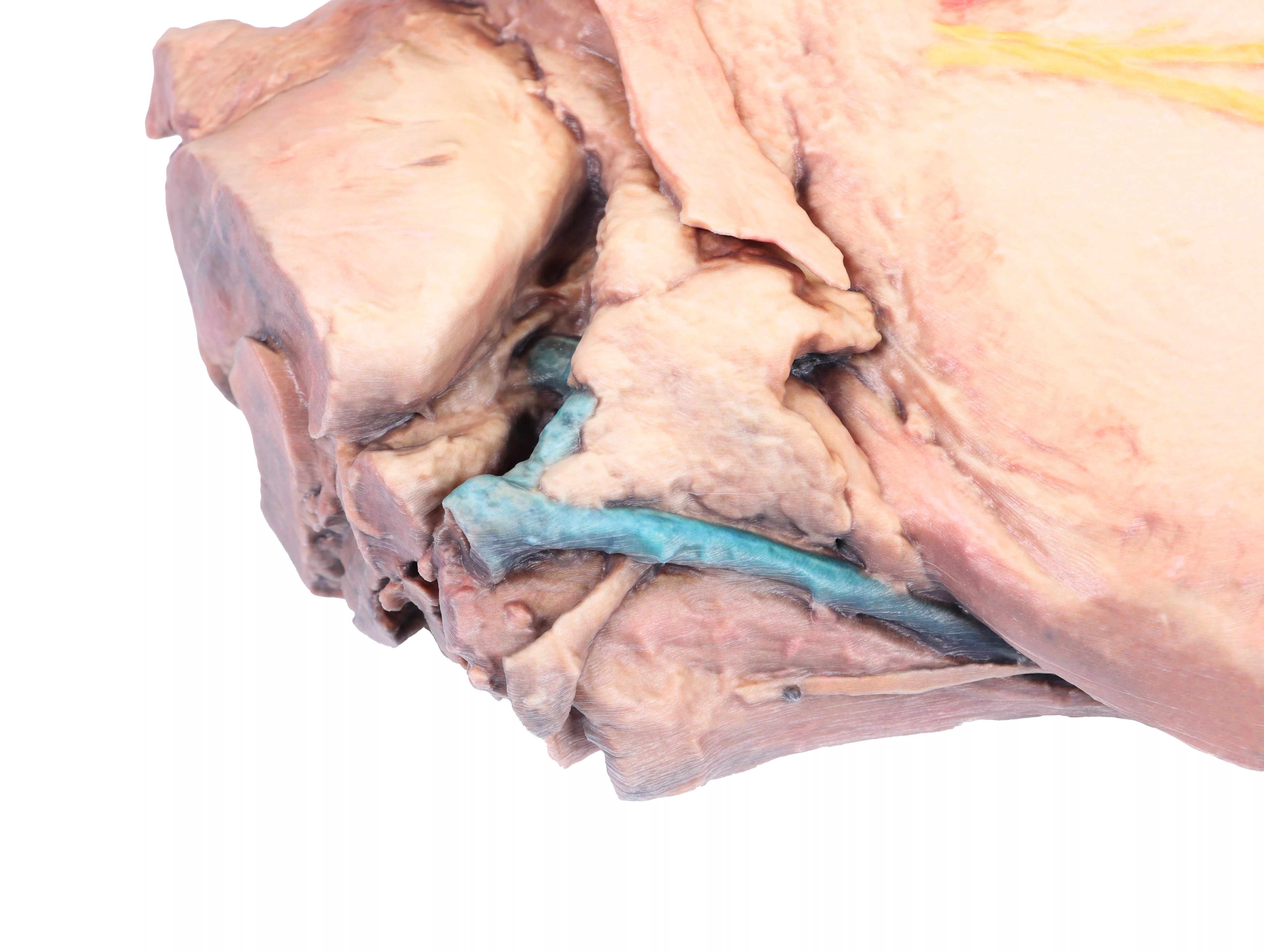
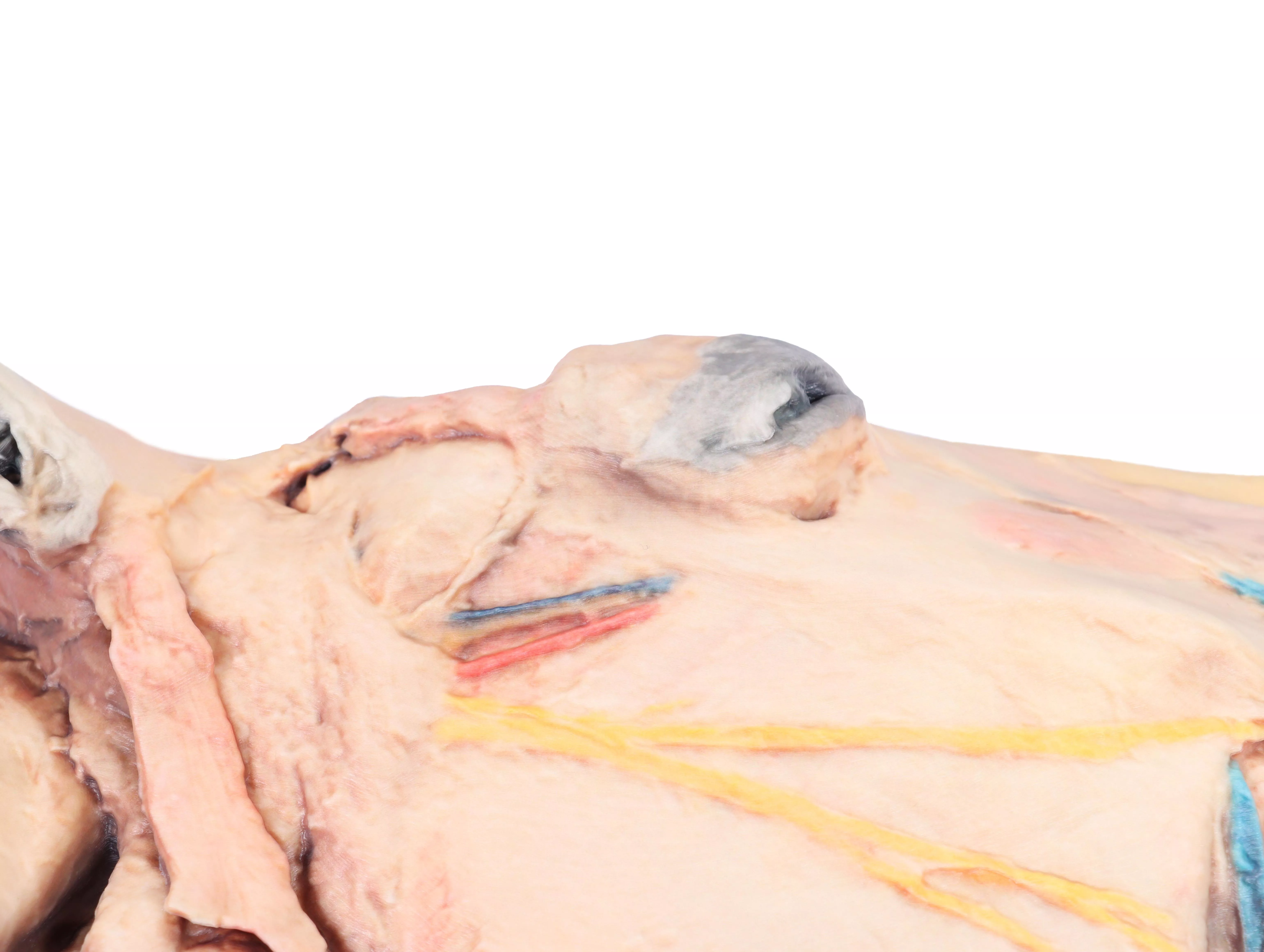
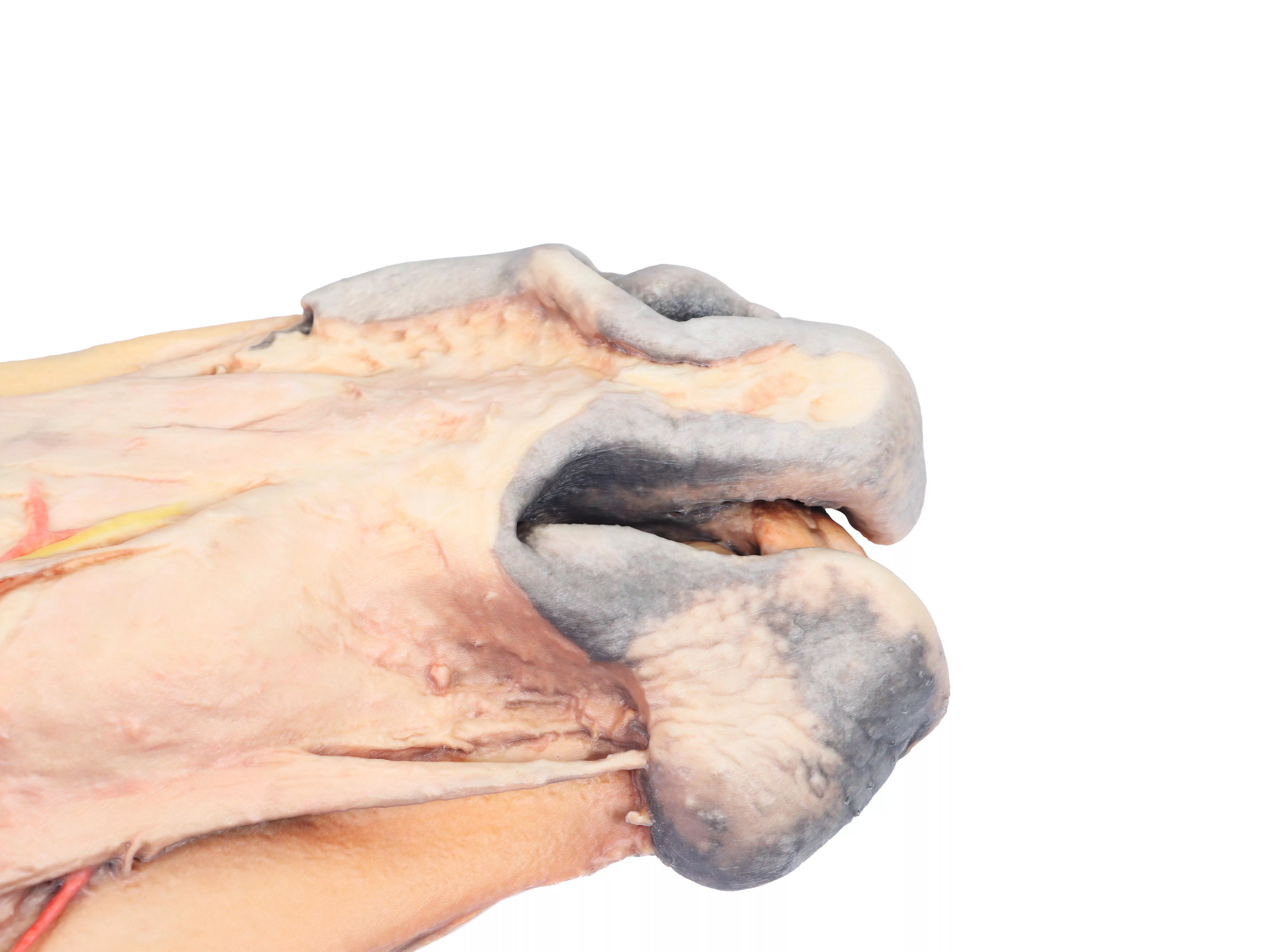
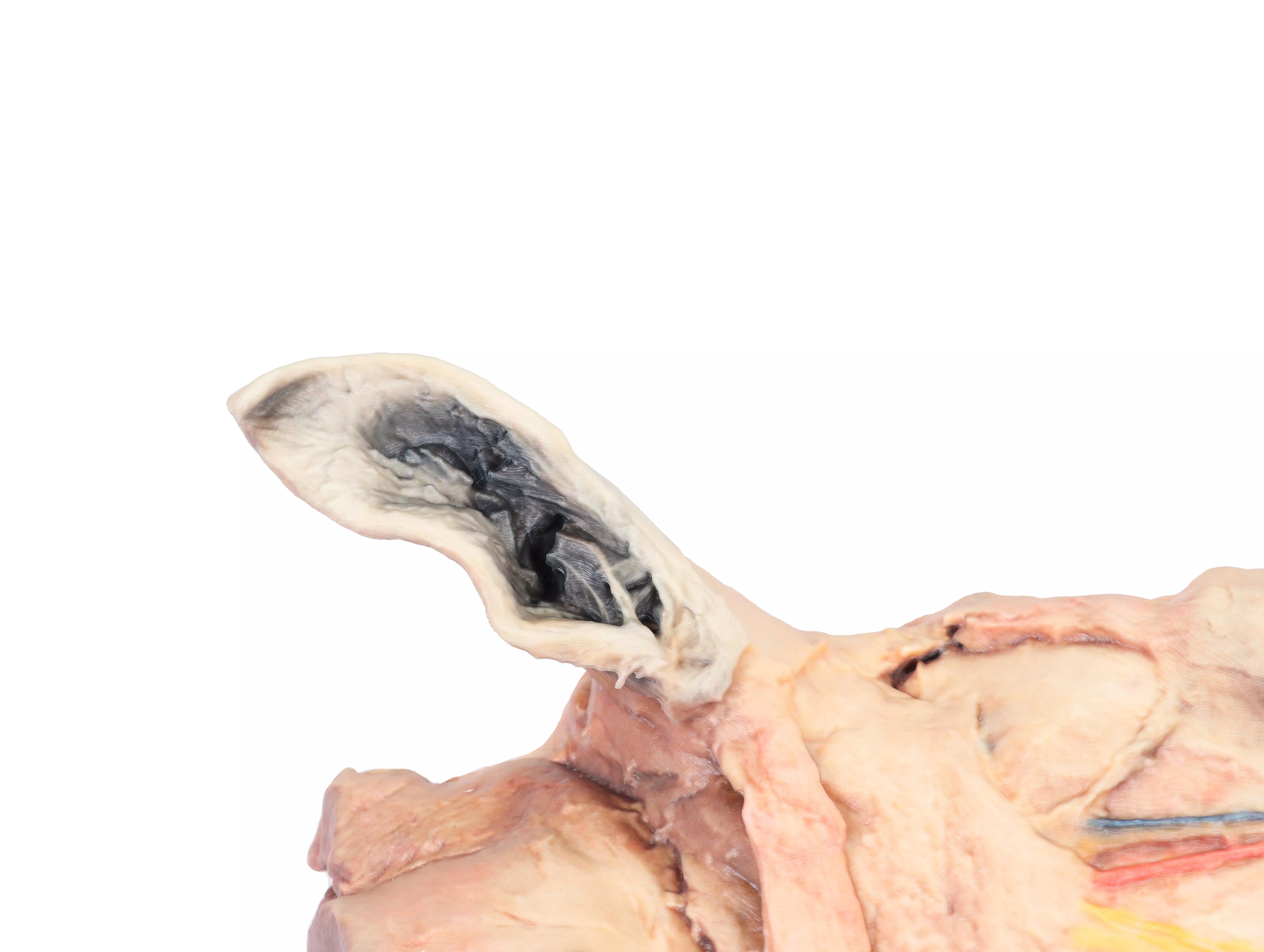
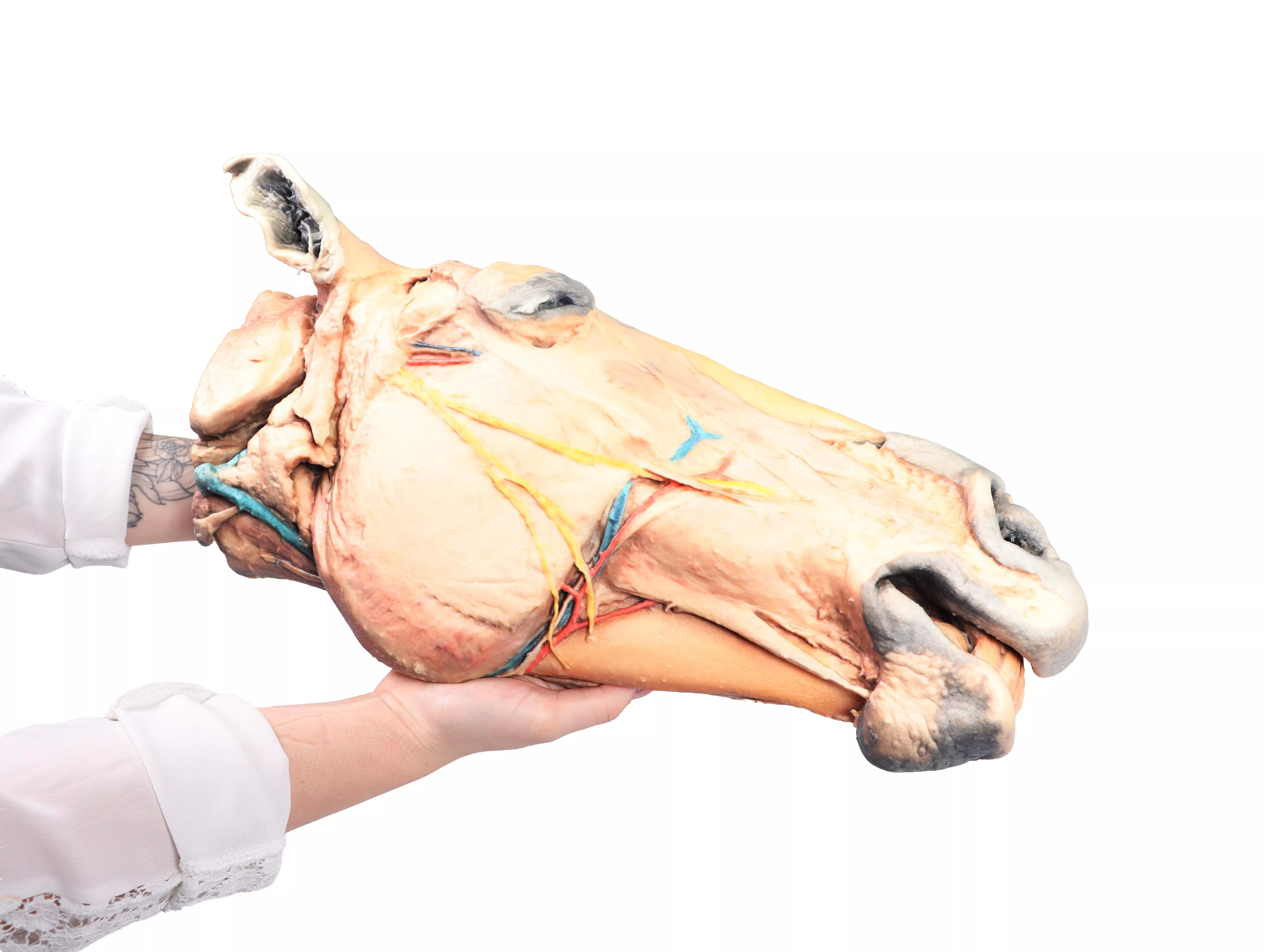
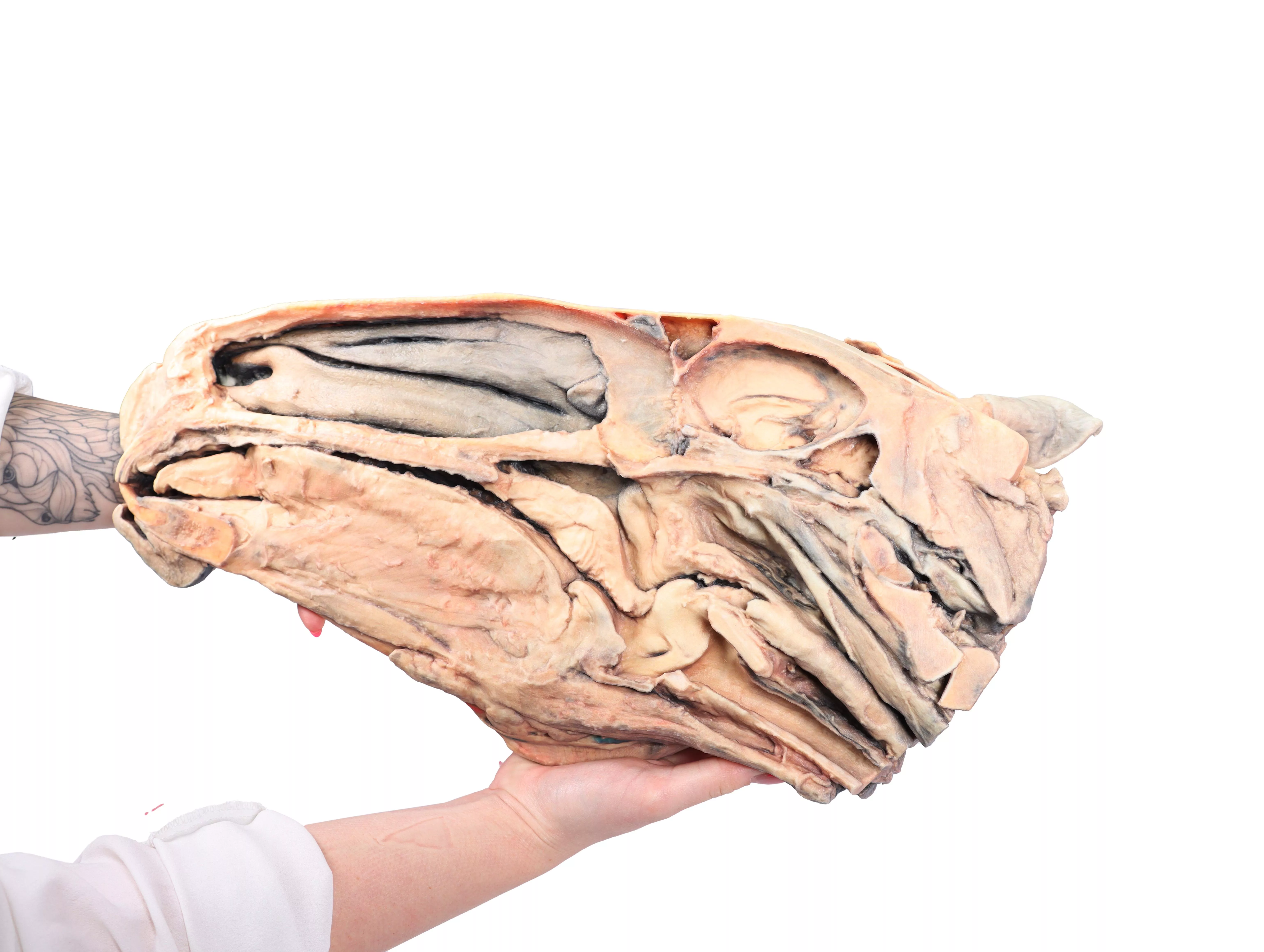
This horse half-head shows a superficial dissection on the right lateral aspect and a middle sagittal section on the medial aspect.
On the lateral side the skin has been removed, except on the nostrils, lips and external ear. In the rostral part of the dissection are the muscles of facial mimicry, the levator nasolabialis, canine, levator labii superioris, zygomaticus, buccinator (buccal portion) and depressor labii inferioris muscles. The caudal part of the dissection is occupied by the masseteric muscle covered by the masseteric fascia from the facial crest of the maxillary bone. This muscle presents on the surface the course of the dorsal and ventral buccal branches of the facial nerve, which end rostrally as buccolabial branches. In the notch of the facial vessels, the facial artery and vein are located together with the parotid duct. In the dorsal and caudal part of the masseter muscle, the transverse facial artery and vein are located together with the auriculotemporal nerve. The parotid-auricular muscle, the parotid gland, and the maxillary and linguofacial veins are located in the cervicofacial transit.
In the middle sagittal section, the nasal cavity is occupied by the ventral nasal concha, dorsal nasal concha, and middle nasal concha, separated by the dorsal, middle, and ventral nasal meatuses. The perpendicular lamina of the ethmoid bone is located in the caudal area of the nasal cavity. This bone is the rostral limit of the cerebral fossa. The cerebellar fossa is caudal to the anterior one. The vomer bone establishes the choanae as transit to the nasopharynx where the pharyngeal opening of the auditory tube is located. The oral cavity is limited dorsally by the hard palate, which continues caudally as the soft palate. The sagittal section of the tongue shows the proper lingual muscle as well as the genioglossus and genihyoid muscles. The oropharynx is occupied by the root of the tongue and the soft palate. The epiglottis, arytenoid cartilage, and cricoid cartilage limit the laryngeal cavity with the lateral ventricle, vocal fold, and infraglottic cavity. The laryngopharynx is occupied by the esophageal vestibule. The esophagus is located dorsal to the trachea and ventral to the long cervical and long capitis muscles. The mandibular lymph nodes are in the intermandibular space.
Erler-Zimmer
Erler-Zimmer GmbH & Co.KG
Hauptstrasse 27
77886 Lauf
Germany
info@erler-zimmer.de
Achtung! Medizinisches Ausbildungsmaterial, kein Spielzeug. Nicht geeignet für Personen unter 14 Jahren.
Attention! Medical training material, not a toy. Not suitable for persons under 14 years of age.


