Produktinformationen "Spleen and pancreas"
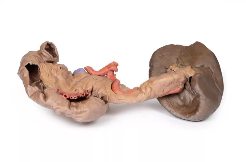
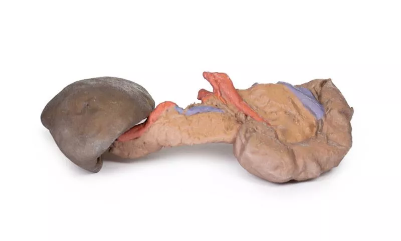
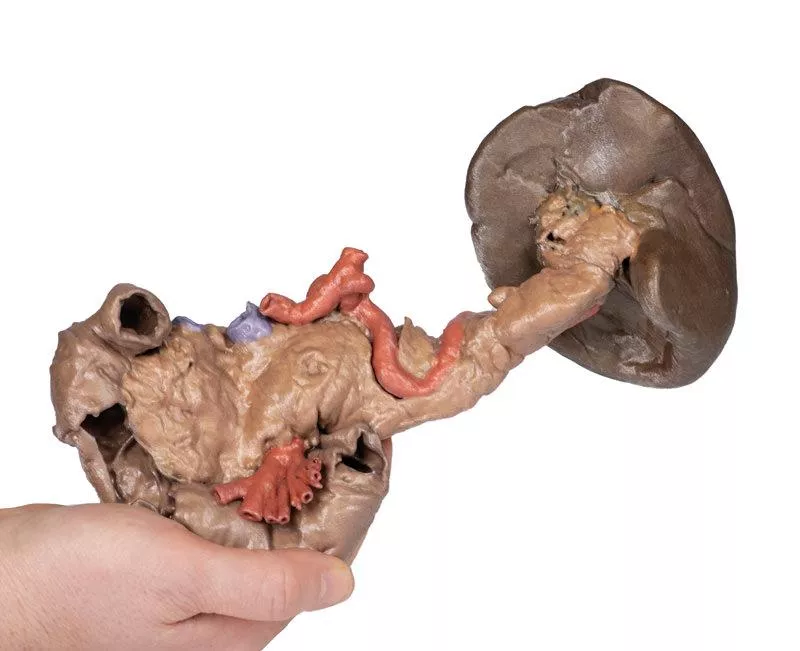
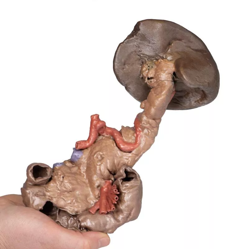
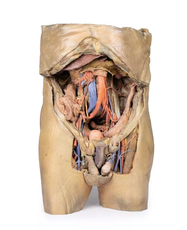
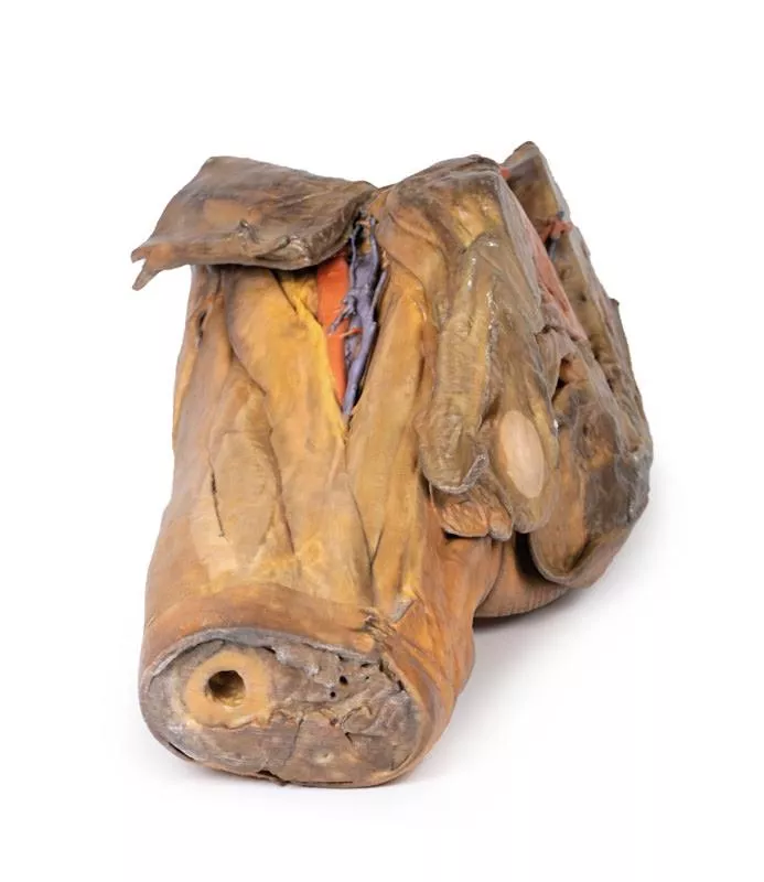
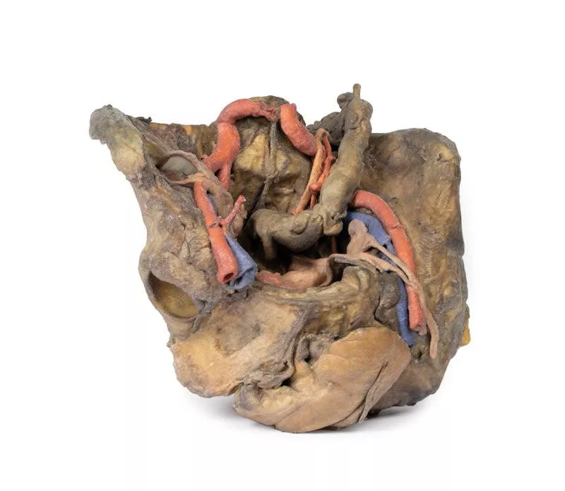
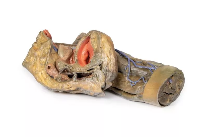
Dieses detaillierte anatomische 3D-Modell zeigt die tief liegenden Organe des Vorderdarms, darunter den absteigenden, horizontalen und aufsteigenden Teil des Zwölffingerdarms, die Bauchspeicheldrüse und die Milz.
Es bietet einen einzigartigen und aufschlussreichen Einblick in die komplexe Anatomie dieser Region.
Zwölffingerdarm
Im Zwölffingerdarm ist ein kleines Fenster geöffnet, um die Plicae circularis, die charakteristischen kreisförmigen Falten des proximalen Dünndarms, sichtbar zu machen. Dies steht im Kontrast zu den markanten Falten des Magens und ermöglicht einen lehrreichen Vergleich der Schleimhautmuster im oberen Magen-Darm-Trakt.
Bauchspeicheldrüse
Die Bauchspeicheldrüse ist in ihrer natürlichen anatomischen Position erhalten, eingebettet in die Krümmung des Zwölffingerdarms. Der Kopf der Bauchspeicheldrüse ist deutlich sichtbar, einschließlich des ausgeprägten Uncus an seinem distalen Rand, der an den Ursprung der A. mesenterica superior (SMA) angrenzt. In diesem Modell ist die SMA bereits in ihre wichtigsten benannten Äste unterteilt, wodurch ihre vaskuläre Komplexität hervorgehoben wird.
Der Körper der Bauchspeicheldrüse weist den oberen Rand auf, an dem sich der aus der absteigenden Bauchaorta abgezweigte Truncus coeliacus befindet. Die gesamte Milzarterie ist mit ihrem gewundenen Verlauf vom Truncus coeliacus zur Milz dargestellt. Das Modell zeigt auch die Ursprünge der linken Magenarterie und der gemeinsamen Leberarterie, die vom Truncus coeliacus abzweigen.
Angrenzend an den Truncus coeliacus ist ein Abschnitt der Milzvene zu sehen, der aus der Pankreaskapsel austritt. Diese Vene verläuft entlang der Milzarterie auf ihrem Weg zur Milz. Zusätzlich ist ein Teil der Vena mesenterica superior zu sehen, der an der hinteren Bauchspeicheldrüse anliegt und ihren Verlauf darstellt, bevor sie mit der Milzvene zusammenfließt, um die Pfortader zu bilden.
Der Schwanz der Bauchspeicheldrüse ist in die Milzkapsel eingebettet und verdeckt teilweise die Äste der Milzarterie, bevor diese in die Milz eintritt. Für detailliertere Ansichten dieser Region verweisen wir auf unsere anderen Milzmodelle (MP1130 und MP1134), die weitere anatomische und räumliche Zusammenhänge veranschaulichen.
Erler-Zimmer
Erler-Zimmer GmbH & Co.KG
Hauptstrasse 27
77886 Lauf
Germany
info@erler-zimmer.de
Achtung! Medizinisches Ausbildungsmaterial, kein Spielzeug. Nicht geeignet für Personen unter 14 Jahren.
Attention! Medical training material, not a toy. Not suitable for persons under 14 years of age.


