


")


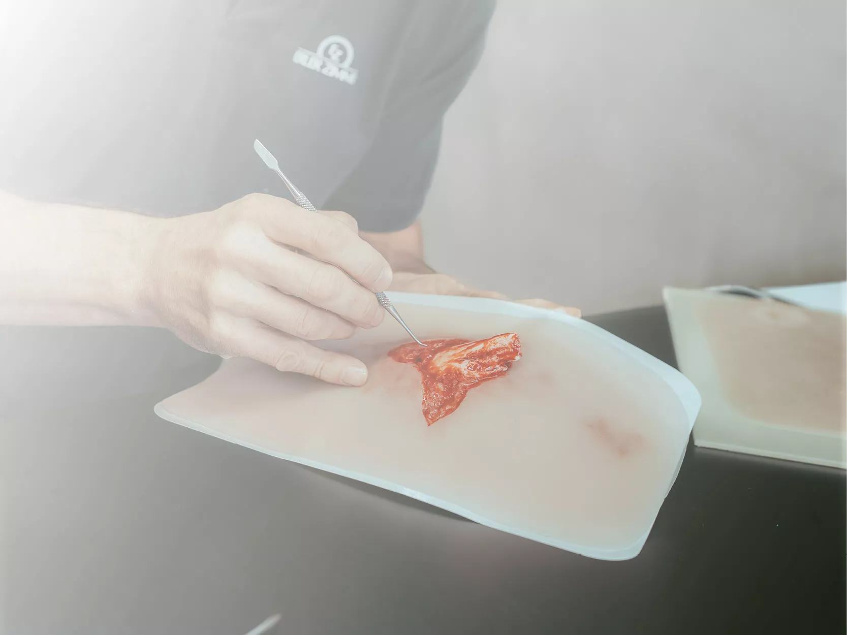
Produktinformationen "Female hemipelvis and thigh"
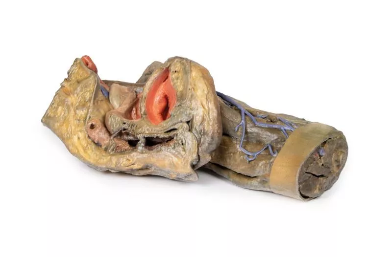
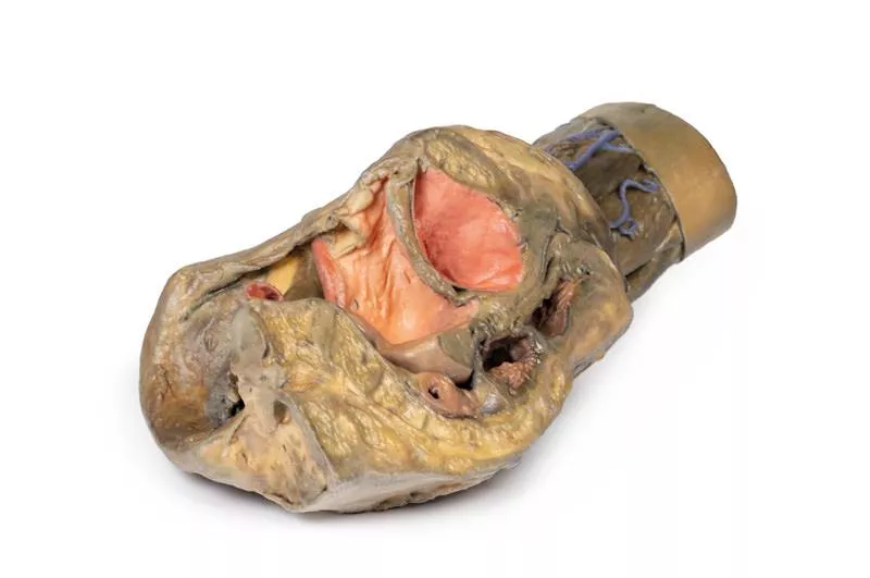
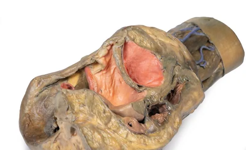
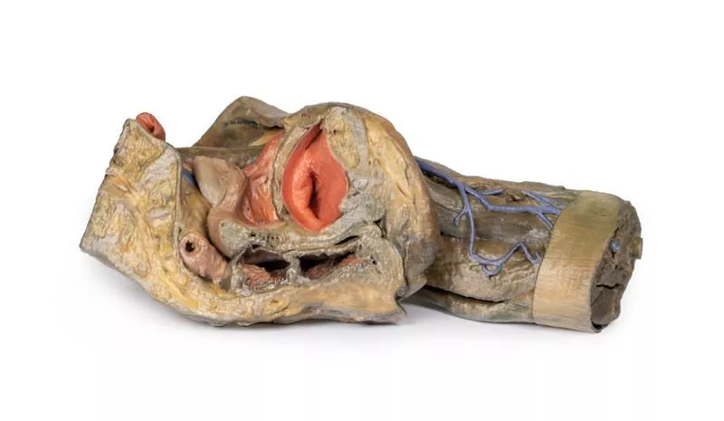
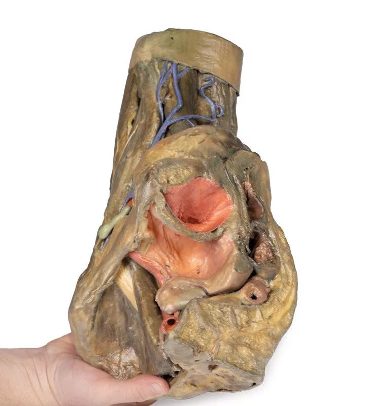
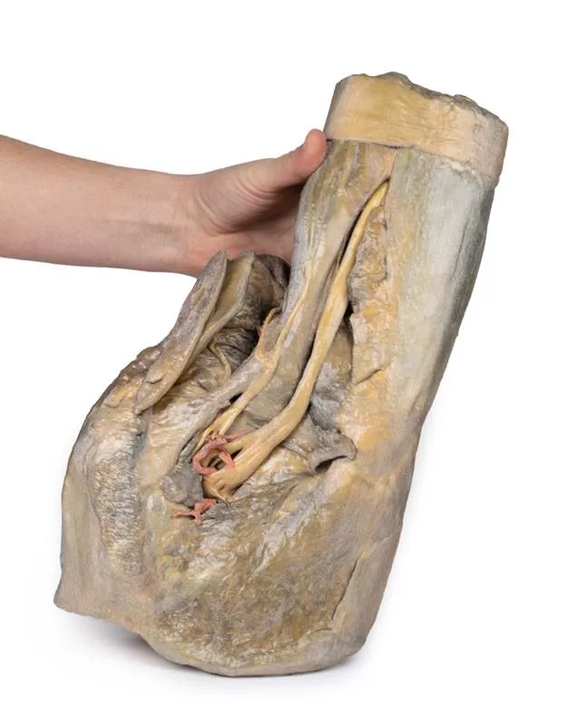
This 3D model preserves a left pelvis divided at the midsagittal plane, and the proximal thigh to approximately the midthigh.
In the midsagittal section, the urinary bladder, uterus and vagina, and rectum can be seen in sequence between the pubic symphysis (anteriorly) and the sacrum (posteriorly). The retention of the peritoneum draped across the superior surface of these organs allows for view of the vesicouterine and rectouterine pouches. The reflection of peritoneum off the uterus forms the broad ligament, with the uterine tube, fimbrae, and closely associated left ovary in position near the pelvic brim. Lateral to the true pelvis contents the common and external iliac arteries can be viewed passing towards the subinguinal space between the common iliac vein and the psoas major muscle. The descending course of the ureter can be traced across these vessels, and the femoral nerve is visible between the psoas major and iliacus muscles.
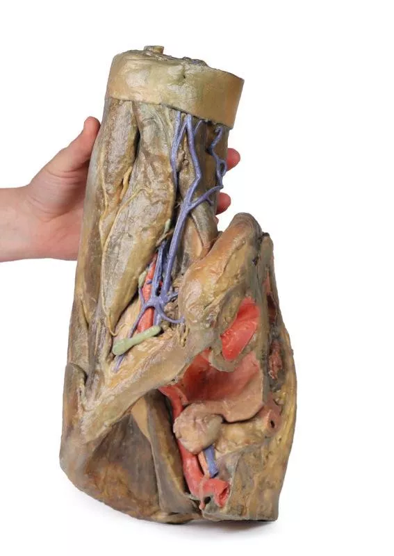
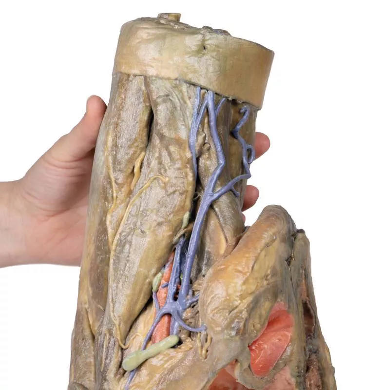
The superficial fascia has been removed across the entire thigh to the lateral margin of the perineum and near the inferior sectioning of the model itself. Anteriorly, the femoral triangle region has been dissected to expose the content as well as the horizontal group of inguinal lymph nodes immediately inferior to the inguinal ligament. Medially, the femoral vein receives drainage through the great saphenous vein and regional veins (including the superficial circumflex iliac, the superficial external pudendal, and the deep pudendal veins). The femoral artery can be seen immediately lateral to the vein, with parts of the femoral nerve descending just lateral to the artery and near the tendon of the iliopsoas muscle. Although somewhat disturbed by dissection, the anterior cutaneous nerves of the thigh and a small part of the lateral cutaneous nerve of the thigh can be seen on the superficial aspect of the sartorius muscle.
Posteriorly, the gluteal region has been dissected with removal of the gluteus maximus to expose the underlying gluteal muscles, with reflection of the piriformis muscle revealing the neurovascular structures in the region. The sciatic nerve can be seen forming through contributions by the tibial and common peroneal nerves around the preserved portions of the superior and inferior gluteal arteries. Medially the posterior cutaneous nerve of the thigh runs in parallel with the sciatic nerve, with both resting on the obturator internus tendon and gemelli muscles before descending into the thigh on top of the quadratus femoris and common hamstring origin, respectively. Deep to the sacrotuberous ligament the course of the internal pudendal artery and pudendal nerve can be followed towards the ischioanal fossa, where the internal pudendal artery arcs anteriorly and the inferior rectal branch of the pudendal nerve can be seen reaching the pelvic diaphragm and external anal sphincter muscle.
In the midsagittal section, the urinary bladder, uterus and vagina, and rectum can be seen in sequence between the pubic symphysis (anteriorly) and the sacrum (posteriorly). The retention of the peritoneum draped across the superior surface of these organs allows for view of the vesicouterine and rectouterine pouches. The reflection of peritoneum off the uterus forms the broad ligament, with the uterine tube, fimbrae, and closely associated left ovary in position near the pelvic brim. Lateral to the true pelvis contents the common and external iliac arteries can be viewed passing towards the subinguinal space between the common iliac vein and the psoas major muscle. The descending course of the ureter can be traced across these vessels, and the femoral nerve is visible between the psoas major and iliacus muscles.
The superficial fascia has been removed across the entire thigh to the lateral margin of the perineum and near the inferior sectioning of the model itself. Anteriorly, the femoral triangle region has been dissected to expose the content as well as the horizontal group of inguinal lymph nodes immediately inferior to the inguinal ligament. Medially, the femoral vein receives drainage through the great saphenous vein and regional veins (including the superficial circumflex iliac, the superficial external pudendal, and the deep pudendal veins). The femoral artery can be seen immediately lateral to the vein, with parts of the femoral nerve descending just lateral to the artery and near the tendon of the iliopsoas muscle. Although somewhat disturbed by dissection, the anterior cutaneous nerves of the thigh and a small part of the lateral cutaneous nerve of the thigh can be seen on the superficial aspect of the sartorius muscle.
Posteriorly, the gluteal region has been dissected with removal of the gluteus maximus to expose the underlying gluteal muscles, with reflection of the piriformis muscle revealing the neurovascular structures in the region. The sciatic nerve can be seen forming through contributions by the tibial and common peroneal nerves around the preserved portions of the superior and inferior gluteal arteries. Medially the posterior cutaneous nerve of the thigh runs in parallel with the sciatic nerve, with both resting on the obturator internus tendon and gemelli muscles before descending into the thigh on top of the quadratus femoris and common hamstring origin, respectively. Deep to the sacrotuberous ligament the course of the internal pudendal artery and pudendal nerve can be followed towards the ischioanal fossa, where the internal pudendal artery arcs anteriorly and the inferior rectal branch of the pudendal nerve can be seen reaching the pelvic diaphragm and external anal sphincter muscle.
Anmelden
Erler-Zimmer
Erler-Zimmer GmbH & Co.KG
Hauptstrasse 27
77886 Lauf
Germany
info@erler-zimmer.de
Achtung! Medizinisches Ausbildungsmaterial, kein Spielzeug. Nicht geeignet für Personen unter 14 Jahren.
Attention! Medical training material, not a toy. Not suitable for persons under 14 years of age.
Andere Kunden kauften auch

Stetige Innovationskraft

Soziale Verantwortung

Gelebte Kundenorientierung

Verständnis für Qualität

Nachhaltiges Handeln

Zertifizierung ISO 9001