Becken
3
Produkte
Sortierung:
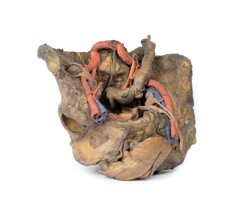
Female pelvis deep dissection
5.472,81 €*
This 3D model presents a deep dissection and isolation of the pelvis from surrounding regions, particularly demonstrating visceral and neurovascular structures relative to deep ligaments and osseous features.Within the false pelvis, the sigmoid colon descends on the left side of the specimen to the rectum, passing superficially across the pelvic brim and the passage of the common and external iliac artery and vein. Adjacent to the sigmoid colon are parts of the sigmoid arteries and superior rectal artery, resting superficial to the common iliac vessels and near the descending ureter. Anterior in the true pelvis is the collapsed urinary bladder, and between the bladder and rectum rests the uterus. The organ is partially covered in the broad ligament, with both the suspensory ligament of the ovary and round ligament have been separated and pulled away from the peritoneum on both sides to expose surrounding blood vessels. While the ovarian ligaments, round ligaments, uterine tubes and ovaries are trapped within the peritoneal fold of the broad ligament, the reduction in ovary size (common with advanced age) has rendered these indistinguishable in the model.Lateral to these organs, branches of the internal iliac artery can be identified – as well as a retained median sacral artery in the midline between the two common iliac arteries. On the left side only the uterine artery can be seen laterally. On the right side, the obturator, superior vesical, and uterine arteries can be observed. In addition, the origins of the inferior epigastric artery and vein can be seen arising from the external iliac vessels just prior to exiting the inferior abdominal cavity.On the right side of the preserved pelvis, the entire femur and thigh musculature has been removed to demonstrate the obturator membrane, the articular cartilage of the acetabulum and the transverse ligament of the acetabulum. Posteriorly the entire gluteal region has been dissected to expose the superior gluteal foramen and the origin of the superior gluteal artery. The sacrotuberous ligament has been removed to demonstrate the sacrospinous ligament, with some branches of the inferior rectal artery retained within the exposed ischoanal fossa.On the left side of the preserved pelvis the sciatic nerve has been maintained within the greater sciatic foramen, as has the sacrotuberous ligament. The ischioanal fossa mirrors that of the right side, where branches of the inferior rectal artery have been retained relative to the fibres of the pelvic diaphragm, and the integration of the external anal sphincter on the projecting external rectal surface.
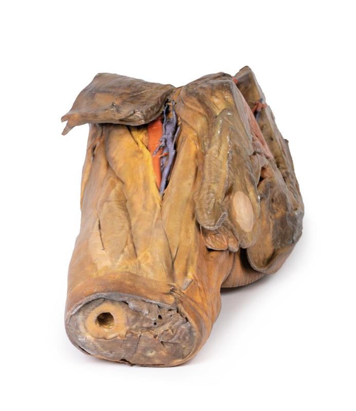
Male hemipelvis and thigh
5.853,61 €*
This 3D model preserves a right male pelvis sectioned just superior to the L5 vertebra and sectioned at the midsagittal plane, with the thigh preserved to near the midshaft of the femur. This specimen compliments our LW 91 female hemipelvic specimen and thigh.The common iliac artery is preserved with several key branches visible, particularly the distribution of the internal iliac within the true pelvis. Several major vessels including the obturator artery and the partially obliterated umbilical artery passes towards the anterior abdominal wall (to form the medial umbilical ligament) and gives off the superior vesicle artery; while the roots of the iliolumbar, superior gluteal, inferior gluteal and internal pudendal artery are visible lateral to the urinary bladder. The ureter descends superficial to these vessels to approach the urinary bladder which is covered with peritoneum in this model. The ductus deferens is exposed from the entry into the space via the deep inguinal ring and passing posteriorly (though sectioned from its normal insertion pathway and resting on the internal iliac artery). Adjacent to the ureter and on the superficial surface of the psoas major muscle is an enlarged iliac lymph node and part of the lymphatic vasculature ascending along the external iliac artery. The majority of the pelvis has been left undissected, allowing for an appreciation of the rectovesicular pouch and the exposed superior rectal artery and vein approaching the preserved portion of rectum. In cross section, the rectum, seminal vesicle and prostate are visible (the section plane preserves parts of both the prostatic urethra and ejaculatory duct).In the anterior thigh the borders and contents of the femoral triangle are well-preserved, with partial coverage by the flap of the anterior abdominal wall. Posteriorly the skin over the gluteal region and the gluteus maximus muscle have been removed as sequential windows to expose the gluteus medius and minimum muscles, the piriformis, the obturator internus with gemelli muscles, and the quadratus femoris muscle. The superior and inferior gluteal arteries are maintained superior and inferior to the piriformis, respectively; with the sciatic nerve exiting inferior to piriformis before passing deep to the retained portion of the gluteus maximus.
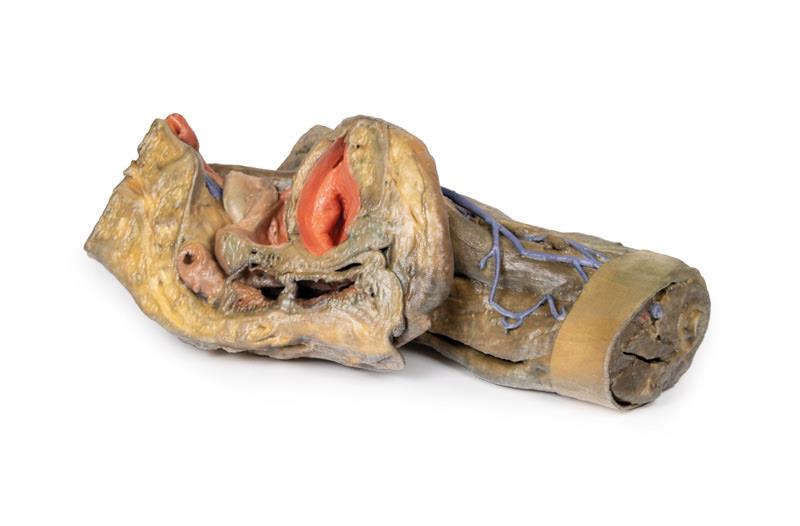
Female hemipelvis and thigh
3.292,73 €*
This 3D model preserves a left pelvis divided at the midsagittal plane, and the proximal thigh to approximately the midthigh.In the midsagittal section, the urinary bladder, uterus and vagina, and rectum can be seen in sequence between the pubic symphysis (anteriorly) and the sacrum (posteriorly). The retention of the peritoneum draped across the superior surface of these organs allows for view of the vesicouterine and rectouterine pouches. The reflection of peritoneum off the uterus forms the broad ligament, with the uterine tube, fimbrae, and closely associated left ovary in position near the pelvic brim. Lateral to the true pelvis contents the common and external iliac arteries can be viewed passing towards the subinguinal space between the common iliac vein and the psoas major muscle. The descending course of the ureter can be traced across these vessels, and the femoral nerve is visible between the psoas major and iliacus muscles.The superficial fascia has been removed across the entire thigh to the lateral margin of the perineum and near the inferior sectioning of the model itself. Anteriorly, the femoral triangle region has been dissected to expose the content as well as the horizontal group of inguinal lymph nodes immediately inferior to the inguinal ligament. Medially, the femoral vein receives drainage through the great saphenous vein and regional veins (including the superficial circumflex iliac, the superficial external pudendal, and the deep pudendal veins). The femoral artery can be seen immediately lateral to the vein, with parts of the femoral nerve descending just lateral to the artery and near the tendon of the iliopsoas muscle. Although somewhat disturbed by dissection, the anterior cutaneous nerves of the thigh and a small part of the lateral cutaneous nerve of the thigh can be seen on the superficial aspect of the sartorius muscle.Posteriorly, the gluteal region has been dissected with removal of the gluteus maximus to expose the underlying gluteal muscles, with reflection of the piriformis muscle revealing the neurovascular structures in the region. The sciatic nerve can be seen forming through contributions by the tibial and common peroneal nerves around the preserved portions of the superior and inferior gluteal arteries. Medially the posterior cutaneous nerve of the thigh runs in parallel with the sciatic nerve, with both resting on the obturator internus tendon and gemelli muscles before descending into the thigh on top of the quadratus femoris and common hamstring origin, respectively. Deep to the sacrotuberous ligament the course of the internal pudendal artery and pudendal nerve can be followed towards the ischioanal fossa, where the internal pudendal artery arcs anteriorly and the inferior rectal branch of the pudendal nerve can be seen reaching the pelvic diaphragm and external anal sphincter muscle.
Menschliche Körperrepliken, um die Lehre zu verbessern!
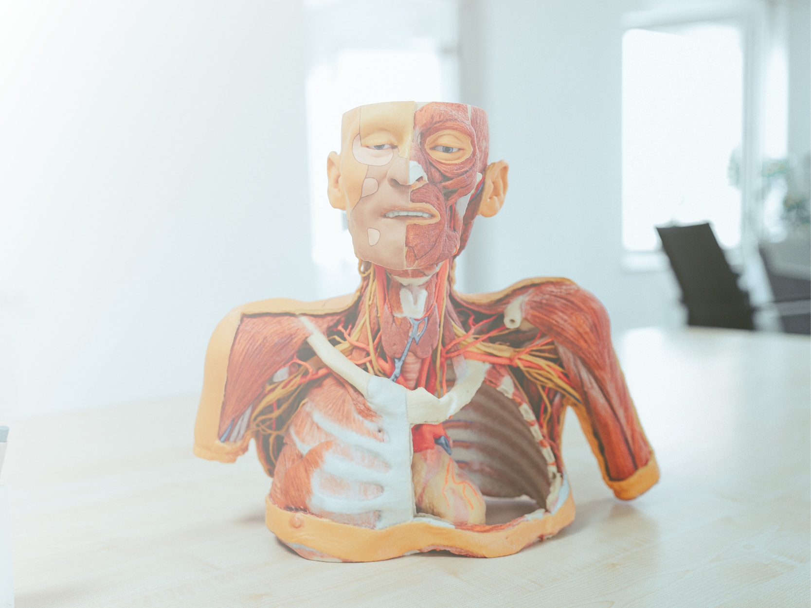
Die bahnbrechende Anatomie Serie von Erler- Zimmer beinhaltet eine einzigartige und unerreichte Sammlung von kolorierten menschlichen Körperrepliken welche speziell entworfen wurden, um die Lehre und das Lernen zu verbessern. Diese Premiumkollektion von höchst akkurater humaner Anatomie wurde direkt aus radiologischen Daten oder echten Präparaten mit neuesten Bildgebenden Verfahren erzeugt. Die 3D menschliche Anatomie Serie bietet einen kosteneffektiven Weg, um Ihrem speziellen Unterrichts- und Demonstrationsbedarf im gesamten curricularen Bereich der Medizin, Gesundheitswissenschaften und der Biologie gerecht zu werden. Eine detaillierte Beschreibung der Anatomie, welche in jedem 3D-gedruckten Präparat dargestellt wird, wir mitgeliefert. Welche Vorteile bietet die Monash 3D Anatomie Serie im Vergleich zu Plastikmodellen oder echten menschlichen Plastinaten?
Jede Körperreplik wurde sorgfältig entwickelt aus ausgewählten radiologischen Patientendaten oder präparierten menschlichen Körpern höchster Qualität, welche von einem hochqualifizierten Anatomenteam im Lehrzentrum für menschliche Anatomie der Monash Universität ausgewählt wurden, um klinisch wichtige Bereiche der Anatomie in einer Qualität und Detailtreue darzustellen, wie es mit konventionellen Modellen nicht möglich ist – es handelt sich um echte Anatomie, nicht um stilisierte.
Jede Körperreplik wurde strengstens überprüft vom hochqualifizierten Anatomenteam im Lehrzentrum für menschliche Anatomie der Monash Universität, um die anatomische Genauigkeit des Endprodukts zu gewährleisten. Die Körperrepliken sind kein echtes menschliches Gewebe und unterliegen deshalb keinen Einschränkungen beim Transport, Import oder der Verwendung in Bildungseinrichtungen, die keine Erlaubnis zur Verwendung von Leichen haben. Die
Die exklusive 3D Anatomie Serie vermeidet diese und andere ethische Probleme, welche auftreten, wenn man mit plastinierten menschlichen Überresten umgeht.
