Cholelithiasis (Gallstones)
Artikel in Produktion, lieferbar vorauss. in 2-3 Wochen
Produktnummer:
MP2075
Artikelnummer: MP2075
Produktinformationen "Cholelithiasis (Gallstones)"
Clinical History
A middle-aged woman was investigated for recurrent bouts of epigastric pain. Endoscopy failed to reveal any peptic ulceration. A cholangiogram demonstrated a non-functioning gallbladder. She died from a myocardial infarction Unfortunately, the patient dies at a later stage after this procedure from a myocardial infarction”.
Pathology
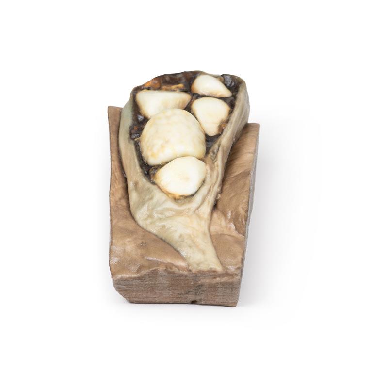
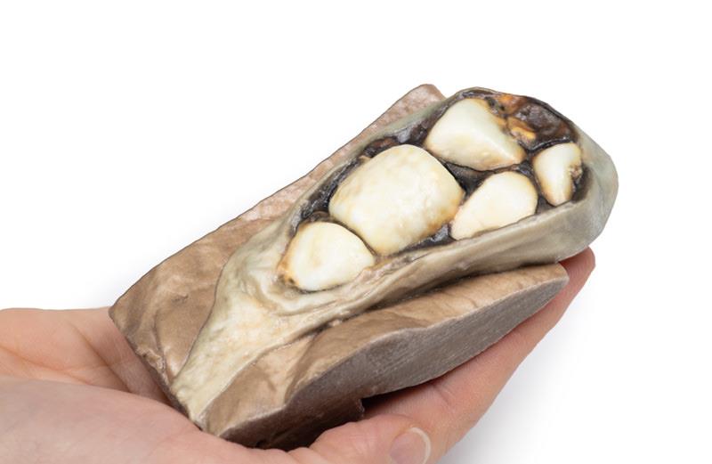
The specimen is a portion of liver with attached gallbladder, which has been opened to display six large faceted mixed calculi. This is an example of cholelithiasis (gallstones).
Further Information
Gallstones contain a mixture of cholesterol, calcium salts, bilirubin, proteins, and mucin. There is a high prevalence in fair-skinned populations. Risk factors are age (greater than 50) and female sex, along with genetic factors1, pregnancy, diabetes mellitus and dyslipidaemia. Lifestyle factors, such as rapid weight loss and certain medications (e.g. erythromcyin, ampicillin, octreotide, cephalosporin), can also promote gallstone formation.
Gallstones may be asymptomatic or may present with a spectrum of disease ranging from uncomplicated biliary colic through to infection, cholecystitis, pancreatitis or gallstone ileus. The typical symptoms include bouts of epigastric or right upper quadrant pain, sometimes associated with eating, and often with sweating, nausea and vomiting. Pain is usually caused by the gallbladder or biliary tract contracting forcefully against a stone, thereby causing increased pressure in the gallbladder, and pain. The risk of developing complications of gallstones is approximately 2-3 percent per year once biliary colic develops. Diagnosis is generally via transabdominal ultrasound, which has largely replaced oral cholecystography studies. Cholescintigraphy (HIDA Scan) can be used distinguish biliary colic from acute cholecystitis. Treatment of attacks are initially with simple analgesia, and subsequently definitive management usually includes elective laparoscopic cholecystectomy. Very severe cases can be life threatening, but deaths from gallstone disease are rare.
It should be noted that epigastric pain can also be caused by myocardial ischemia, particularly in females, who do not necessarily present with the classic ‘left shoulder tip pain’ of an acute myocardial infarct. Therefore, should gallbladder stones be excluded in a patient presenting with epigastric pain, an ECG should always be performed, to help rule out cardiac disease[1].
References
1. Portincasa P; Moscheta A; Palasciano G (2006) Cholesterol gallstone disease. Lancet. 2006; 368(9531):230-9
Erler-Zimmer
Erler-Zimmer GmbH & Co.KG
Hauptstrasse 27
77886 Lauf
Germany
info@erler-zimmer.de
Achtung! Medizinisches Ausbildungsmaterial, kein Spielzeug. Nicht geeignet für Personen unter 14 Jahren.
Attention! Medical training material, not a toy. Not suitable for persons under 14 years of age.
