Produktinformationen "Ruptured Thoracic Aortic Aneurysm"
Klinische Vorgeschichte
Für dieses Präparat sind keine klinischen Angaben verfügbar.
Pathologie
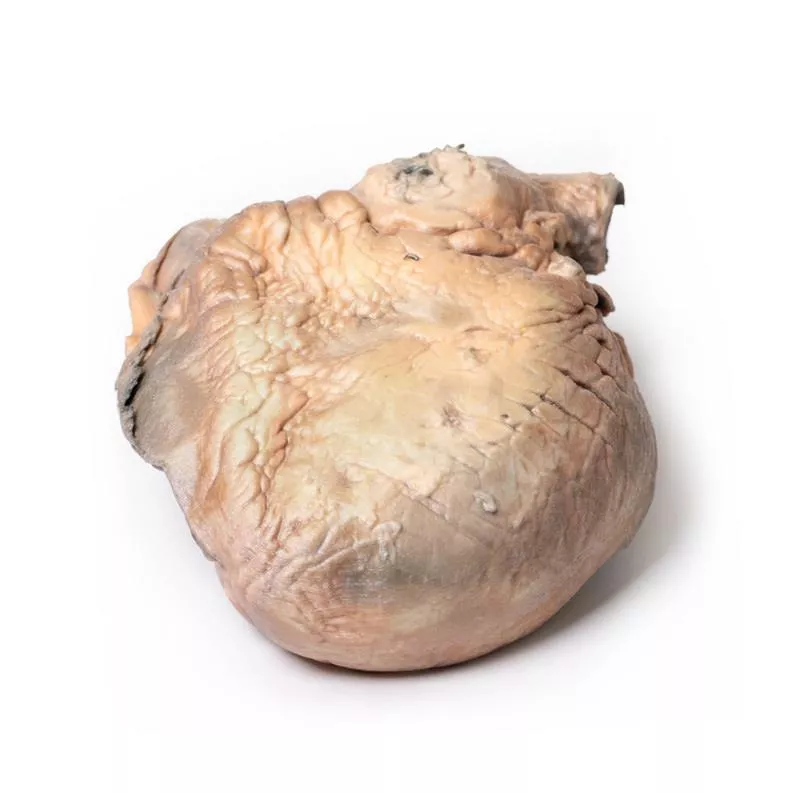
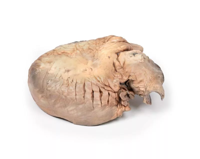
Das Herz wird von der Rückseite gezeigt, beide Ventrikel sind sichtbar. Die aufsteigende thorakale Aorta zeigt eine deutliche sackförmige Erweiterung mit mehreren atherosklerotischen Plaques. Die Aorta ist hinten rupturiert, erkennbar an der dunklen Färbung. Beide Ventrikel sind hypertrophiert, die Koronararterien sowie die Aorten- und Trikuspidalklappen sind normal. Dies stellt ein rupturiertes Aneurysma der aufsteigenden Aorta dar.
Weitere Informationen
Eine Erweiterung der aufsteigenden Aorta wird häufig zufällig bei transthorakaler Echokardiografie entdeckt. Die thorakale Aorta besteht aus drei Abschnitten: aufsteigend, Bogen und absteigend. Die aufsteigende Aorta beginnt direkt oberhalb der Aortenklappe und endet vor dem Brachiocephalicus, ist etwa 5 cm lang. Sie umfasst die Aortenwurzel (mit Koronarsinusen und Sinotubulärem Übergang) und den röhrenförmigen aufsteigenden Abschnitt. Über 50 % der thorakalen Aortenaneurysmen liegen hier und betreffen entweder die Wurzel oder den röhrenförmigen Abschnitt. Ein Aneurysma ist eine örtlich begrenzte Erweiterung der Aorta um mehr als 50 % des erwarteten Durchmessers (Verhältnis beobachtet/erwartet = 1,5) und unterscheidet sich von der Ektasie, einer diffusen Erweiterung unter 50 %. Die Inzidenz der aufsteigenden thorakalen Aortenaneurysmen beträgt etwa 10 pro 100.000 Personenjahre.
Referenz: Saliba et al. (2015). Int J Cardiol Heart Vasc. 6: 91–100.
Erler-Zimmer
Erler-Zimmer GmbH & Co.KG
Hauptstrasse 27
77886 Lauf
Germany
info@erler-zimmer.de
Achtung! Medizinisches Ausbildungsmaterial, kein Spielzeug. Nicht geeignet für Personen unter 14 Jahren.
Attention! Medical training material, not a toy. Not suitable for persons under 14 years of age.


