Produktinformationen "Traumatic Oesophageal-aortic fistula"
Klinische Vorgeschichte
Eine Frau verschluckte beim Mittagessen einen Chop-Knochen und brach später zusammen, begleitet von massiver Hämatemesis. Bei der Laparotomie war der Magen mit frischem Blut gefüllt, die Ursache wurde jedoch nicht gefunden. Am Folgetag verstarb sie. Die Autopsie zeigte eine Verbindung zwischen Aorta und Speiseröhre.
Pathologie
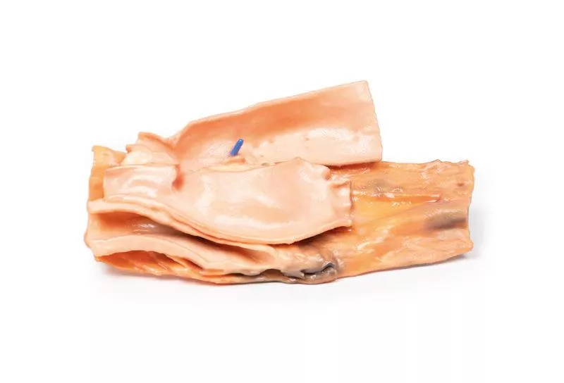
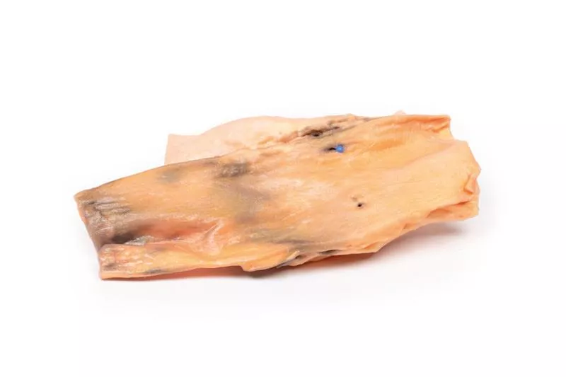
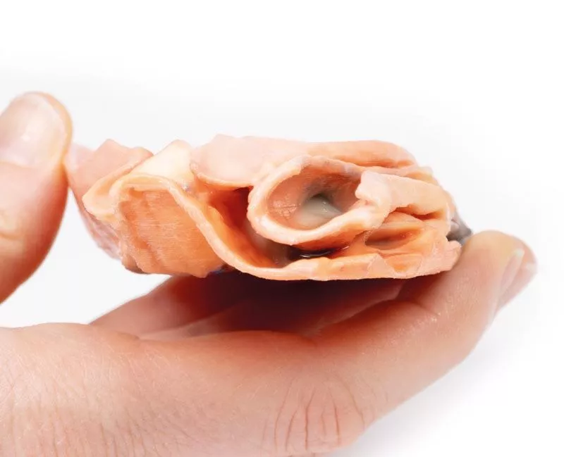
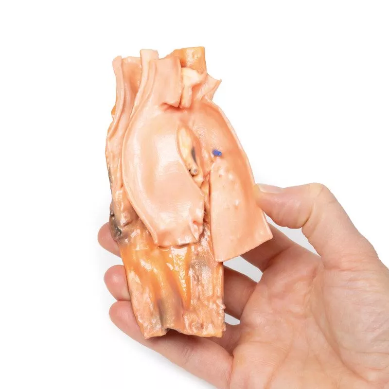
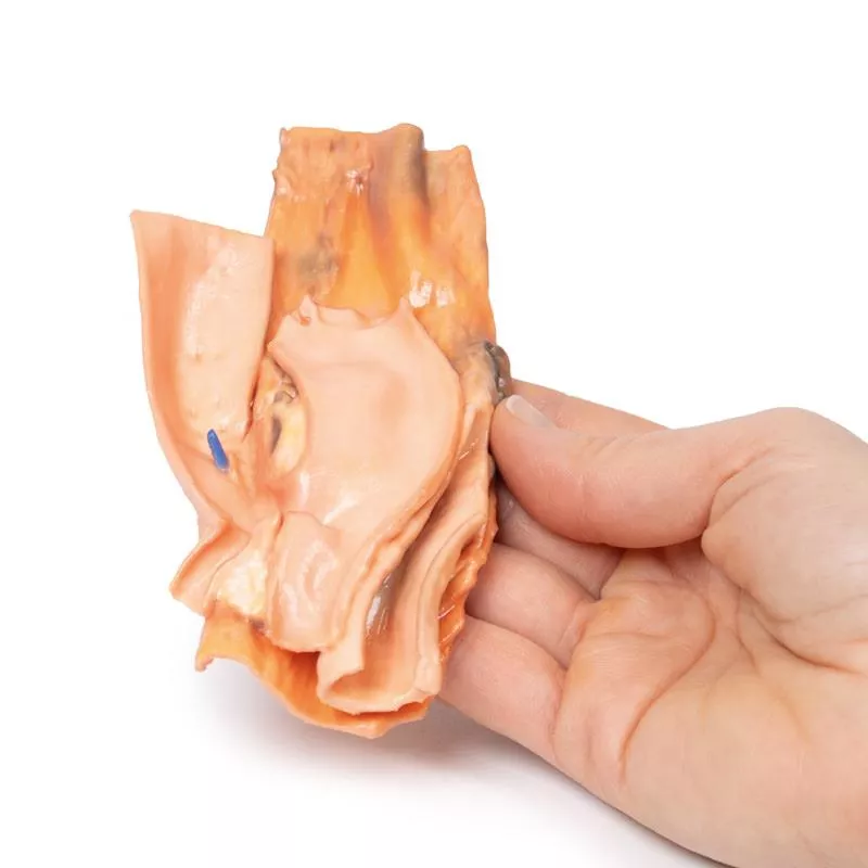
Das Präparat umfasst den distalen Tracheaabschnitt, den Aortenbogen (koronaler Schnitt, von vorne betrachtet) und die Speiseröhre (längs geöffnet). Die Speiseröhren-Schleimhaut ist ulzeriert und blutig. Eine kleine Sonde zeigt eine Fistel zwischen Speiseröhre und der hinteren Wand der thorakalen absteigenden Aorta.
Weitere Informationen
Obwohl dieser Fall traumatisch verursacht wurde, können aorto-ösophageale Fisteln auch durch nicht-traumatische Ursachen entstehen, etwa durch Druck von Aortenaneurysmen, fortgeschrittene Tumoren oder Erosion von Aortengrafts in den Verdauungstrakt. Diese Fisteln sind lebensbedrohlich und äußern sich meist durch gastrointestinale Blutungen, die von kleinen Blutungen bis zu lebensbedrohlichen Massenblutungen mit Kreislaufversagen reichen. Symptome sind Meläna, offensichtliche Blutungen im Stuhl oder Hämatemesis wie hier. Kleinere Fisteln können auch Unwohlsein oder Durchblutungsstörungen der Beine verursachen. Die Diagnose ist oft schwierig und richtet sich nach Stabilität des Patienten: stabile Patienten werden endoskopisch oder per CT-Angiografie untersucht, instabile benötigen häufig eine sofortige Laparotomie und Bluttransfusionen.
Erler-Zimmer
Erler-Zimmer GmbH & Co.KG
Hauptstrasse 27
77886 Lauf
Germany
info@erler-zimmer.de
Achtung! Medizinisches Ausbildungsmaterial, kein Spielzeug. Nicht geeignet für Personen unter 14 Jahren.
Attention! Medical training material, not a toy. Not suitable for persons under 14 years of age.


