




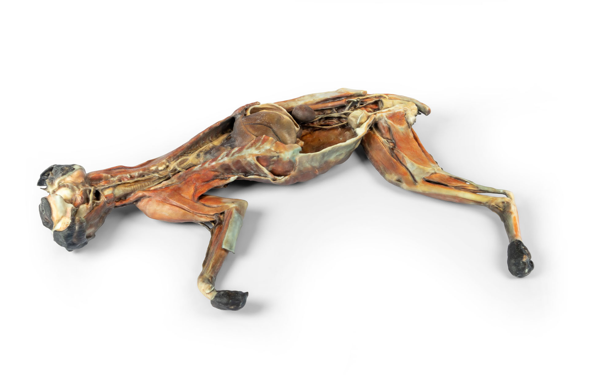
Produktinformationen "Katzenpräparat (Ganzkörper)"
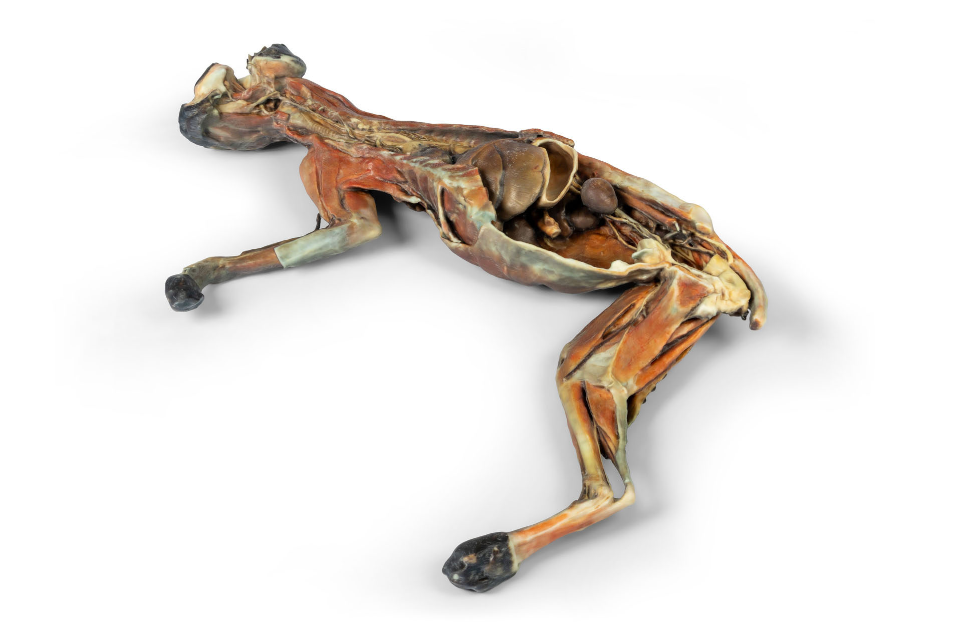
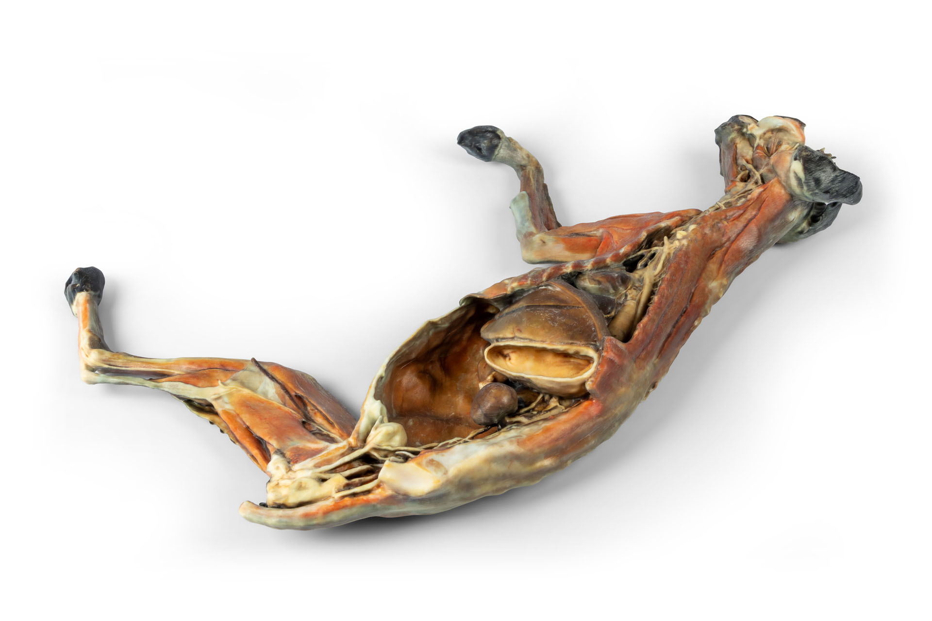
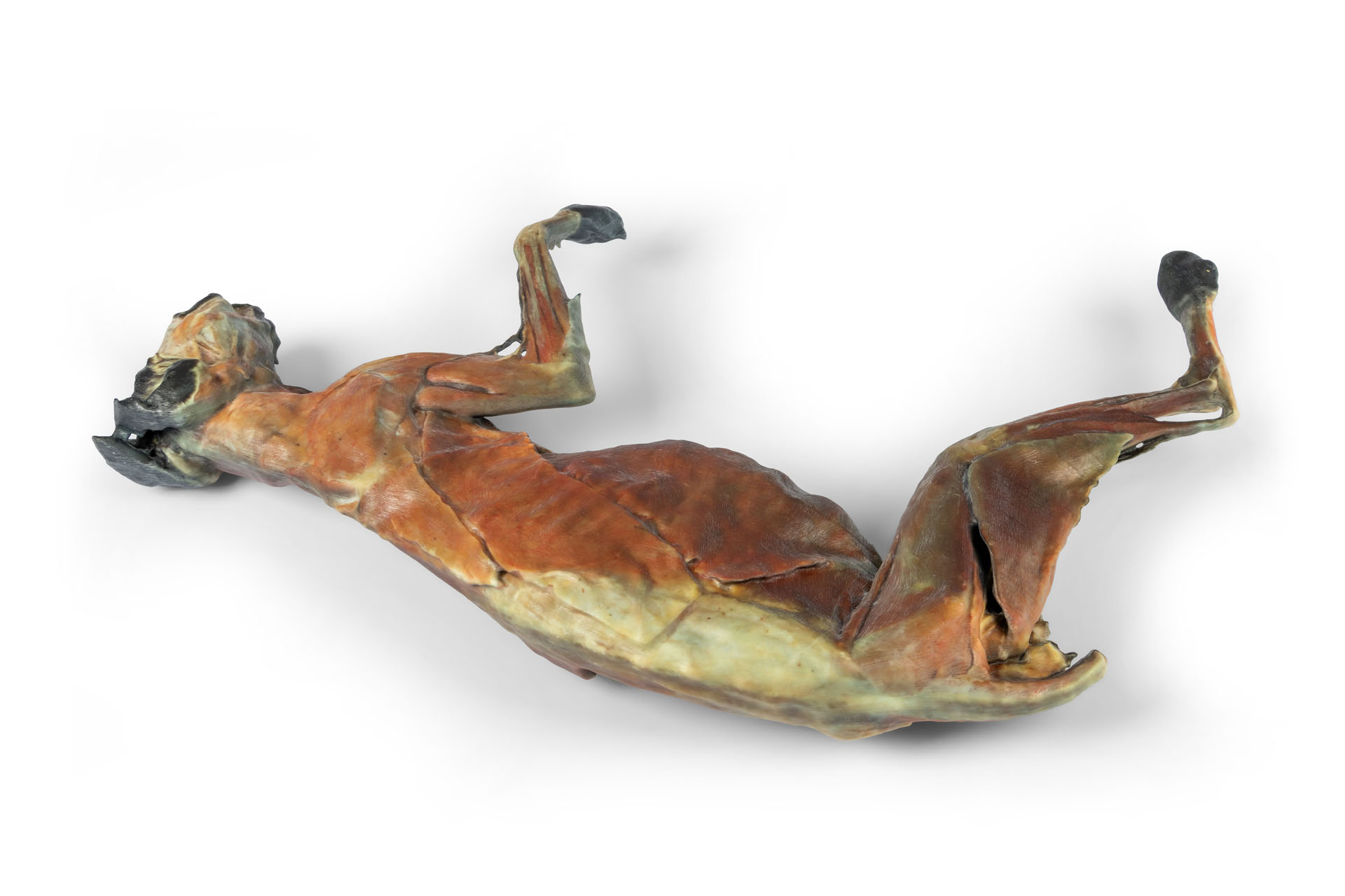
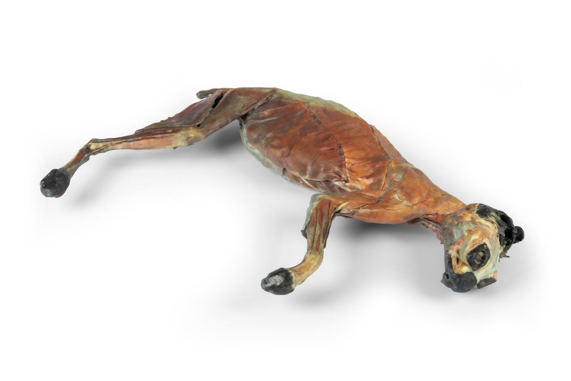
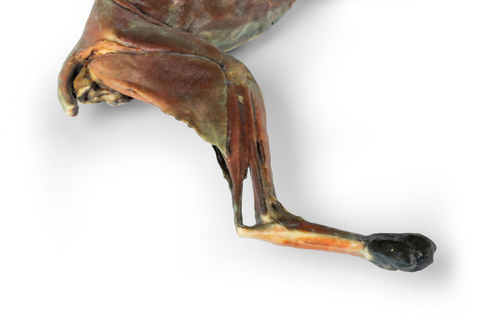
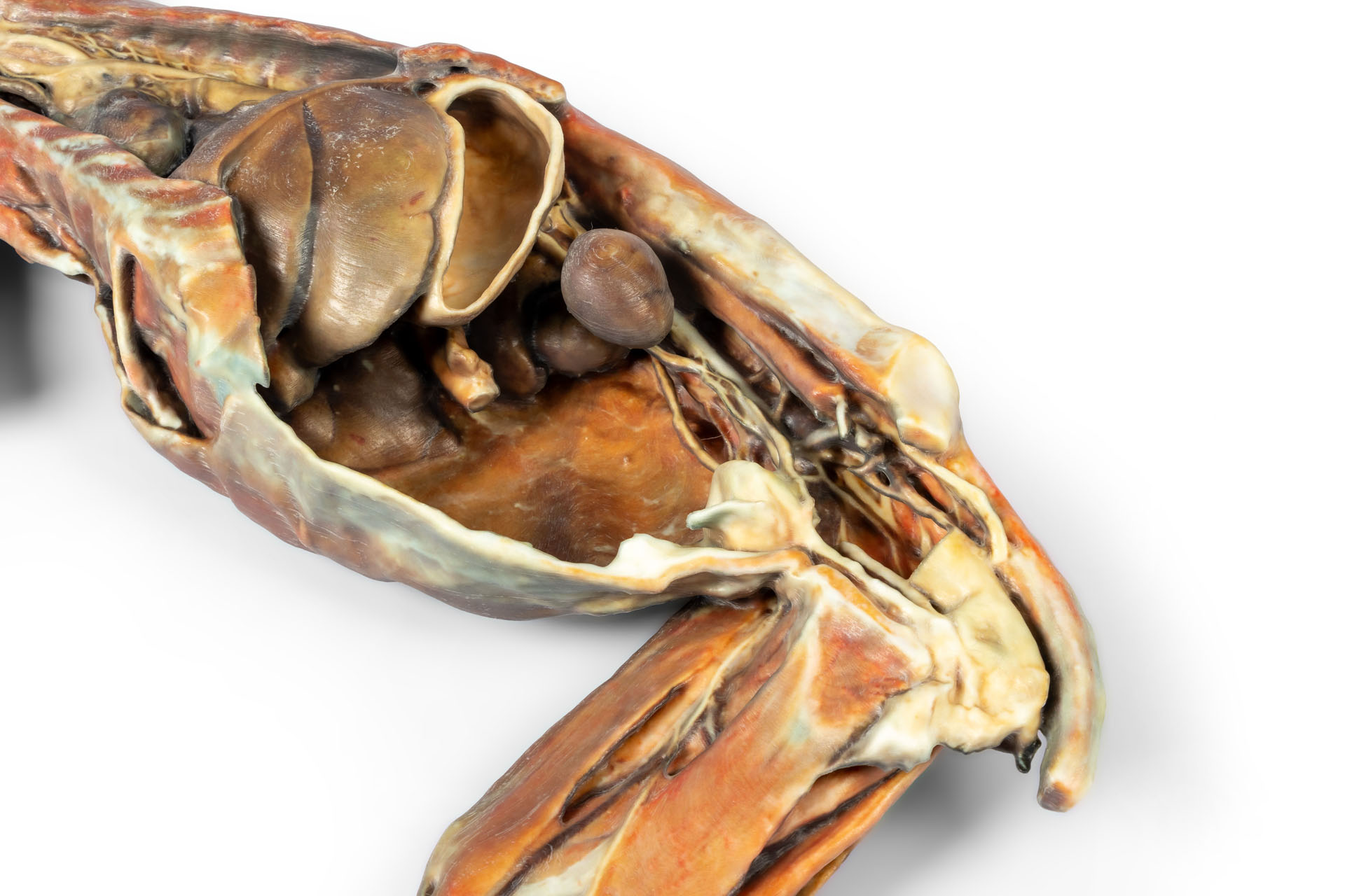
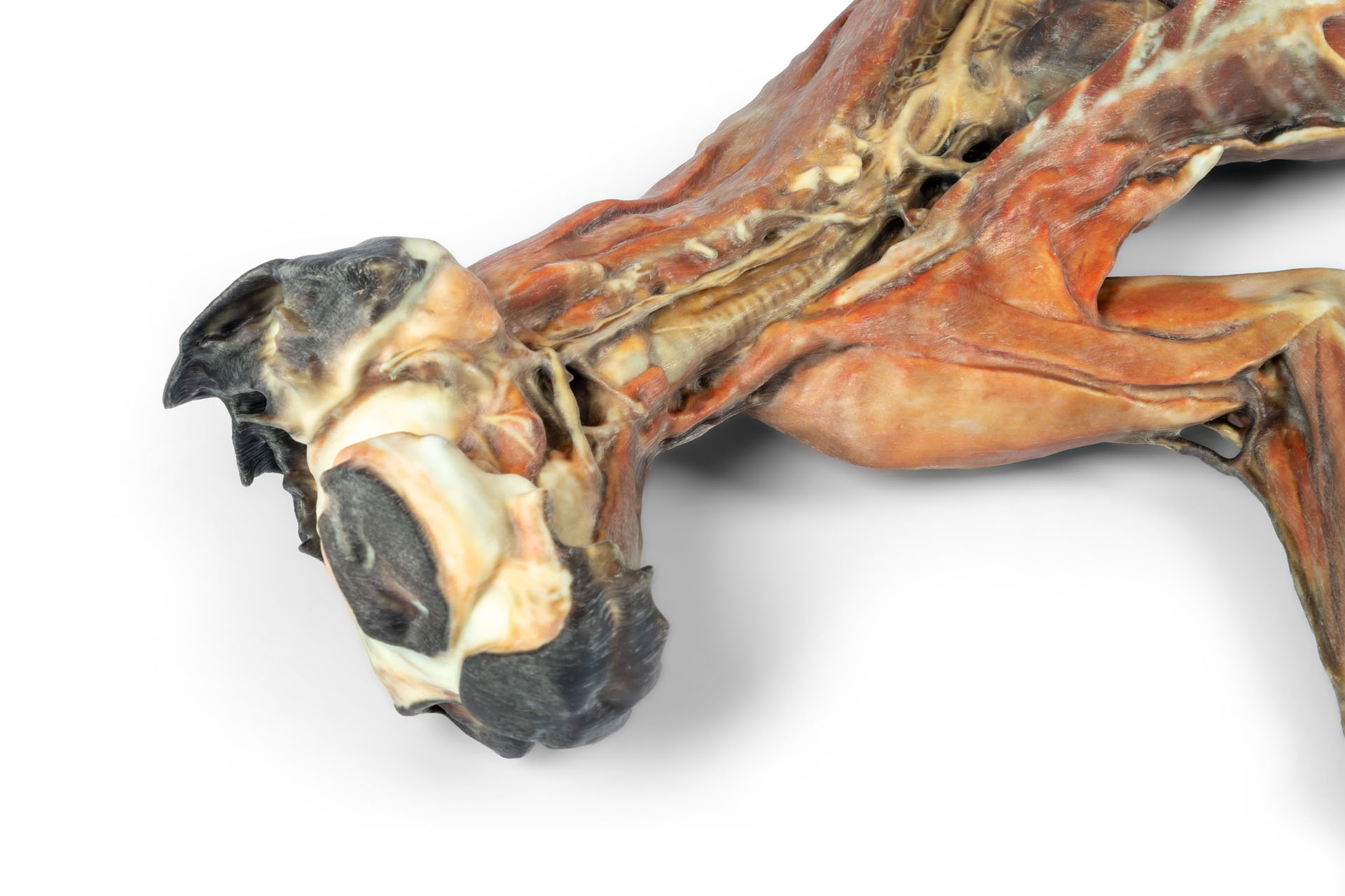
Dieses Präparat zeigt eine vollständige Katzensektion, die zwei sich ergänzende Ansichten des Körpers ermöglicht. Auf der einen Seite ist die Sezierung von Kopf, Hals, Brust-, Bauch- und Beckenhöhle dargestellt. Diese Ansicht bietet eine integrierte Darstellung der Topografie zahlreicher Organe und wichtiger anatomischer Strukturen im Kontext des gesamten Körpers. Auf der gegenüberliegenden Seite ist die oberflächliche Sezierung der Muskulatur zu sehen, die einen wertvollen funktionellen Überblick über den Bewegungsapparat bietet, insbesondere über die Verbindung zwischen Rumpf und Vorder- und Hintergliedmaßen.
Dokumente
Anleitung / Manual VP12000 Herunterladen

0 von 0 Bewertungen

Bewerten Sie dieses Produkt!

Teilen Sie Ihre Erfahrungen mit anderen Kunden.


Erler-Zimmer

Erler-Zimmer Medical GmbH
Hauptstrasse 27
77886 Lauf
Germany
info@erler-zimmer.de


Achtung! Medizinisches Ausbildungsmaterial, kein Spielzeug. Nicht geeignet für Personen unter 14 Jahren.

Attention! Medical training material, not a toy. Not suitable for persons under 14 years of age.


Dokumente
Anleitung / Manual VP12000 Herunterladen

Andere Kunden kauften auch

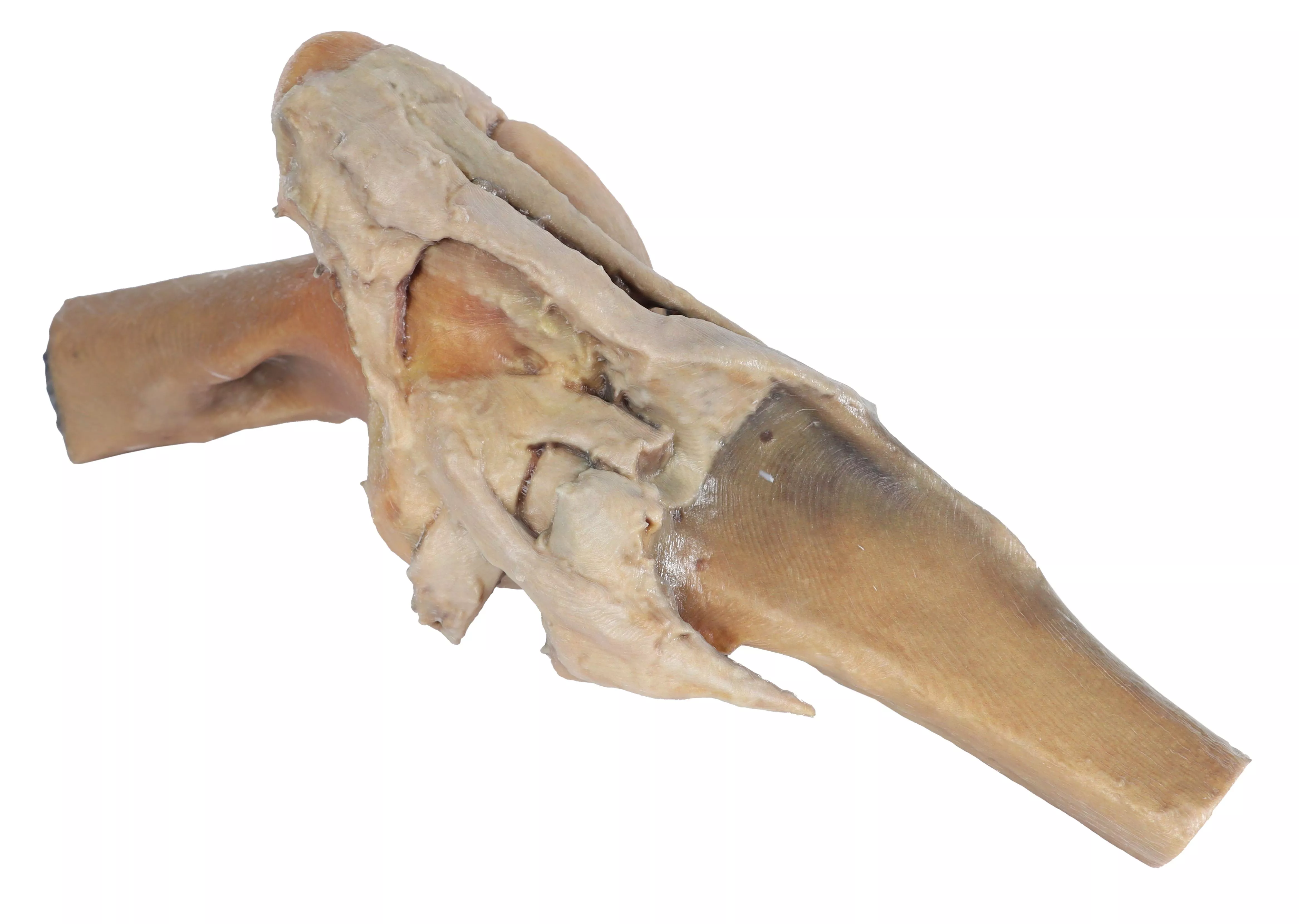
Kniegelenk Pferd - Präparation
This specimen demonstrates the ligaments of the stifle joint with the leg in extension. In the anterior view, with the patella and the three patellar ligaments.The medial and lateral menisci, the caudal cruciate, the femoropatellar and the collateral ligaments are visible.

1.391,11 €*
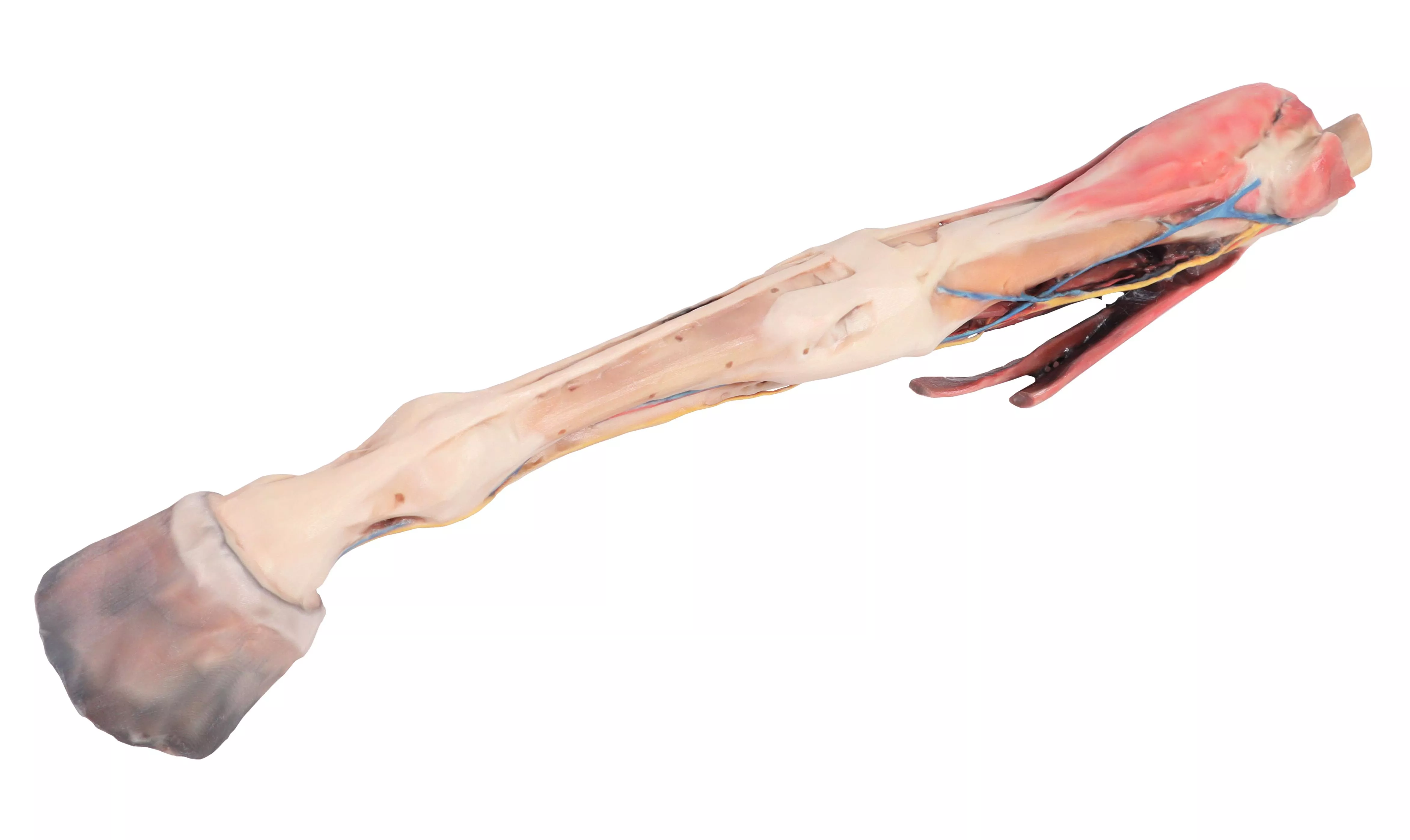
Vorderbein des Pferdes – Muskeln, Sehnen, Bänder, Gefäße und Nerven, distal zum Kniegelenk - 1/3 lebensgröße
This specimen demonstrates the superficial anatomy of a right forelimb from the elbow joint to the hoof. The carpus and digital extensor and flexor muscles are located in the forearm related to the pathway of the median and cubital nerves. The main branches of the median artery as well as the median and cephalic veins have been maintained. Distal to the carpus the specimen shows the topographic relation of the palmar nerves with the digital tendon flexors. Distal to the metacarpophalangeal joint it is possible to see at both sides the palmar digital neurovascular bundle.

2.790,55 €*
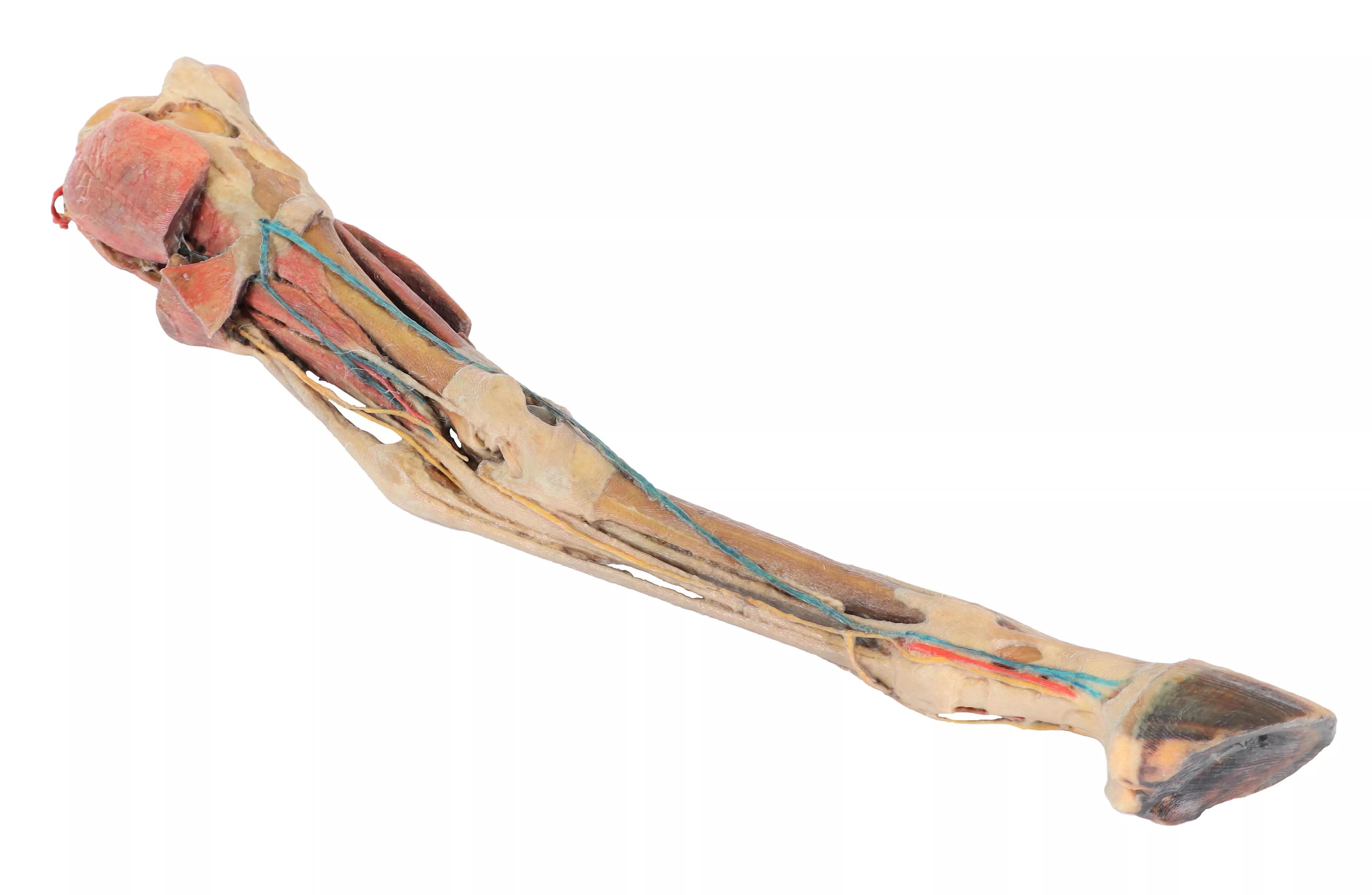
Hinterbein des Pferdes – Muskeln, Sehnen, Bänder, Gefäße und Nerven distal des Kniegelenks - 1/3 lebensgröße
This specimen preserves a superficial dissection of the lower limb musculature from the midthigh to the hoof. The capsule of the stifle and tarsus joints has been opened to demonstrate the menisci, the main ligaments and the topography of the tendons. Muscles extensor and flexor of these joints as well as the foot joints have been dissected from the origin in the femur and tibia bones, to the insertions tendons in the tarsus, metatarsus and phalanges bones. Nerves and vessels have been dissected in the popliteal fossa, tarsus and digit regions. Detailed anatomical description on request.

2.300,27 €*
Sagittaler Kopfschnitt eines Pferdes
This horse half-head shows a superficial dissection on the right lateral aspect and a middle sagittal section on the medial aspect.On the lateral side the skin has been removed, except on the nostrils, lips and external ear. In the rostral part of the dissection are the muscles of facial mimicry, the levator nasolabialis, canine, levator labii superioris, zygomaticus, buccinator (buccal portion) and depressor labii inferioris muscles. The caudal part of the dissection is occupied by the masseteric muscle covered by the masseteric fascia from the facial crest of the maxillary bone. This muscle presents on the surface the course of the dorsal and ventral buccal branches of the facial nerve, which end rostrally as buccolabial branches. In the notch of the facial vessels, the facial artery and vein are located together with the parotid duct. In the dorsal and caudal part of the masseter muscle, the transverse facial artery and vein are located together with the auriculotemporal nerve. The parotid-auricular muscle, the parotid gland, and the maxillary and linguofacial veins are located in the cervicofacial transit.In the middle sagittal section, the nasal cavity is occupied by the ventral nasal concha, dorsal nasal concha, and middle nasal concha, separated by the dorsal, middle, and ventral nasal meatuses. The perpendicular lamina of the ethmoid bone is located in the caudal area of the nasal cavity. This bone is the rostral limit of the cerebral fossa. The cerebellar fossa is caudal to the anterior one. The vomer bone establishes the choanae as transit to the nasopharynx where the pharyngeal opening of the auditory tube is located. The oral cavity is limited dorsally by the hard palate, which continues caudally as the soft palate. The sagittal section of the tongue shows the proper lingual muscle as well as the genioglossus and genihyoid muscles. The oropharynx is occupied by the root of the tongue and the soft palate. The epiglottis, arytenoid cartilage, and cricoid cartilage limit the laryngeal cavity with the lateral ventricle, vocal fold, and infraglottic cavity. The laryngopharynx is occupied by the esophageal vestibule. The esophagus is located dorsal to the trachea and ventral to the long cervical and long capitis muscles. The mandibular lymph nodes are in the intermandibular space.

5.253,85 €*
Hundekopf – oberflächliche und tiefe Präparationen-
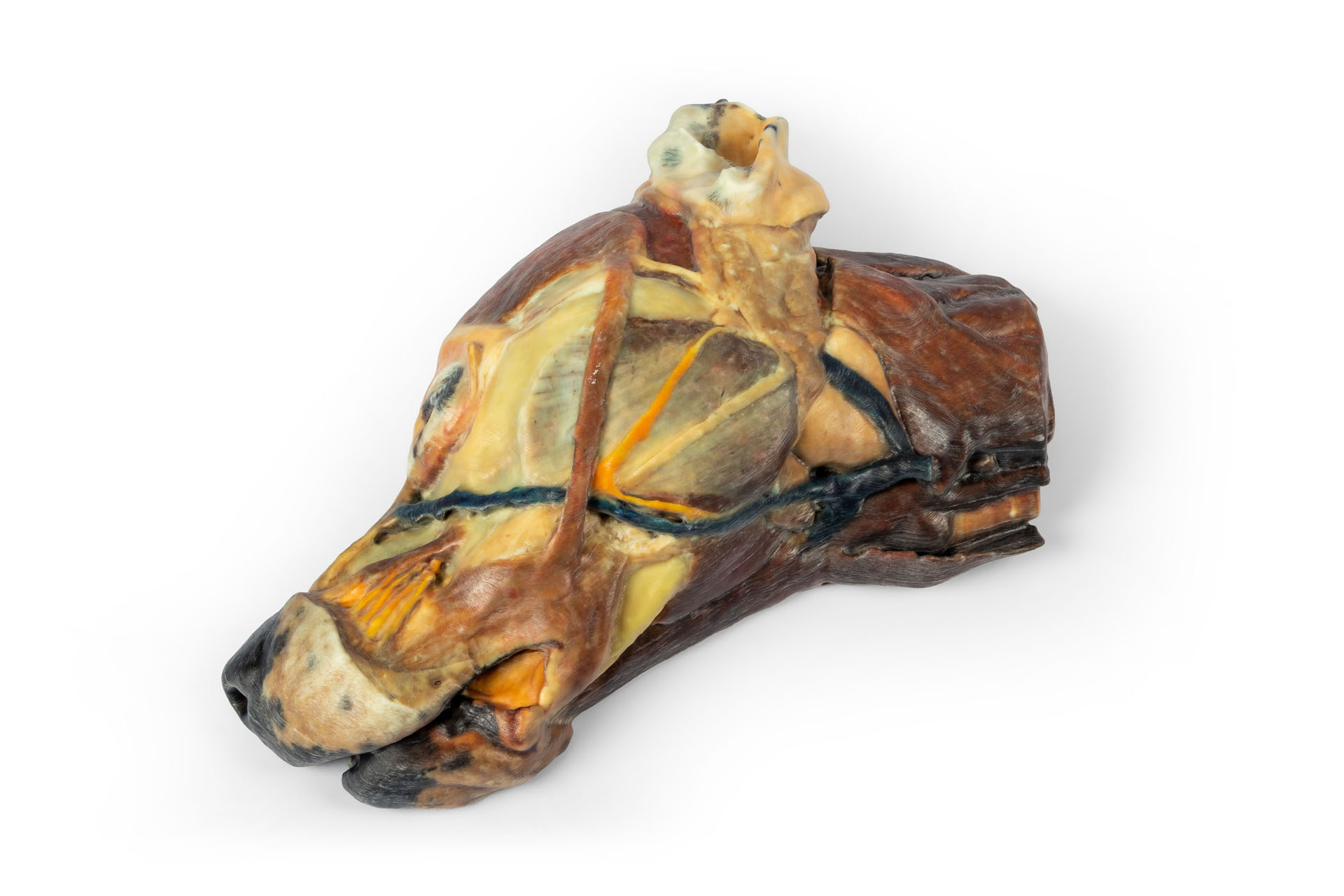
Superficial anatomical structures:Tip of the nose (Apez nasi)Right and left wings of the nose (Alae nasi)Nonglandular skin on the tip of the nose (Planun nasale)Superficial disscetion on the left side:On the left side, the skin has been removed to identify the main anatomical structures,which are described grouped below.Muscles of the facial neuromuscular system:• M. nasolabial levator (M. levator nasolabialis)• M. canine (M. caninus)• M. buccinator (M. buccinator)• M. Zygoma9c (M. zygoma:cus)• M. parodidoauricularisMuscles of the mandibular neuromuscular system (mas9cators):• M. masseter (M. masseter)• M. Temporary (M.temporalis)Nerves:• Facial nerve: (N . facialis)• Dorsal buccal Branch (Rami buccales)• Ventral buccal Branch (Rami buccales)• Bucolabial branches (Rami buccolabiales)• N. Auriculopalpebral (N. Auriculopalpebralis)Vascular:• Facial artery (Arteria facialis)• External jugular vein: (V. Jugularis externa)o Maxillary vein (V. Maxillaris)o Linguofacial vein (V. Linguofacialis)Salivary glands:• Parotid gland and paro9d duct (Glandula parotis) (Ductus parotideus)• Mandibular gland Glandula mandibularisLymph nodes:• N. L. mandibular (Lymphonodi mandubulares)Deep dissection on the right side:On this right side mandible has been removed to see deeper anatomical structures. You can see the surfaces of the temporal bone for the temporomandibular joint (Articulatio Temporomandibularis). The medial pterygoid muscle (M. pterygoideus medialis) is identified and sectioned in its insertion area to the ramus of the mandible (Ramus mandibulae). Next to this muscle, the branches of the mandibular nerve (N. mandibularis) are identified, as well as the maxillary nerve (nevus maxillaris) and the maxillary artery (arteria maxillaris). Near the external acoustic meatus (Meatus acusticus externus), the facial nerve (N . facialis) has been maintained, with one of its branches,the auriculopalpebral nerve (N. Auriculopalpebralis), running parallel to the zygomatic arch (Arcus zygomaticus), visible atier removing the parotd salivary gland (Glandula parotis). The tongue is identified in its entire caudal extension, and the styloglossus (M. stylogossus), genioglossus (M. genioglossus) and genihyoid (M. geniohydeus) muscles reach it. Next to these muscles, the hypoglossal nerve (N. hypoglossus) is identified. In relation to the pharynx (Pharynx), the constrictor muscles of the pharynx (Mm. constrictors phyngis caudalis) are identified. In the most caudal area, in relation to the neck, the course of the common caro9d artery (arteria caarotis communis) and the vagosympathetic trunk nerve (Truncus vagosympathicus) are identified.

4.338,74 €*
Vorderbein des Hundes – Muskeln, Sehnen, Bänder, Gefäße und Nerven, distal bis zum Ellenbogen
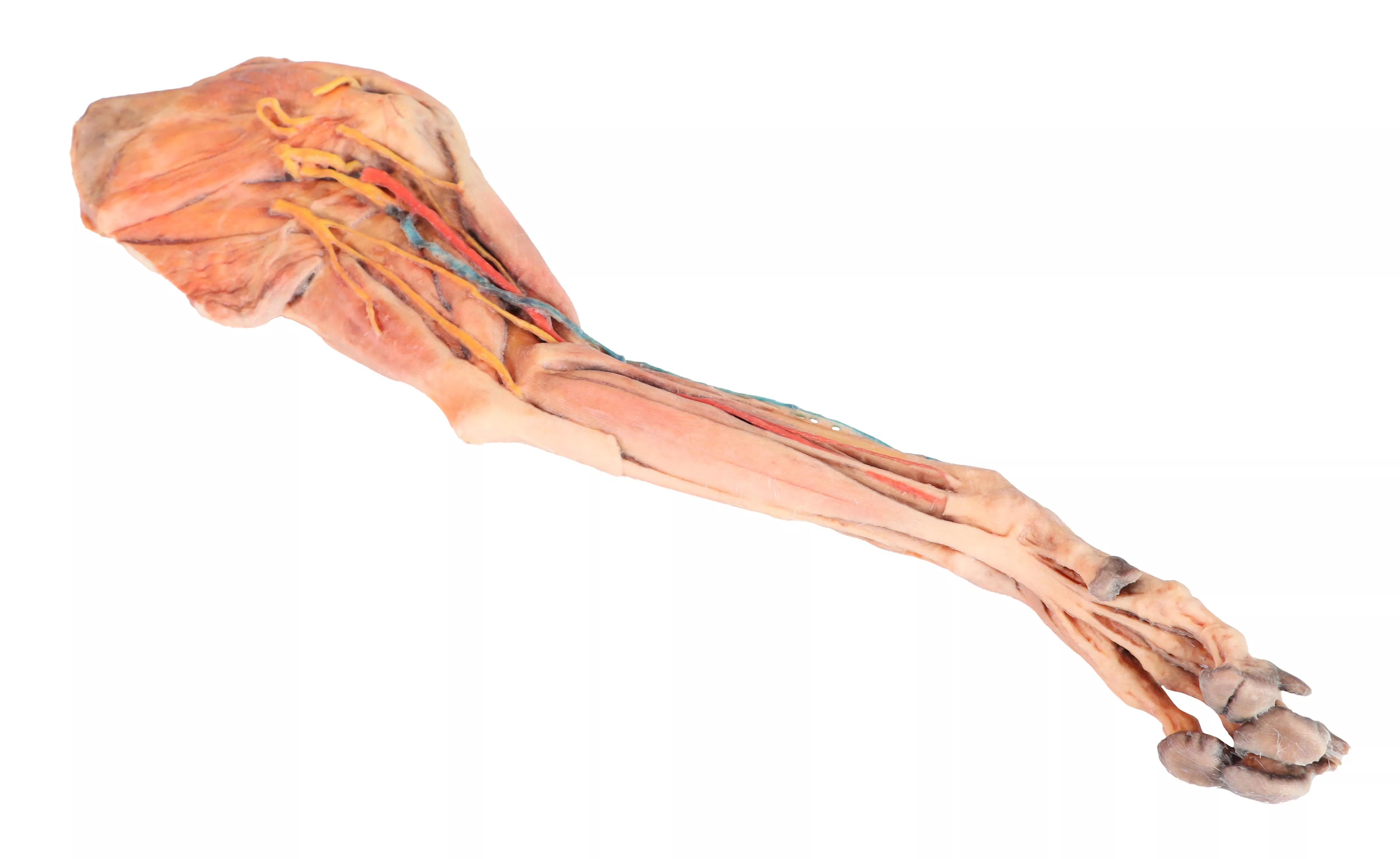
This specimen demonstrates the superficial anatomy of a dog's right thoracic limb from the scapula to the hand. The shoulder flexor and extensor muscles of the scapular region have been preserved, along with the arm flexor and extensor muscles of the elbow. In the forearm and hand are the flexor and extensor muscles of the carpus and fingers. On the medial side of the axillary region the main nerves of the brachial plexus have been preserved. Similarly, the paths of the brachial and median arteries and their respective veins are identified. In a superficial position, the cephalic vein has been preserved together with the antebrachialis lateral cutaneous nerve of the forearm. Detailed anatomical description on request.

2.114,63 €*
Hunde-Halbkopf – Sagittalschnitt
Dieses hochwertige, anatomische 3D-Druckmodell eines Hundekopfes bietet eine detaillierte und realitätsnahe Darstellung komplexer anatomischer Strukturen des Kopf- und Halsbereichs. Das Modell zeigt eine präzise Gesichtspräparation sowie die mediale Ansicht mit freigelegtem Rachen und Kehlkopf und ermöglicht dadurch ein vertieftes Verständnis der topographischen Anatomie des Hundes. Durch die sorgfältige Ausarbeitung der anatomischen Details werden relevante Strukturen des Gesichts, der Muskulatur sowie des oberen Atemwegs anschaulich visualisiert. Farblich hervorgehobene Bereiche erleichtern die Orientierung und unterstützen die anatomische Zuordnung einzelner Strukturen. Gefertigt im hochwertigen 3D-Druckverfahren, vereint das Modell Präzision, Langlebigkeit und eine hohe Detailtiefe. Es eignet sich ideal für den Einsatz in der veterinärmedizinischen Ausbildung, Lehre, Demonstration sowie für wissenschaftliche Sammlungen und anatomische Studien. Besondere Merkmale: • Detailliertes anatomisches 3D-Druckmodell eines Hundekopfes • Darstellung einer Gesichtspräparation mit freigelegten Strukturen • Mediale Ansicht von Rachen und Kehlkopf zur Veranschaulichung der Atemwegsanatomie • Farblich differenzierte anatomische Bereiche für bessere Orientierung • Robuste und präzise Ausführung durch modernes 3D-Druckverfahren

2.796,50 €*

Stetige Innovationskraft

Soziale Verantwortung

Gelebte Kundenorientierung

Verständnis für Qualität

Nachhaltiges Handeln

Zertifizierung ISO 9001

Ihre zuletzt angesehenen Produkte