Product information "
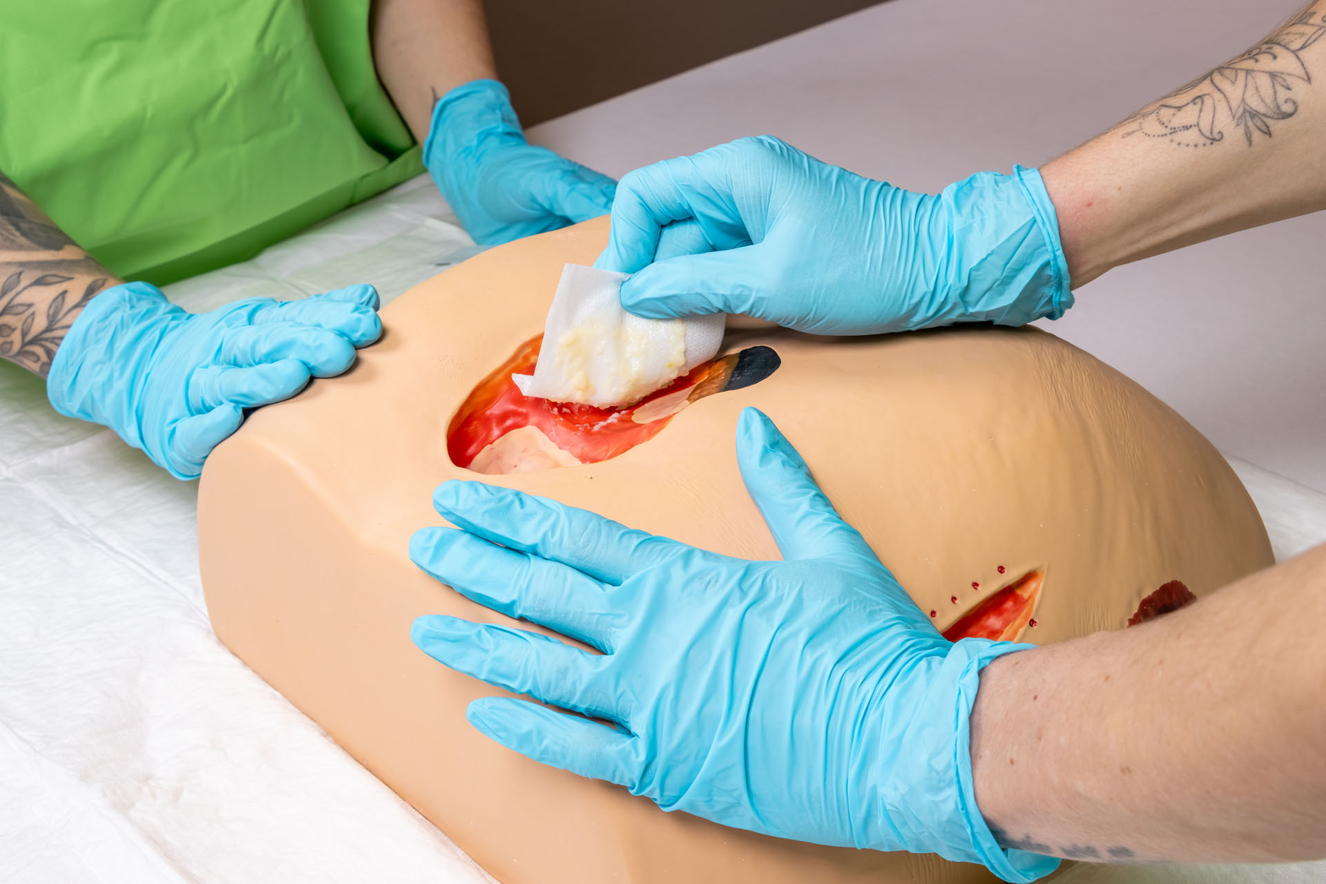
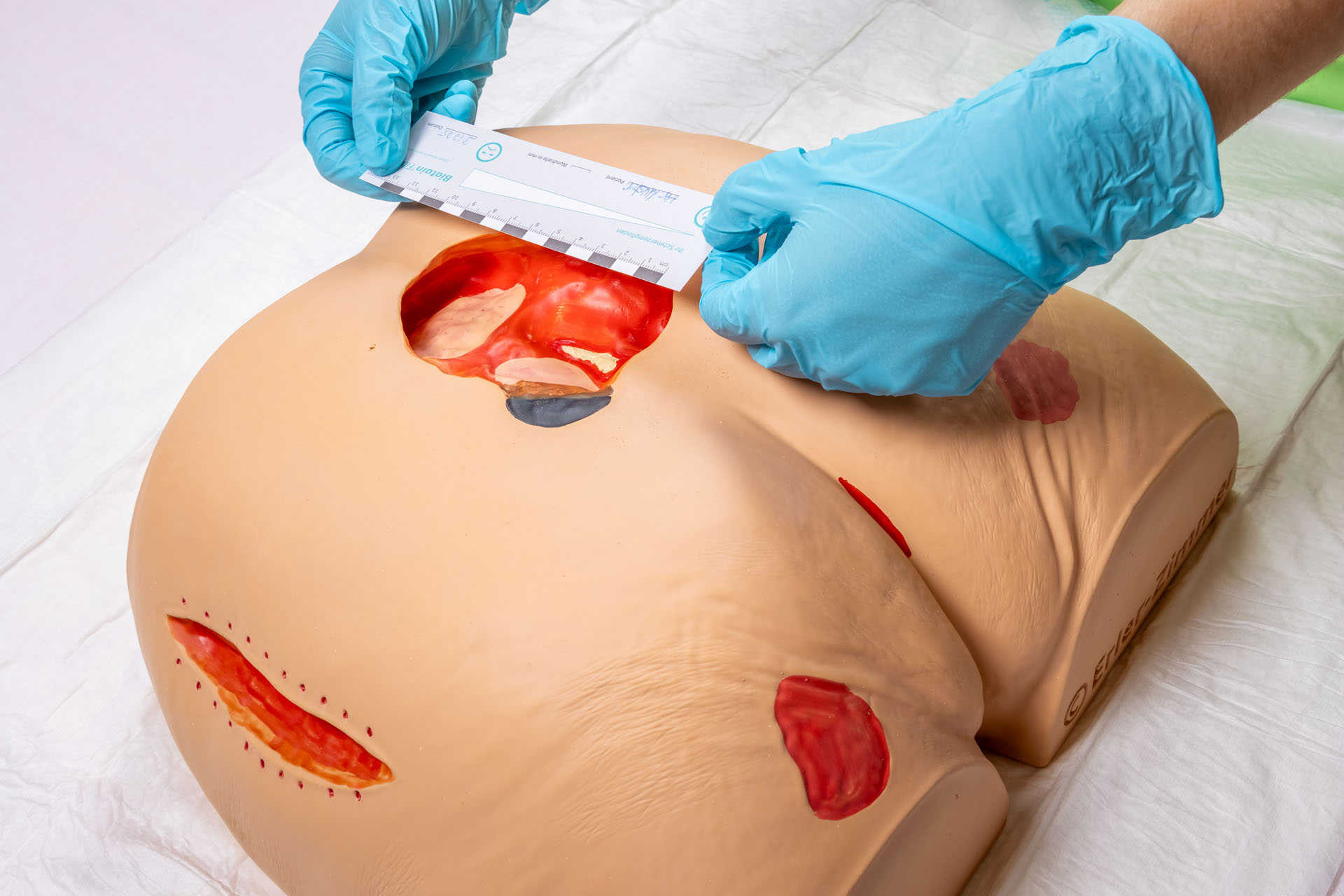
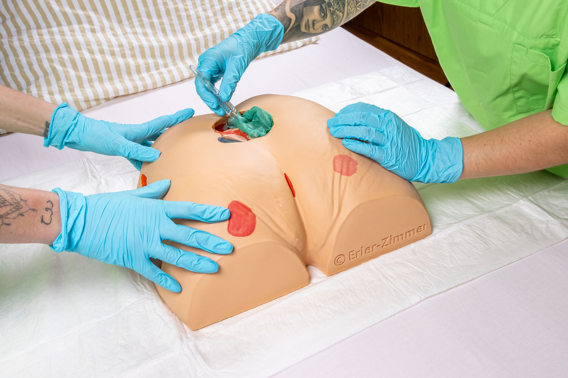
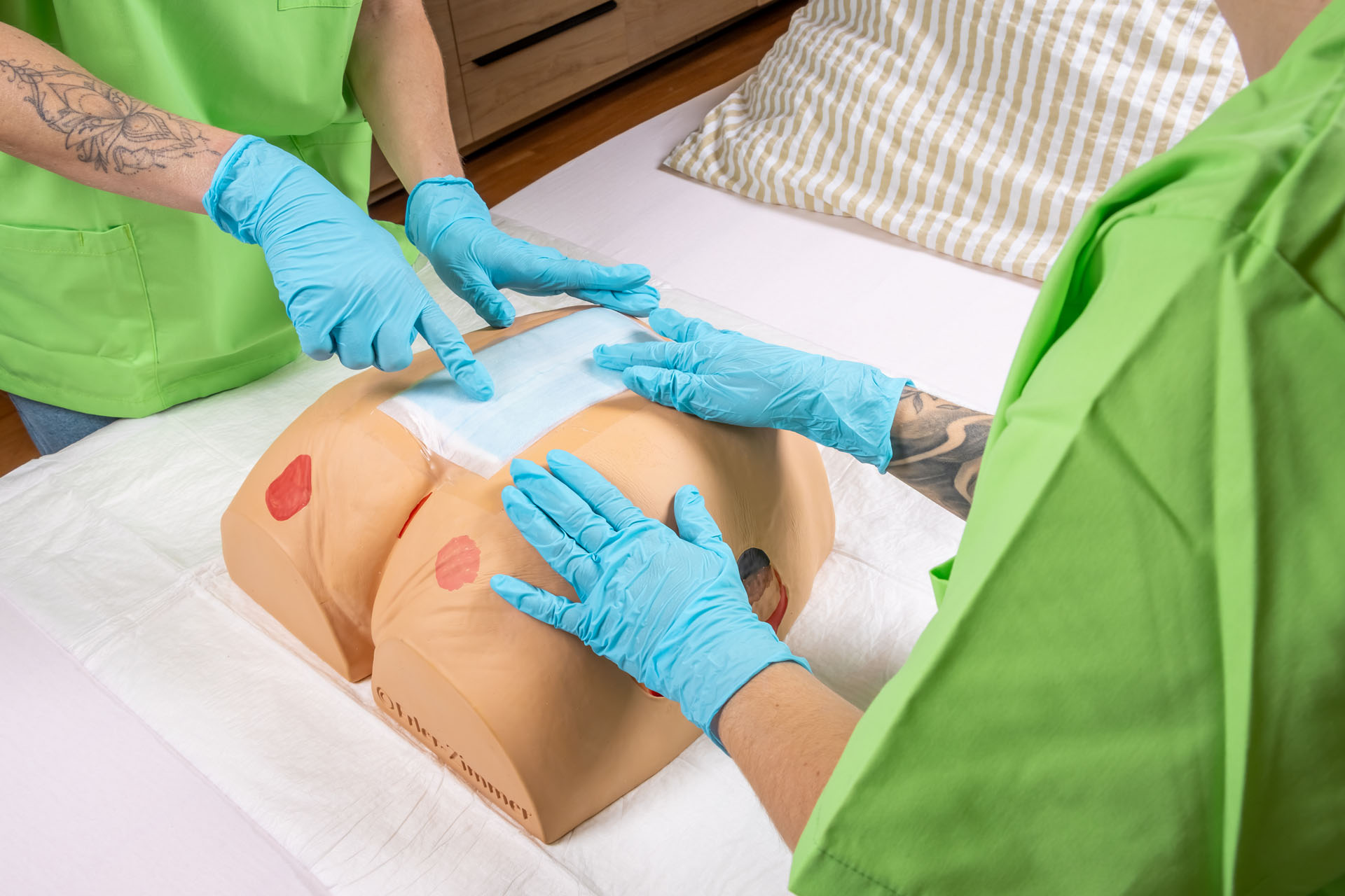
"Decubitus Dan" care and dressing simulator"
This universal pressure ulcer simulator shows stage 1, stage 2, stage 3 and stage 4 pressure ulcers. The buttocks of a geriatric patient also show a deep tissue injury, unclassifiable necrosis and a deshisced wound measuring approximately 12 cm. The life-size model is made of soft plastic and is ideal for dressing exercises, for vacuum wound therapy and for all other care measures.
Size: 30x34x14cm, weight 1.3kg
Documents
Instruction 7600 Download

0 of 0 reviews

Leave a review!

Share your experiences with other customers.


First Distributor meeting in Thailand was an absolute success!

Pattaya, Thailand – The first Erler-Zimmer distributor meeting in Thailand took place with great response and was a complete success.
Erler-Zimmer

Erler-Zimmer GmbH & Co.KG
Hauptstrasse 27
77886 Lauf
Germany
info@erler-zimmer.de


Achtung! Medizinisches Ausbildungsmaterial, kein Spielzeug. Nicht geeignet für Personen unter 14 Jahren.

Attention! Medical training material, not a toy. Not suitable for persons under 14 years of age.


Documents
Instruction 7600 Download

Other customers also bought

Wound moulage sacral decubitus, cleaned
This moulage shows the sacral area with a pressure ulcus after a surgical cleaning. The necrotic tissue has been removed, wound pockets are present. The wound is available either freshly cleaned or with wound exsudate. The moulage can be used for demonstration of vacuum wound therapy. The moulage adheres on simulated patients or manikins, it can be fixed with special skin glue for longer training sessions. The anal fold is represented to demonstrate the typical challenges when dressing the wound.Supplied with storage box.

€427.21*
Wound moulage sacral decubitus, cleaned including stand
This moulage shows the sacral area with a pressure ulcus after a surgical cleaning. The necrotic tissue has been removed, wound pockets are present. The wound is available either freshly cleaned or with wound exsudate. The moulage can be used for demonstration of vacuum wound therapy. The moulage adheres on simulated patients or manikins, it can be fixed with special skin glue for longer training sessions. The anal fold is represented to demonstrate the typical challenges when dressing the wound. The moulage is delivered in a holder and can also be removed.

€412.93*
Wound moulage sacral decubitus, untreated
This moulage shows the sacral area with a pressure ulcus in an unattended stage. The tissue has died under continuous pressure and necrotic tissue is visible. The actual size of the necrotic area can not be defined and needs surgical therapy. The moulage adheres on simulated patients or manikins, it can be fixed with special skin glue for longer training sessions. The anal fold is represented to demonstrate the typical challenges when dressing the wound.Supplied with storage box.

€427.21*
Wound Moulage Colostoma under skin level, incl. stand
This moulage shows a colostomy, the most common type of stoma creation. The terminal colostoma is usually found in the middle of the body or on the left side of the body and shows the colon sewn in. The colon is under skin level. Due to the use of a special plastic compound, all common stoma bags adhere to the moulage. The model is suitable for sizing using the stoma template, cleaning the skin with water and attaching the base plate and stoma bag. The moulage is in a holder and can also be removed.

€365.33*
Wound Moulage Colostoma with inflammation, incl. stand
This moulage shows a colostomy, the most common type of stoma creation. The terminal colostoma is usually found in the middle of the body or on the left side of the body and shows the colon sewn in above skin level. The area around the colon is inflammed Due to the use of a special plastic compound, all common stoma bags adhere to the moulage. The model is suitable for sizing using the stoma template, cleaning the skin with water and attaching the base plate and stoma bag. The moulage is in a holder and can also be removed.

€365.33*
Wound Moulage Ileostoma, incl. stand
This moulage shows an ileostomy, the second most common type of ostomy. The terminal ileostoma is usually found on the right side of the body and shows the small intestine sewn in above skin level. Due to the use of a special plastic compound, all common stoma bags adhere to the moulage. The model is suitable for sizing using a stoma template, cleaning the skin with water and attaching the base plate and stoma bag. The moulage is in a holder and can also be removed.

€365.33*
Wound Moulage Colostoma, incl. stand
This moulage shows a colostomy, the most common type of stoma creation. The terminal colostoma is usually found in the middle of the body or on the left side of the body and shows the colon sewn in above skin level. Due to the use of a special plastic compound, all common stoma bags adhere to the moulage. The model is suitable for sizing using the stoma template, cleaning the skin with water and attaching the base plate and stoma bag. The moulage is in a holder and can also be removed.

€365.33*
Wound Moulage Colostoma in abdominal crease, incl. stand
This moulage shows a colostomy, the most common type of stoma creation. The terminal colostoma is usually found in the middle of the body or on the left side of the body and shows the colon sewn in above skin level. The Stoma is located in a abdominal crease. Due to the use of a special plastic compound, all common stoma bags adhere to the moulage. The model is suitable for sizing using the stoma template, cleaning the skin with water and attaching the base plate and stoma bag. The moulage is in a holder and can also be removed.

€365.33*
Elderly Pressure Ulcer Foot
Chronic foot ulcers affect elderly the most, we have designed the Elderly Pressure Ulcer Foot for instruction on care and cleaning of pressure ulcers in various stages. Pressure ulcers are more likely to appear over pressure points such as heels, tips of toes, between toes, or anywhere the bones may protrude and rub against socks, shoes, or bed sheets. This replica contains all four severity stages:- Stage I: Located on the bottom of the toe. Surface of the skin is red.- Stage II: Located on the bottom of the foot just under the toe. Surface of the skin is red and deeper into the skin layers.- Stage III: Located on the side of the foot. Surface of the skin is red, looks more like a crater, and reaches the bottom layer of the skin.- Stage IV: Located on the heel of the foot. Surface of the skin is red. A great amount of tissue has been damaged, including muscle, bone, joints, and tendons.

€408.17*

Continuous innovation

Social responsibility

Active customer orientation

Understanding quality

Sustainable actions

ISO 9001 certification

Your last viewed products