Veterinary

11 Products
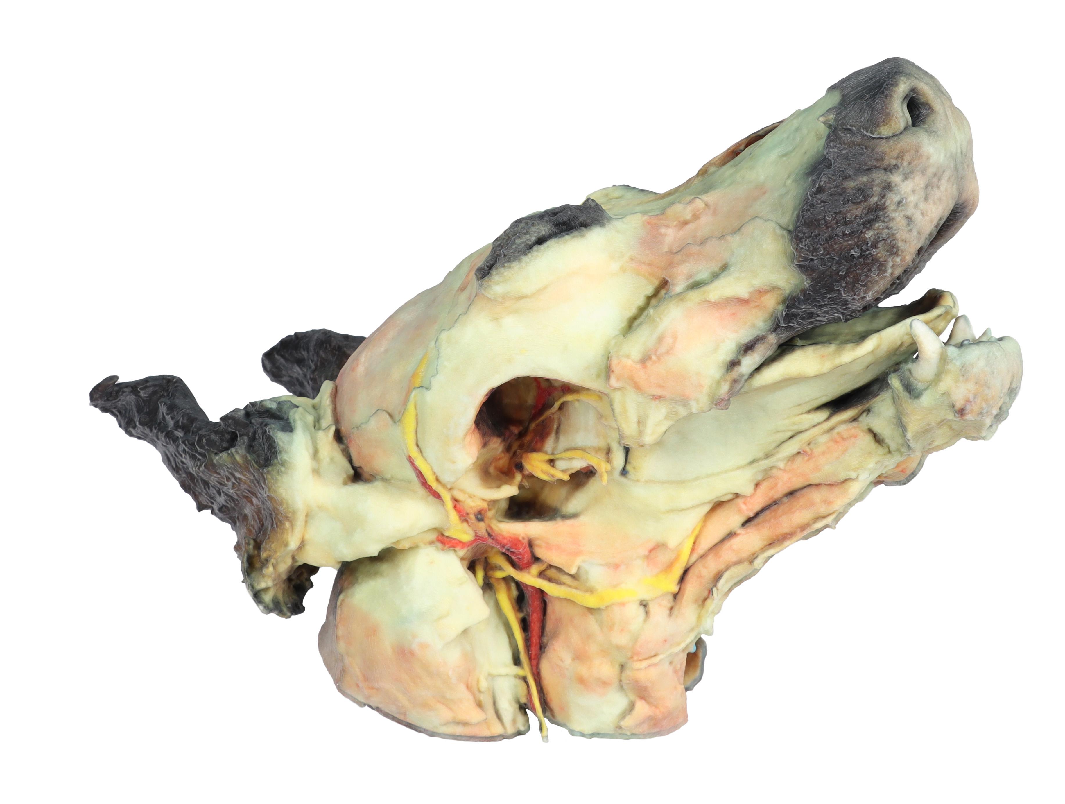
Dog head - superficial and deep dissections
Superficial anatomical structures:Tip of the nose (Apez nasi)Right and left wings of the nose (Alae nasi)Nonglandular skin on the tip of the nose (Planun nasale)Superficial disscetion on the left side:On the left side, the skin has been removed to identify the main anatomical structures,which are described grouped below.Muscles of the facial neuromuscular system:• M. nasolabial levator (M. levator nasolabialis)• M. canine (M. caninus)• M. buccinator (M. buccinator)• M. Zygoma9c (M. zygoma:cus)• M. parodidoauricularisMuscles of the mandibular neuromuscular system (mas9cators):• M. masseter (M. masseter)• M. Temporary (M.temporalis)Nerves:• Facial nerve: (N . facialis)• Dorsal buccal Branch (Rami buccales)• Ventral buccal Branch (Rami buccales)• Bucolabial branches (Rami buccolabiales)• N. Auriculopalpebral (N. Auriculopalpebralis)Vascular:• Facial artery (Arteria facialis)• External jugular vein: (V. Jugularis externa)o Maxillary vein (V. Maxillaris)o Linguofacial vein (V. Linguofacialis)Salivary glands:• Parotid gland and paro9d duct (Glandula parotis) (Ductus parotideus)• Mandibular gland Glandula mandibularisLymph nodes:• N. L. mandibular (Lymphonodi mandubulares)Deep dissection on the right side:On this right side mandible has been removed to see deeper anatomical structures. You can see the surfaces of the temporal bone for the temporomandibular joint (Articulatio Temporomandibularis). The medial pterygoid muscle (M. pterygoideus medialis) is identified and sectioned in its insertion area to the ramus of the mandible (Ramus mandibulae). Next to this muscle, the branches of the mandibular nerve (N. mandibularis) are identified, as well as the maxillary nerve (nevus maxillaris) and the maxillary artery (arteria maxillaris). Near the external acoustic meatus (Meatus acusticus externus), the facial nerve (N . facialis) has been maintained, with one of its branches,the auriculopalpebral nerve (N. Auriculopalpebralis), running parallel to the zygomatic arch (Arcus zygomaticus), visible atier removing the parotd salivary gland (Glandula parotis). The tongue is identified in its entire caudal extension, and the styloglossus (M. stylogossus), genioglossus (M. genioglossus) and genihyoid (M. geniohydeus) muscles reach it. Next to these muscles, the hypoglossal nerve (N. hypoglossus) is identified. In relation to the pharynx (Pharynx), the constrictor muscles of the pharynx (Mm. constrictors phyngis caudalis) are identified. In the most caudal area, in relation to the neck, the course of the common caro9d artery (arteria caarotis communis) and the vagosympathetic trunk nerve (Truncus vagosympathicus) are identified.

€3,885.35*
Dog forelimb - muscles, tendons, ligaments, vessels and nerves distal to the elbow
This specimen demonstrates the superficial anatomy of a dog‘s right thoracic limb from the scapula to the hand. The shoulder flexor and extensor muscles of the scapular region have been preserved, along with the arm flexor and extensor muscles of the elbow. In the forearm and hand are the flexor and extensor muscles of the carpus and fingers. On the medial side of the axillary region the main nerves of the brachial plexus have been preserved. Similarly, the paths of the brachial and median arteries and their respective veins are identified. In a superficial position, the cephalic vein has been preserved together with the antebrachialis lateral cutaneous nerve of the forearm. Detailed anatomical description on request.

€1,892.10*
Dog hindlimb- muscles, tendons, ligaments, vessels and nerves distal to the stifle
This specimen preserves a superficial dissection of the lower hindlimb musculature from the mid-thigh to the foot, as well as nerves and vessels of the femoral canal and popliteal fossa.

€2,082.50*
Horse sagittal half-head
This horse half-head shows a superficial dissection on the right lateral aspect and a middle sagittal section on the medial aspect.

€4,700.50*
Horse forelimb - muscles, tendons, ligaments vessels and nerves distal to the stifle - 1/3 life-size
This specimen demonstrates the superficial anatomy of a right forelimb from the elbow joint to the hoof. The carpus and digital extensor and flexor muscles are located in the forearm related to the pathway of the median and cubital nerves. The main branches of the median artery as well as the median and cephalic veins have been maintained. Distal to the carpus the specimen shows the topographic relation of the palmar nerves with the digital tendon flexors. Distal to the metacarpophalangeal joint it is possible to see at both sides the palmar digital neurovascular bundle.

€2,499.00*
Horse stifle joint dissection
This specimen demonstrates the ligaments of the stifle joint with the leg in extension. In the anterior view, with the patella and the three patellar ligaments.

€1,243.55*
Horse (foal) pelvis dissection - ligaments
This specimen is focused on the sacroiliac joint, the two coxal bones (os coxae) are exposed in the dissection: Ilium, pubis and ischium, together with lumbar vertebrae and sacrum. At the sacroiliac joint, the dorsal sacroiliac ligament can be identified. The broad sacrotuberous ligament of the pelvis extends from the lateral sacral crest to the sciaticspine and ischial tuberosity. The greater sciatic foramen occupied by the lumbosacral trunk. From the vertebral bodies and part of the transverse processes of the lumbarvertebrae, the tendons of insertion of the psoas minor muscles are identified. The ventral longitudinal ligament connects the ventral aspect of most cranial lumbar vertebral bodies.

€5,353.81*
Horse foot - tendons, ligaments, vessels and nerves - 1/3 life-size
This dissected right-foot specimen displays a mixture of the vascular, nervous, ligaments and tendons. The plantar digital neurovascular bundles both sides of the flexor tendons. The two digital extensor tendons are identified in the proximal dorsal aspect of the specimen. Metatarsophalangeal and proximal interphalangeal joints show all ligaments. Between the plantar aspect of the metatarsal bones and the digital tendon flexors is the interosseus ligament with its sesamoid and extensor branches.

€2,082.50*
Horse foot - deep palmar dissection
This dissected left-foot specimen displays the plantar digital neurovascular bundle both sides of the flexor tendons. The two digital extensor tendons are identified in the dorsal aspect of the specimen. The plantar aspect of the metacarpophalangeal and proximal interphalangeal joints show a dissection of the deep ligaments and their relationship with the neurovascular bundle.

€1,773.10*
Horse hindlimb - muscles, tendons, ligaments, vessels and nerves distal to the stifle - 1/3 life-size
This specimen preserves a superficial dissection of the lower limb musculature from the midthigh to the hoof. The capsule of the stifle and tarsus joints has been opened to demonstrate the menisci, the main ligaments and the topography of the tendons. Muscles extensor and flexor of these joints as well as the foot joints have been dissected from the origin in the femur and tibia bones, to the insertions tendons in the tarsus, metatarsus and phalanges bones. Nerves and vessels have been dissected in the popliteal fossa, tarsus and digit regions. Detailed anatomical description on request.

€2,058.70*
Ox foot - tendons and ligaments
This specimen records the anatomy of an ox‘s right foot from the metatarsus to the distal phalanges. In the dorsal aspect, the insertion of the tendons of the common, lateral and medial digital extensor muscles are the main structures. In the plantar aspect, the relation of the tendons of the superficial and deep digital flexor muscles with deep structures such as the interosseous ligament can be detected. Capsular and extra-capsular ligaments of the metatarsophalangeal and interphalangeal joints are also preserved.

€3,045.21*

High-quality 3D printed anatomical specimens for the veterinary medicine

High-quality 3D printed anatomical specimens play a crucial role in enhancing the teaching and learning of clinical veterinary anatomy. Traditional methods of studying anatomy often rely on two-dimensional illustrations or preserved cadavers, which may not provide the depth and detail needed for a comprehensive understanding. 3D printing technology allows for the creation of accurate and intricate anatomical models that closely resemble real animal structures. These printed specimens offer a hands-on and tangible experience, enabling students to explore complex anatomical structures in a three-dimensional space. This tactile approach enhances spatial awareness, fosters better comprehension of anatomical relationships, and facilitates a more immersive learning experience. The accessibility of these high-quality 3D printed specimens make them an invaluable tool in advancing the education and training of veterinary professionals, ultimately contributing to improved clinical skills and better patient care.


Your last viewed products