


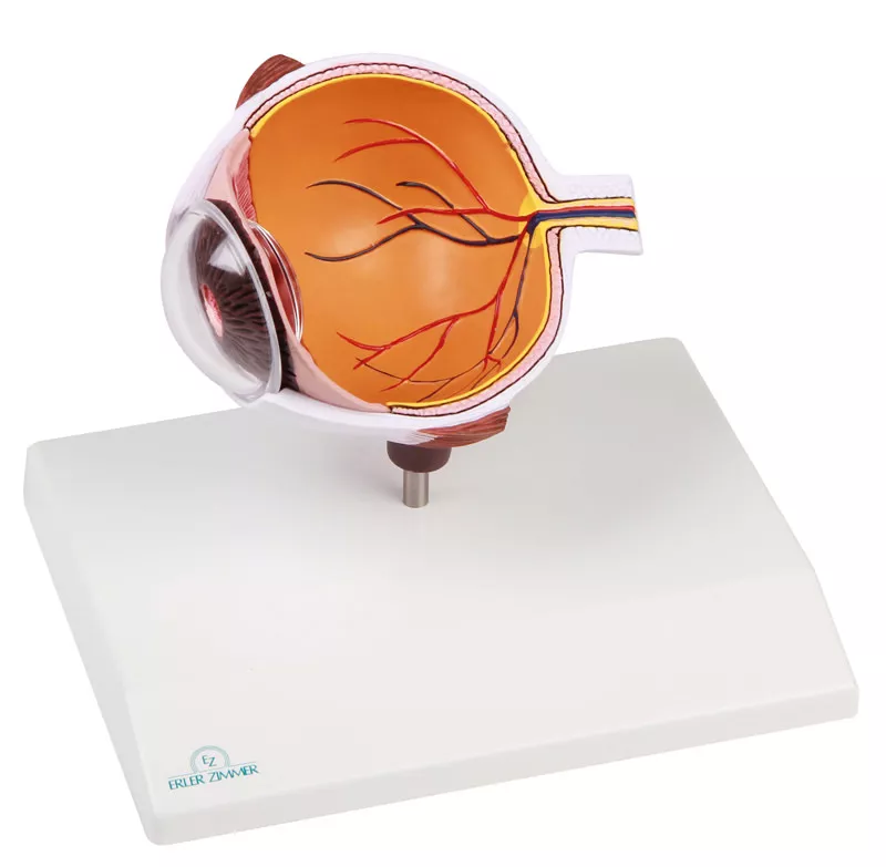
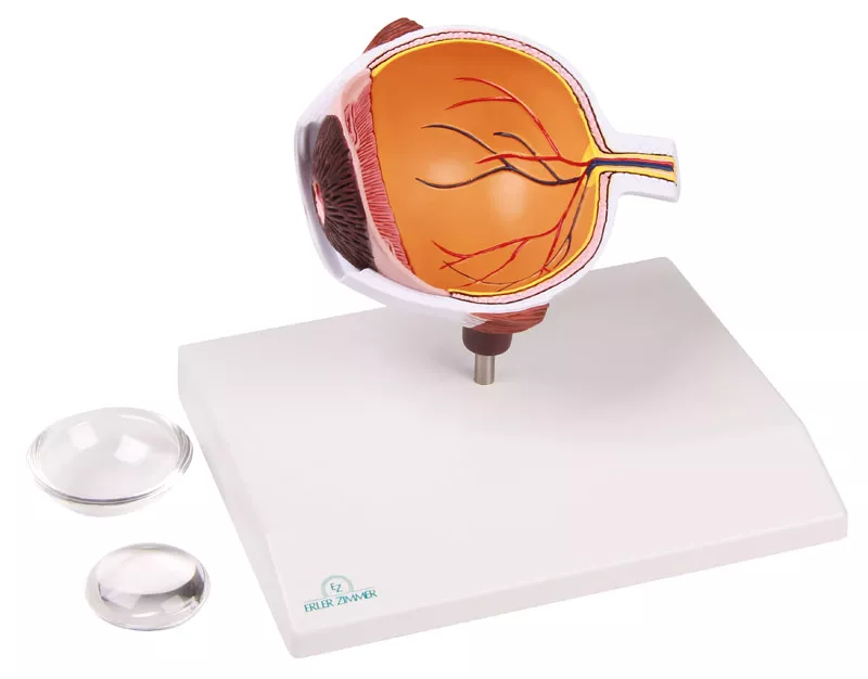

Eye half, enlarged - EZ Augmented Anatomy
Question regarding article:€108.29*
Available, within 1-3 working days
Item number: F50
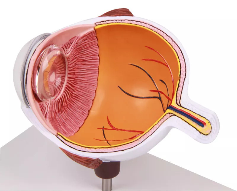
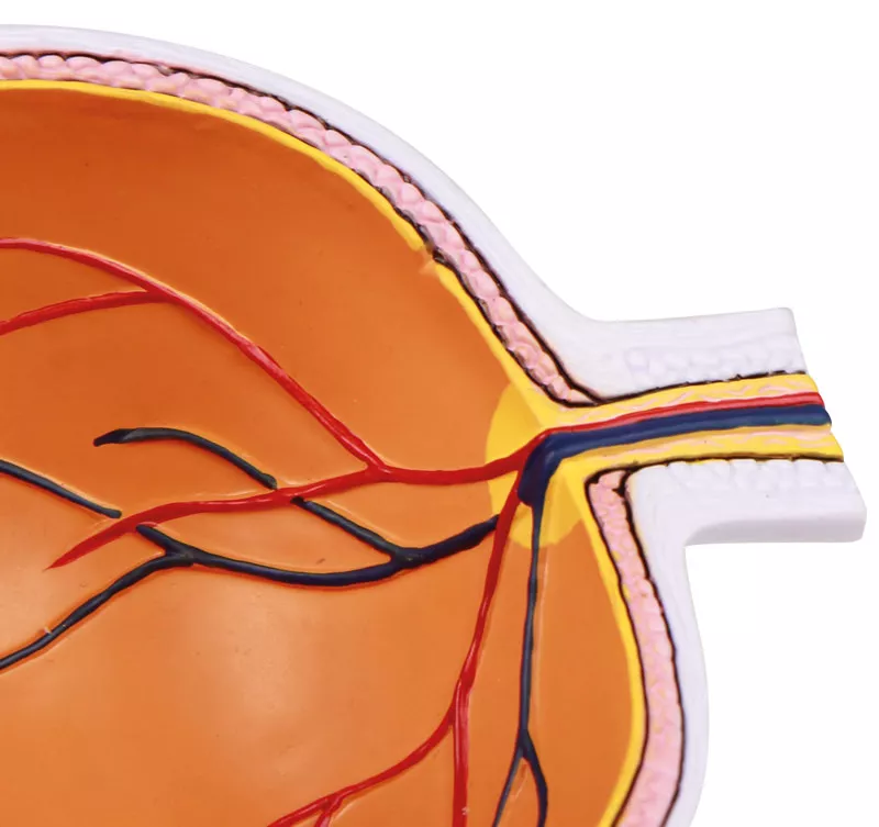
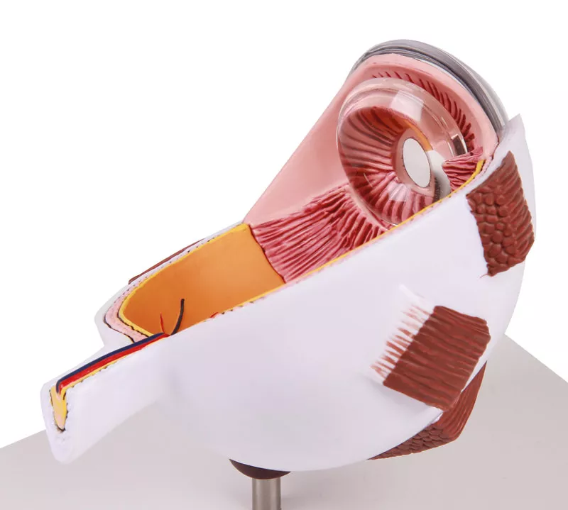
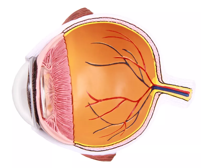
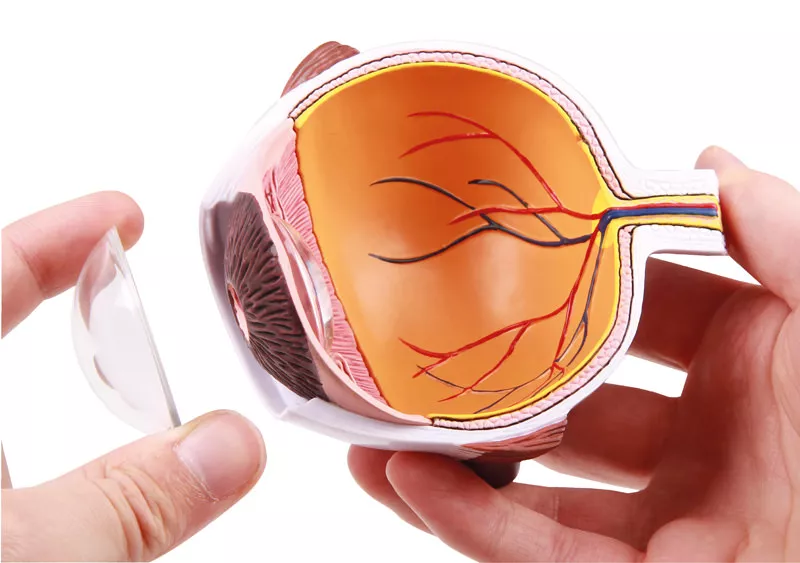
The following anatomy is shown:
Choroid, Retina, Macula, Optic disc, Optic nerve, central retinal artery and vein, retinal blood vessels, superior and inferior rectus muscle, ora serrata, lens, iris, cornea and sclera.
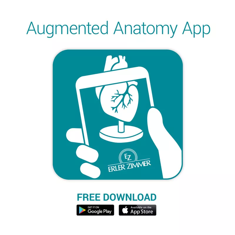
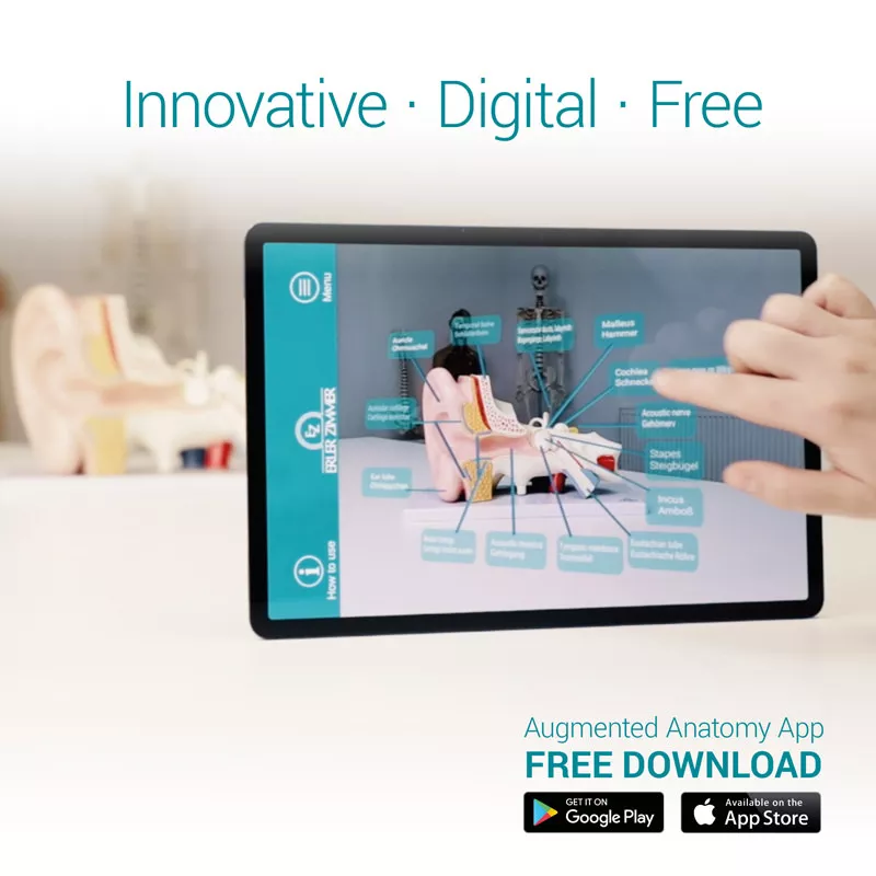
Augmented- Anatomy- App
The new Augmented Anatomy App in combination with this high quality anatomical model makes learning now even easier and more efficient!
This app automatically recognises our anatomical models and displays the nomenclature in augmented reality. As an Erler Zimmer customer, you can use it completely free of charge and for an unlimited period of time.
- High-quality Augmented Reality learning app
- Free of charge and without registration
- Nomenclature always and everywhere available
- Further online links in the learning lexicon
Our augmented anatomy app works on all common smartphones or tablets. Click here to Download the App
Login
Erler-Zimmer Medical GmbH
Hauptstrasse 27
77886 Lauf
Germany
info@erler-zimmer.de
Achtung! Medizinisches Ausbildungsmaterial, kein Spielzeug. Nicht geeignet für Personen unter 14 Jahren.
Attention! Medical training material, not a toy. Not suitable for persons under 14 years of age.
Other customers also bought


Continuous innovation

Social responsibility

Active customer orientation

Understanding quality

Sustainable actions

ISO 9001 certification