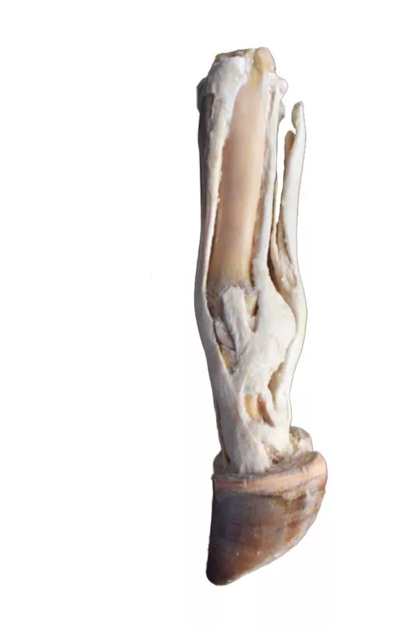
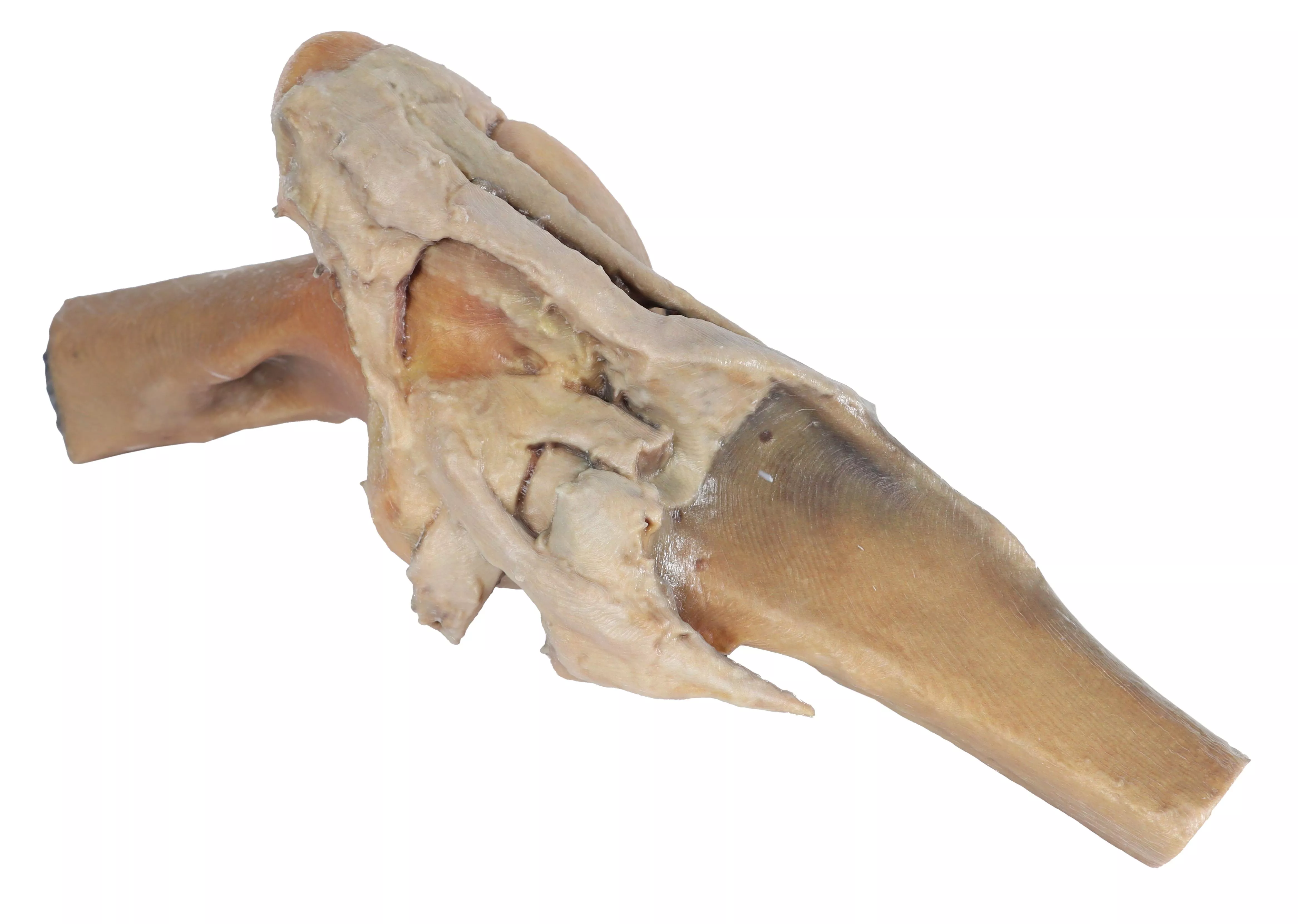
Product information "Ox foot - tendons and ligaments"
This specimen records the anatomy of an ox‘s right foot from the metatarsus to the distal phalanges. In the dorsal aspect, the insertion of the tendons of the common, lateral and medial digital extensor muscles are the main structures. In the plantar aspect, the relation of the tendons of the superficial and deep digital flexor muscles with deep structures such as the interosseous ligament can be detected. Capsular and extra-capsular ligaments of the metatarsophalangeal and interphalangeal joints are also preserved.

0 of 0 reviews

Leave a review!

Share your experiences with other customers.


Erler-Zimmer

Erler-Zimmer GmbH & Co.KG
Hauptstrasse 27
77886 Lauf
Germany
info@erler-zimmer.de


Achtung! Medizinisches Ausbildungsmaterial, kein Spielzeug. Nicht geeignet für Personen unter 14 Jahren.

Attention! Medical training material, not a toy. Not suitable for persons under 14 years of age.


Other customers also bought

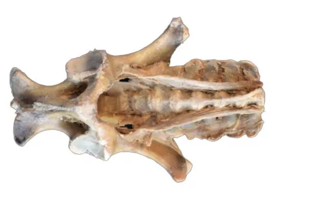
Horse (foal) pelvis dissection - ligaments
This specimen is focused on the sacroiliac joint, the two coxal bones (os coxae) are exposed in the dissection: Ilium, pubis and ischium, together with lumbar vertebrae and sacrum. At the sacroiliac joint, the dorsal sacroiliac ligament can be identified. The broad sacrotuberous ligament of the pelvis extends from the lateral sacral crest to the sciaticspine and ischial tuberosity. The greater sciatic foramen occupied by the lumbosacral trunk. From the vertebral bodies and part of the transverse processes of the lumbarvertebrae, the tendons of insertion of the psoas minor muscles are identified. The ventral longitudinal ligament connects the ventral aspect of most cranial lumbar vertebral bodies.

€5,353.81*
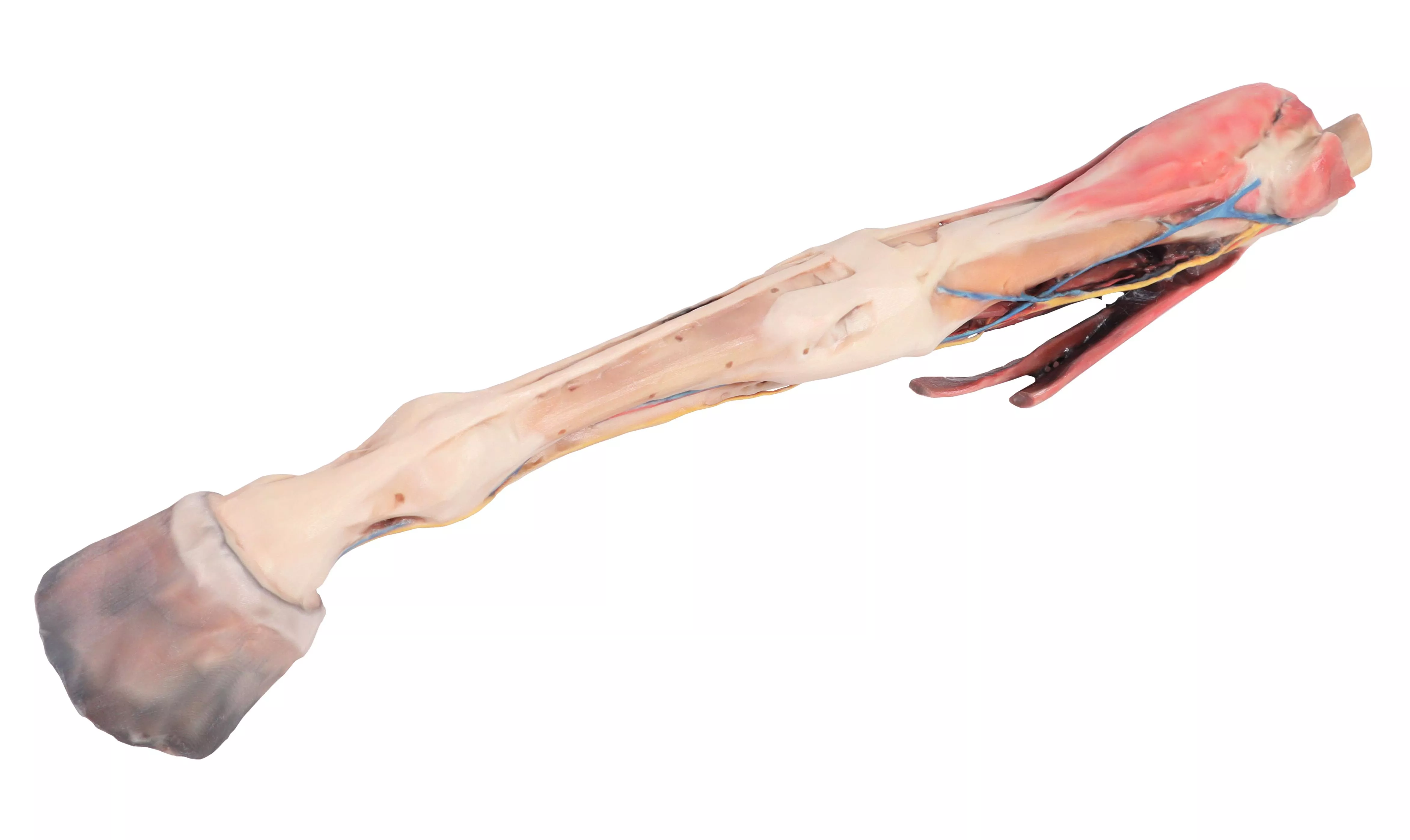
Horse forelimb - muscles, tendons, ligaments vessels and nerves distal to the stifle - 1/3 life-size
This specimen demonstrates the superficial anatomy of a right forelimb from the elbow joint to the hoof. The carpus and digital extensor and flexor muscles are located in the forearm related to the pathway of the median and cubital nerves. The main branches of the median artery as well as the median and cephalic veins have been maintained. Distal to the carpus the specimen shows the topographic relation of the palmar nerves with the digital tendon flexors. Distal to the metacarpophalangeal joint it is possible to see at both sides the palmar digital neurovascular bundle.

€2,499.00*
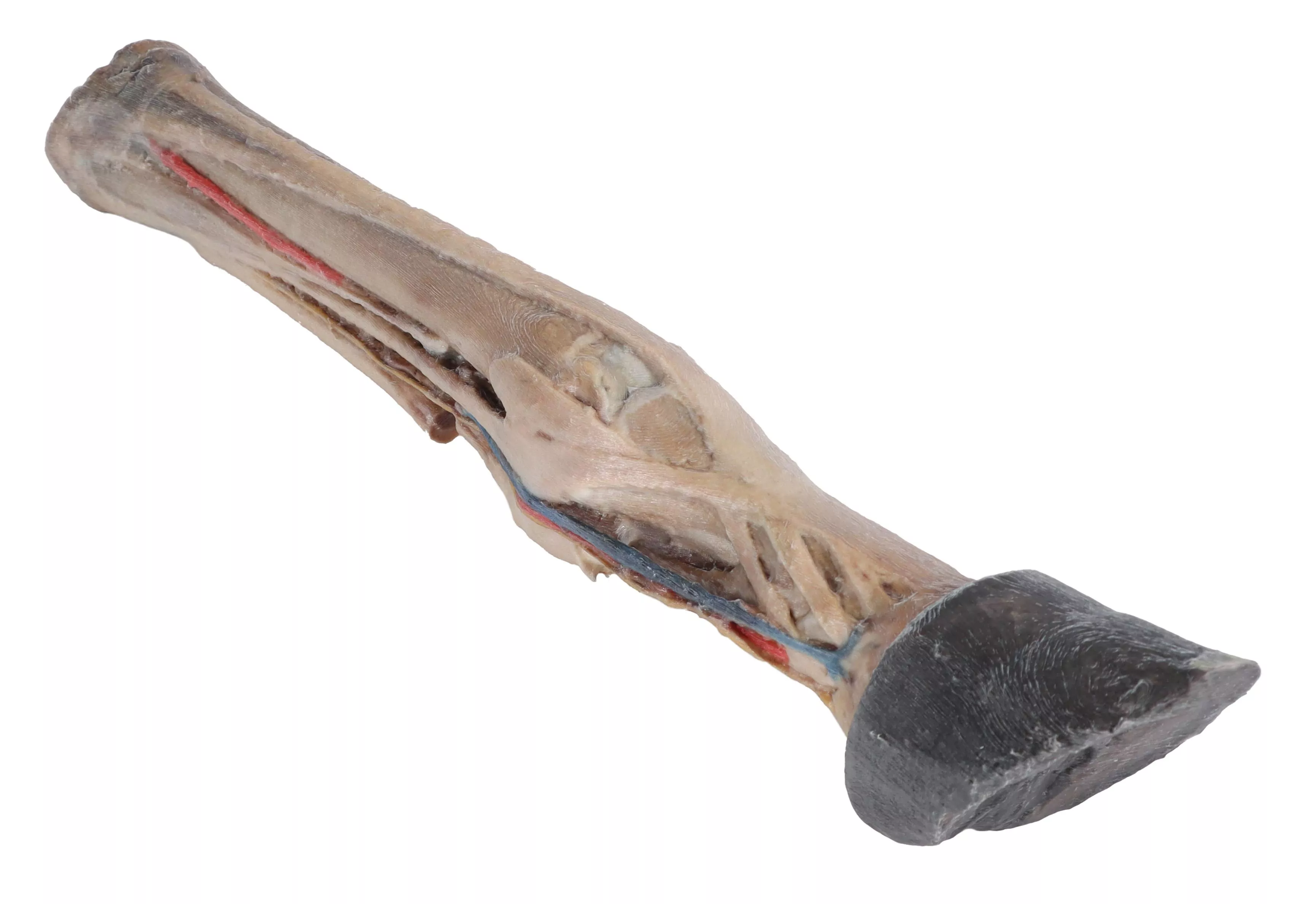
Horse foot - tendons, ligaments, vessels and nerves - 1/3 life-size
This dissected right-foot specimen displays a mixture of the vascular, nervous, ligaments and tendons. The plantar digital neurovascular bundles both sides of the flexor tendons. The two digital extensor tendons are identified in the proximal dorsal aspect of the specimen. Metatarsophalangeal and proximal interphalangeal joints show all ligaments. Between the plantar aspect of the metatarsal bones and the digital tendon flexors is the interosseus ligament with its sesamoid and extensor branches.

€2,082.50*
Horse stifle joint dissection
This specimen demonstrates the ligaments of the stifle joint with the leg in extension. In the anterior view, with the patella and the three patellar ligaments.

€1,243.55*
Horse sagittal half-head
This horse half-head shows a superficial dissection on the right lateral aspect and a middle sagittal section on the medial aspect.

€4,700.50*

Continuous innovation

Social responsibility

Active customer orientation

Understanding quality

Sustainable actions

ISO 9001 certification

Your last viewed products