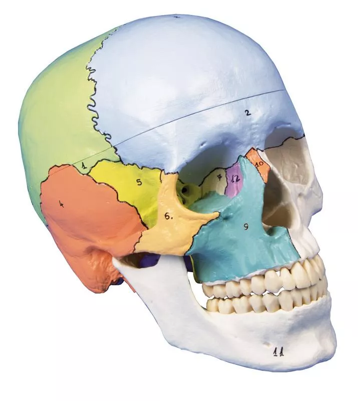
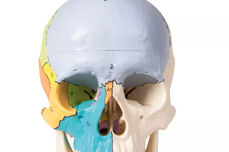
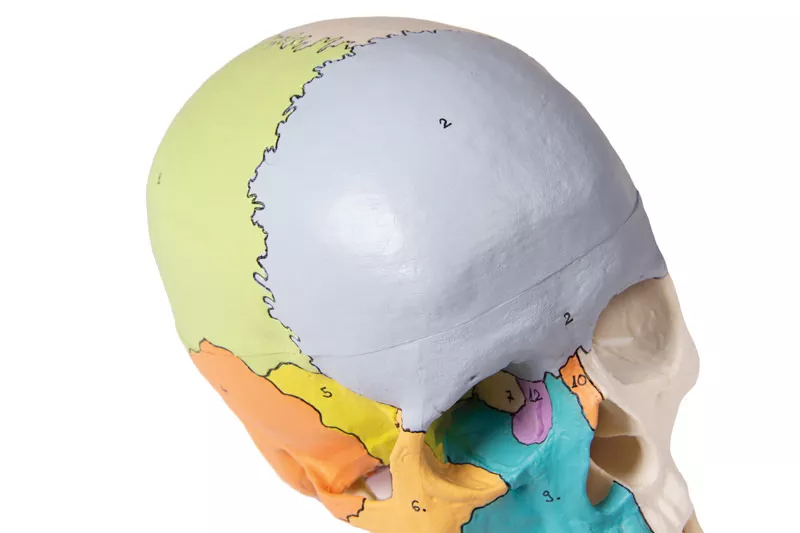
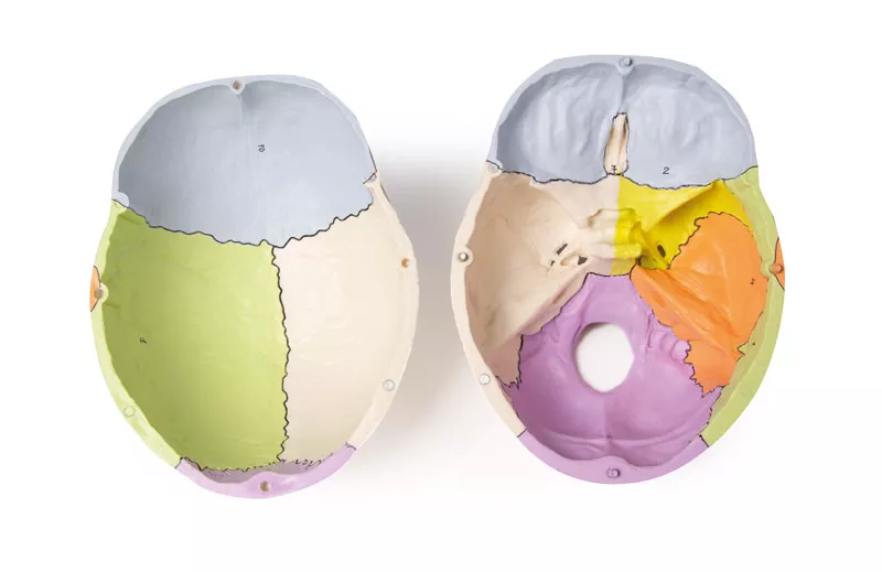
Product information "Skull model, 3-part, didactical painted"
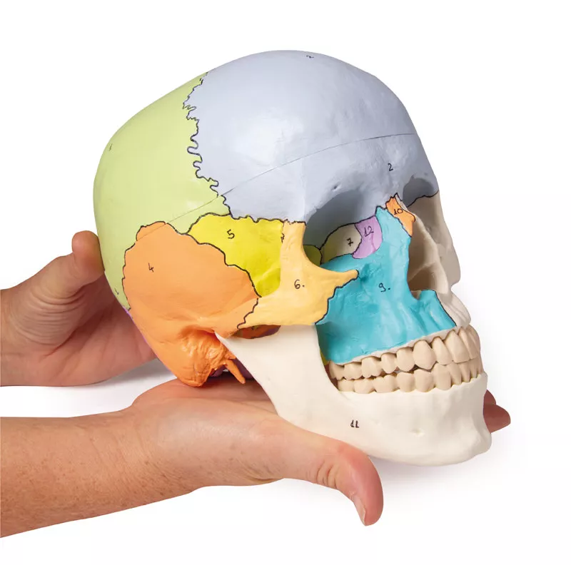
Skull model like ref.no. 4500, additionally with didactical painting of individual bones on one side of the skull. The bones are numbered referring to the included nomenclature.
Size: 18 x 19 x 12 cm, weight: 0.7 kg
0 of 0 reviews
Leave a review!
Share your experiences with other customers.
Login
No reviews found. Share your insights with others.
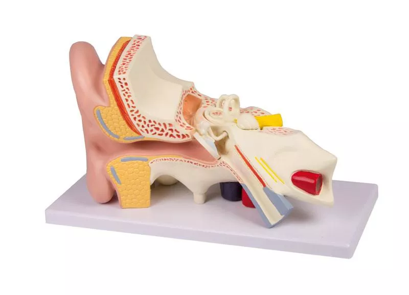
Enlarged approx. 15 times. The model shows instructive the organs of the middle and inner ear. The bony and membranous labyrinths are shown and the cochlea can be opened. On stand.
High Quality anatomical chart, made of 200g/sqm art paper, size 50x70cm, incl. attachable plastic rods with hanger. Nomenclature in German and English.
This life-size brain model shows the major anatomical landmarks and cortical areas of the brain in carefully painted color. The right half is painted to show the four lobes of the brain: parietal, temporal, occipital, and frontal. The left half comes apart in four pieces- the frontal and parietal, the temporal and occipital, the brainstem, and the cerebellum. It is painted to show 5 cortical areas of the brain: the primary motor cortex, the sensory cortex, the visual cortex, Broca‘s area (motor/speech), the auditory cortex, and Wernicke‘s speech area. This model fits perfectly in our skull models ref.no. 4500, 4505, 4508, 4509, 4512, 4514, 4701 and 4708 and is compatible with the Falx cerebri model ref.no. C925.Augmented- Anatomy- AppThe new Augmented Anatomy App in combination with this high quality anatomical model makes learning now even easier and more efficient!This app automatically recognises our anatomical models and displays the nomenclature in augmented reality. As an Erler Zimmer customer, you can use it completely free of charge and for an unlimited period of time. - High-quality Augmented Reality learning app- Free of charge and without registration- Nomenclature always and everywhere available- Further online links in the learning lexiconOur augmented anatomy app works on all common smartphones or tablets. Click here to Download the App
The front heart wall can be removed to show the inner structures in detail. All important structures are present such as ventricles, atriums, aortic, mitral, pulmonary and tricuspid valves. Heart muscle, fatty tissue, arteries and veins are painted in detail; the structures are shown on the educational card (German/English). The model is made of unbreakable plastic and removable from the stand.Size: 8 x 8 x 14 cm, weight: 0.4 kgAugmented- Anatomy- AppThe new Augmented Anatomy App in combination with this high quality anatomical model makes learning now even easier and more efficient!This app automatically recognises our anatomical models and displays the nomenclature in augmented reality. As an Erler Zimmer customer, you can use it completely free of charge and for an unlimited period of time. - High-quality Augmented Reality learning app- Free of charge and without registration- Nomenclature always and everywhere available- Further online links in the learning lexiconOur augmented anatomy app works on all common smartphones or tablets. Click here to Download the App
This model of two lumbar vertebrae in about double life size has the opportunity to demonstrate a lateral intervertebral disc hernia. If the two vertebrae are pressed together, the inner core of the disc will be protruding to the outside, forming a hernia. The protruding hernia shows graphically the pressure which affects the spinal nerve. With this helpful tool you can demonstrate fast and easily what happens when a patient suffers from a herniated disc. Because of the enlarged size it is easy to handle and also suitable for group teaching.