


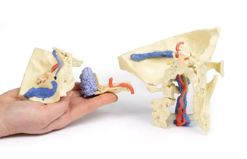


Product information "Temporal Bone Model, Set of 3"
This 3-part 3D printed model, derived from CT data, highlights the complex anatomy of the temporal bone, including ossicles, canals, chambers, foramina, and air spaces. Internal casts (endocasts) reveal the intricate internal structure, aiding visualization of the auditory and vestibular apparatus.
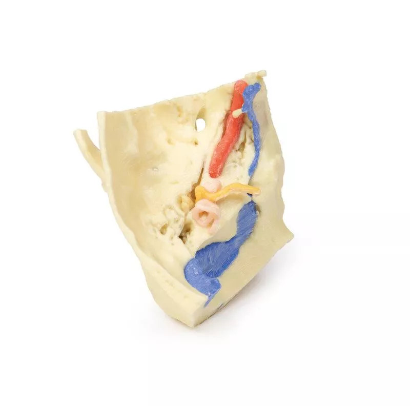
Part 1: Skull Preparation
The model exposes the posterior quadrant of the skull, including the posterior and middle cranial fossa. The temporal bone and its relationship with the sphenoid, parietal, and occipital bones are clearly visible.
The middle ear (orange) shows the tympanum, aditus, antrum, and pharyngotympanic tube. The bony labyrinth of the inner ear (green) displays the semicircular canals and cochlea. The facial nerve (CN VII) is shown in yellow, tracing its course to the stylomastoid foramen. Mastoid air cells (blue) and the temporomandibular joint are also demonstrated.
The model highlights key vascular structures, including the internal carotid artery, transverse and sigmoidal dural venous sinuses, and the jugular foramen, along with the foramen magnum and first three cervical vertebrae.
Part 2: Petrous Part of the Temporal Bone
Enlarged x3, this section emphasizes the detailed internal architecture of the petrous temporal bone, including the middle ear ossicles (malleus, incus, stapes), mastoid air cells, and bony canal of the internal carotid artery.
The facial nerve and chorda tympani nerve are traced through the tympanic cavity, illustrating their relationship with the auditory structures. The model highlights connections between the tympanum and mastoid air cells via the aditus and antrum.
Part 3: Auditory and Vestibular Apparatus
Also enlarged x3, this model section focuses on the auditory and vestibular apparatus and their relation to surrounding otological structures. The bony labyrinth, ossicles, and tympanic connections to the nasopharynx are clearly shown.
The model demonstrates the passage of the facial (CN VII) and vestibulocochlear (CN VIII) nerves through the temporal bone, including the cochlear nerve, geniculate ganglion, and chorda tympani. The spatial relationship between these nerves, the internal carotid artery, and dural venous sinuses is accurately represented.
Login
Erler-Zimmer
Erler-Zimmer Medical GmbH
Hauptstrasse 27
77886 Lauf
Germany
info@erler-zimmer.de
Achtung! Medizinisches Ausbildungsmaterial, kein Spielzeug. Nicht geeignet für Personen unter 14 Jahren.
Attention! Medical training material, not a toy. Not suitable for persons under 14 years of age.
Other customers also bought

Head and visceral column of the neck
This 3D print provides a detailed exploration of the head and visceral column of the neck, showcasing muscles, nerves, arteries, veins, and cranial structures.Facial AnatomyThe right side of the face is dissected to reveal the facial nerve and its branches (temporal, zygomatic, buccal, marginal mandibular, and cervical) following removal of the parotid gland. The relationships of the facial nerve, retromandibular vein, and external carotid artery are clearly visible. Muscles of the head include temporalis, masseter, and posterior belly of digastric, while arteries such as the facial, transverse facial, and superficial temporal arteries are preserved. The facial and transverse facial veins unite to form the common facial vein, which joins the retromandibular vein to form the external jugular vein.From the anterior view, facial muscles around the mouth (buccinator, orbicularis oris, zygomaticus major) and muscles of mastication are displayed. In the left infratemporal fossa, the medial and lateral pterygoids are dissected to show the mandibular division of the trigeminal nerve, including the lingual nerve and inferior alveolar branch. The ophthalmic division branches (supraorbital and supratrochlear nerves) supplying the forehead and scalp are also demonstrated. The submandibular glands and facial vessels coursing over the mandible are clearly visible. Neck AnatomyThe musculoskeletal structures of the neck are removed to expose the pharynx posteriorly, larynx anteriorly, and neurovascular bundles laterally. The suprahyoid and infrahyoid muscles are visible, as are the vocal folds viewed from below the trachea. The cricothyroid muscle is also identifiable. Neurovascular structures include the hypoglossal nerve, vagus nerve, internal jugular vein, common carotid artery with bifurcation into ECA and ICA, and the superior thyroid artery. The ansa cervicalis and internal branch of the superior laryngeal nerve are visible on the anterior neck. Posterior Pharynx and Vertebrobasilar StructuresThe superior, middle, and inferior constrictors of the pharynx are displayed, along with the esophagus and posterior horn of the hyoid bone. The carotid sheath shows the vagus nerve and its pharyngeal branches, and the recurrent laryngeal nerve is briefly visible. Occipital arteries curve around the mastoid, and the vertebral arteries are seen entering the foramen magnum. Removal of the cerebellum exposes the fourth ventricle, cut cerebellar peduncles, and the posterior inferior cerebellar artery on the right. Cranial Cavity and Visual PathwaysThe orbits are opened to reveal orbital nerves, vessels, eyes, and optic nerves. The optic chiasm, optic tracts, and lateral geniculate bodies are retained, showing key visual pathways. The brainstem is cut to demonstrate cranial nerve origins, while the olfactory tracts and bulbs are also displayed.

Lateral Orbit
This 3D print provides a lateral view of the orbit, with the bony lateral wall and part of the calvaria removed to reveal underlying structures. The frontal and temporal lobes of the brain are clearly exposed, allowing detailed observation of cranial and orbital anatomy.Extraocular Muscles and NervesWithin the orbit, the lateral rectus (LR) muscle has been divided to demonstrate the intraconal space. Its insertion is reflected anteriorly to reveal the inferior oblique muscle (IO) insertion, while the portion near its origin from the annulus of Zinn is reflected to show the abducens nerve (VI) entering the bulbar aspect of the muscle belly. Orbital Vessels and StructuresAdditional features visible in this model include the tarsal plate (TP), lacrimal gland (LG), lacrimal artery (LA), lacrimal nerve (LNv), and numerous other nerves and vessels surrounding the optic nerve, providing a comprehensive view of the lateral orbit.

3D Printed Anatomy - Arterial Circulation
This 3D print presents an expanded version of the dataset used for our Circle of Willis model, derived from careful segmentation of angiographic data.Intracranial ArteriesLike the original Circle of Willis print, this model demonstrates the internal carotid and vertebral arteries entering the skull and branching into the intracranial arteries that supply the brain. Expanded Arterial NetworkThis expanded model includes the full branching pattern of the cerebral and cerebellar arteries, showing the internal carotid and vertebral artery anastomoses along with the complete Circle of Willis. Detailed Cerebral Artery BranchesThe model highlights the pericallosal arteries (from the anterior cerebral arteries) with their named branches, the superior and inferior divisions of the middle cerebral artery (including sulcal, temporal, and parietal arteries), and the posterior cerebral artery branches, providing a detailed view of the brain’s arterial network.

3D Printed Anatomy - Arterial and Venous Circulation
This 3D printed specimen integrates segmented angiographic data of both the cranial arterial and venous circulation into a single model. Further description of the visible structures can be found under the ‘Circle of Willis’, ‘Cranial Arterial Circulation’ and ‘Cranial Venous Circulation’ prints.

Head and Neck
This 3D print provides a detailed view of head and neck anatomy, showing different structures depending on the perspective.Lateral Face and Parotid RegionA window exposes the parotid region, with the pinna intact and the mastoid process revealed by reflecting the sternocleidomastoid (SCM). The parotid gland is removed to display hidden structures, including the posterior digastric, masseter muscle, and mandibular condyle in the TMJ. The external carotid artery (ECA) and its branches, internal jugular vein (IJV), hypoglossal, vagus, spinal accessory, and facial nerves are clearly visible. Facial dermatomes are illustrated via the trigeminal nerve branches. Brain and Cranial CavityThe medial cerebrum shows the corpus callosum, thalamus, sulci, and gyri, while parts of the cerebellum are removed to expose the cranial fossae floor and fourth ventricle. The cavernous sinus, sella turcica, and intracranial course of cranial nerves II, III, V, VII–X, XI are displayed, including the facial canal and geniculate ganglion. Medial Surface and Key VesselsThe parasagittal section reveals the lateral ventricle, cerebral peduncle, posterior cerebral artery, and the tentorium cerebelli. The internal carotid artery in the cavernous sinus is shown lateral to the optic chiasm. Additional structures include the mouth, tongue, associated muscles, nasal cavity, nasopharynx, and cervical vertebrae.

Paranasal Sinus model
This unique 3D model is derived from dural skull (Print #8) data, obtained via CT imaging and segmentation of internal spaces. Parts of the skull have been retained, while sections or windows have been removed to expose the paranasal sinuses.Frontal SinusesThe paired frontal sinuses are highlighted in blue. The right frontal sinus is fully exposed, showing the frontonasal ostium, which drains as a funnel-shaped tube into the infundibulum of the middle meatus of the nasal cavity. The left frontal sinus remains partially surrounded by frontal bone, illustrating the natural anatomy. Ethmoid and Maxillary SinusesThe ethmoid sinuses (purple) are visible only on the left side, with the medial wall of the orbit preserved, formed by the orbital plate of the ethmoid bone. The maxillary sinus (green) on the left is partially exposed and partially retained within the maxilla, demonstrating its close relationship to the inferior orbit. The opening of the maxillary sinus into the lateral wall of the nose is subtly indicated in the middle meatus. Sphenoid SinusThe left sphenoid sinus (pink) is embedded within the sphenoid bone, which has been partly digitally removed. Despite this, the impression for the internal carotid artery remains clearly identifiable, highlighting critical anatomical relationships.

3D Printed Anatomy - Venous Circulation
This 3D print is based on the same dataset as our Circle of Willis and cranial arterial circulation models, derived from careful segmentation of angiographic data. The model highlights the dural venous sinus network, segmented according to structures visible via contrast medium circulation.Venous Network and SinusesMost dural venous sinuses are represented; however, due to limited contrast in the anterior venous system, structures such as the cavernous sinus and petrosal sinuses are not included. The model clearly shows the extensive network of dural veins and venous lacunae, which join in the midline to the superior sagittal sinus. Deep Venous StructuresBeneath this network, the great cerebral vein, inferior sagittal sinus, and straight sinus are visible, converging with the superior sagittal sinus at the confluence of sinuses. Transverse and Sigmoid SinusesSeveral dural veins drain into the left and right transverse sinuses, passing anteriorly toward the petrous portion of the temporal bone. The sigmoid sinuses are visible in the posterior cranial fossa before exiting the skull at the jugular foramen, forming the internal jugular vein, which can be seen on the inferior surface of the skull.

Superior Orbit
This 3D printed model provides an in-depth view of the anterior and middle cranial fossae, with the calvaria and cerebrum removed to expose underlying structures.Midbrain and Arterial AnatomyThe midbrain is sectioned at the level of the tentorium cerebelli, revealing the superior colliculi, cerebral peduncles, and substantia nigra. The vertebral arteries are clearly visible as they rise from the posterior cranial fossa and divide into the posterior cerebral arteries. Anteriorly, the internal carotid arteries emerge from the roof of the cavernous sinus, medial to the anterior clinoid processes and lateral to the optic nerves and chiasm. The oculomotor nerves penetrate the roof of the cavernous sinuses posterior to the carotid emergence. Olfactory and Orbital StructuresIn the anterior cranial fossa midline, the crista galli is retained with olfactory bulbs above the cribriform plates. On the right, removal of the frontal orbital plate exposes the frontal nerve, splitting into the supraorbital and supratrochlear nerves, lying above the levator palpebrae superioris. The trochlear nerve enters the superior oblique muscle medially. Ethmoidal air cells are visible in the medial orbital wall after partial removal of the lamina papyracea.On the left, the levator palpebrae and superior rectus muscles are divided along with the frontal nerve to reveal the optic nerve, nasociliary nerve, ophthalmic artery, and superior ophthalmic vein within the intraconal space. Facial AnatomyThe face has been dissected to show facial muscles around the orbit on the right, while the infraorbital nerve is visible on the left. The infratrochlear nerve, as well as facial veins and arteries, are also demonstrated.

Deep face/Infratemporal fossa
This specimen provides a detailed view of the infratemporal fossa and surrounding neck structures.Mandible and Infratemporal FossaThe ramus, coronoid process, and mandibular head have been removed to expose the deep infratemporal fossa. The pterygoid muscles are removed to show the lateral pterygoid plate and posterior surface of the maxilla. The buccinator remains visible, originating from the maxilla, pterygomandibular raphe, and mandible. The superior constrictor muscle arises from the posterior pterygomandibular raphe, and the internal laryngeal nerve is preserved. Neck Muscles and Styloid RegionIdentifiable neck muscles include the mylohyoid, strap muscles, and inferior constrictor. The styloid muscles descend from the styloid process to their insertions, and the internal carotid artery is visible deep to the styloid, near the origins of stylohyoid, styloglossus, and stylopharyngeus. Medial Sagittal ViewThe medial sagittal surface reveals the lateral wall of the nasal cavity (superior, middle, and inferior conchae; sphenoethmoidal recess; superior, middle, and inferior meatuses), nasopharynx, auditory tube opening, hard and soft palate, oropharynx, laryngopharynx, hyoid bone, and laryngeal cartilages. Tongue muscles, laryngeal, and pharyngeal structures are clearly visible. Cervical SpineThe mid-sagittal cut displays the vertebral bodies of C2–C5, the atlas (C1) arch, and the dens of the axis (C2).

Bronchial Tree
This 3D printed specimen preserves a human trachea, carina, and complete right and left bronchial trees down to the level of the tertiary lobar bronchi. Each set of lobar bronchi has been colour-coded to clearly demonstrate the bronchopulmonary segments of the right and left lung lobes.Right Lung Bronchial AnatomyFrom the right primary bronchus, the secondary bronchus to the upper lobe branches into tertiary bronchi supplying the apical (yellow), anterior (brown), and posterior (sienna) segments. The bronchus intermedius divides to supply the middle lobe, including the lateral (lilac) and medial (tan) segments. The lower lobe bronchus gives rise to the tertiary bronchi of the superior (yellow) and basal segments, which include anterior (purple), posterior (sienna), lateral (brown), and medial (tan). Left Lung Bronchial AnatomyFrom the left primary bronchus, the secondary bronchus to the upper lobe gives rise to tertiary bronchi supplying the apical-posterior (sienna), anterior (brown), superior lingual (tan), and inferior lingual (lilac) segments. The lower lobe bronchus divides into tertiary bronchi for the superior (yellow) and basal segments, including anteromedial (brown), lateral (tan), and posterior (sienna).

Circle of Willis
This highly detailed 3D printed model offers a precise representation of the intracranial arteries supplying the human brain, created from segmented angiographic imaging data. The model displays vascular structures that cannot be physically dissected and viewed in situ, making it a unique and valuable teaching tool.Vertebral and Basilar ArteriesThe paired vertebral arteries are shown entering the cranial cavity through the foramen magnum, where they unite to form the basilar artery. The basilar artery travels along the brainstem and terminates by branching into the posterior cerebral arteries. Just before this bifurcation, the superior cerebellar arteries arise, supplying parts of the cerebellum and midbrain. Internal Carotid Arteries and Carotid SiphonThe internal carotid arteries (ICAs) are traced from their entry at the carotid canal within the temporal bone. From there, they course medially and anteriorly through the petrous portion of the bone and emerge via the upper opening of the foramen lacerum. While the cavernous sinus is not shown, the model beautifully captures the characteristic S-shaped carotid siphon on both sides, clearly positioned lateral to the sella turcica and passing medial to the anterior clinoid processes. Major Cerebral Arteries and the Circle of WillisEach internal carotid artery divides into the anterior cerebral artery (ACA) and middle cerebral artery (MCA). The posterior communicating arteries, connecting the posterior cerebral arteries (PCAs) to the MCAs, are distinctly visible, allowing for an excellent demonstration of the posterior portion of the Circle of Willis.The anterior communicating artery, completing the circle between the paired anterior cerebral arteries, is also present—though its visibility is limited due to the close proximity of the ACAs.

Dural Skull
This high-quality 3D print of the skull and cranial cavity preserves key anatomical structures, including the dural folds. A thin strip of the calvaria has been retained in the mid-sagittal plane to maintain the attachment of the falx cerebri.Preserved Tentorium CerebelliThe tentorium cerebelli is carefully preserved in this model, clearly demonstrating the tentorial notch, which normally houses the mid-brain. Highlighted Sella Turcica and Internal Carotid ArteriesIn the region of the sella turcica, the entry points of the internal carotid arteries through the roof of the cavernous sinuses are highlighted in red, providing an intuitive visual guide for students and professionals. Focus on Dural Venous SinusesThe main purpose of this model is to showcase the disposition of the dural folds and the position of the dural venous sinuses, which are highlighted in pale blue. The following sinuses are clearly indicated:- Superior sagittal sinus- Inferior sagittal sinus- Straight sinus- Transverse sinuses- Superior and inferior petrosal sinuses- Sphenoparietal sinuses- Cavernous sinuses

Medial Orbit
This 3D print provides a unique medial perspective of the orbit, with the majority of the lateral wall of the nasal cavity and intervening ethmoidal sinuses carefully removed. This allows detailed visualization of the orbital contents and their close anatomical relations.Orbital Nerves and MusclesThe posterior ethmoidal nerve (PEN), a branch of the nasociliary nerve (V1), is visible passing between the medial rectus (MR) inferiorly and the superior oblique muscle superiorly. A small portion of the orbital plate of the ethmoid bone (EB) has been retained to illustrate the nerve’s path as it enters the posterior ethmoidal foramen. Additional Anatomical FeaturesOther key structures visible in this model include the frontal nerve (FN), sphenoid sinus (SS), pituitary gland (PG), and the frontal sinus mucosal lining, which is exposed after removal of the orbital plate of the frontal bone on the anterior roof of the orbit. The internal carotid artery and optic nerve are also clearly displayed, highlighting the intricate relationships within the medial orbital region.

Continuous innovation

Social responsibility

Active customer orientation

Understanding quality

Sustainable actions

ISO 9001 certification