


Product information "Ultrasound Training model Anatomy / Pathology"
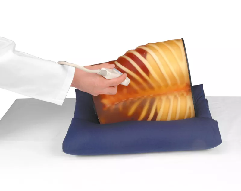
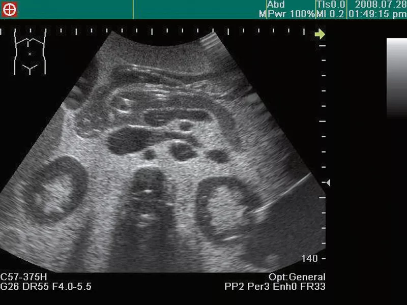
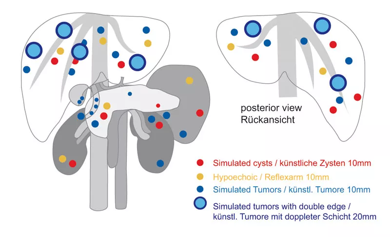
This high fidelity training model allows practice of ultrasound with existing ultrasound – machines. It includes the anatomy of the abdomen as well as many pathologies. The model includes the liver (Couinnaud’s segments are visible), biliary tract, pancreas, spleen, kidneys, detailed vascular structures like aorta, vena cava, celiac artery and ist branches, portal vein and its branches, superior mesenteric vessels, renal vessels etc. Multiple cysts and tumors in this model give various training opportunities for advanced examination. The phantom can be scanned from all sides like a real patient.
Size: 28 x 25 x 18 cm, weight 12 kg
Size: 28 x 25 x 18 cm, weight 12 kg
Login
Kyotokagaku
Kyotokagaku Co. Ltd.
15 Kitanekoya-cho
Fushimi-ku
612-8388 Kyoto
Japan
rw-kyoto@kyotokagaku.co.jp
Verantwortlich/Responsible:
Erler-Zimmer Medical GmbH
Hauptstrasse 27
77886 Lauf
Germany
info@erler-zimmer.de
Achtung! Medizinisches Ausbildungsmaterial, kein Spielzeug. Nicht geeignet für Personen unter 14 Jahren.
Attention! Medical training material, not a toy. Not suitable for persons under 14 years of age.
Other customers also bought

Anatomical Model for Ultrasound education
€4,057.90*
This 20 part model of the upper abdominal organs represents exactly the anatomy that is inside the training models R16560 and R16570. This allows you to see the structures and organs three dimensional in front of you while you are scanning them in the training model. The single parts are: liver (can be separated into 8 segments), gall bladder, spleen, left kidney, vena cava, spine, large and small intestine, portalvein, bile duct and hepatic artery, pancreas, right kidney, abdominal aorta, hepatic vein and stomach.

Anatomy/pathology set Ultrasound model and anatomy model
This upper class training model enables you to practise ultrasound with the available ultrasound equipment. It includes the anatomy of the upper abdominal organs and many pathologies. The model contains in detail the liver (segments to be recognised), bile duct system, pancreas, spleen, kidneys and many vascular structures such as aorta, vena cava, upper abdominal vessels with secondary branches, portal vein with secondary branches, upper mesenteric vessels and renal vessels. A large number of cysts and tumours in the model offer comprehensive training opportunities, even for advanced diagnostics. The phantom can be scanned from all sides like a human body.Set consisting of ultrasound training model R16560 and anatomy model R16580.

Continuous innovation

Social responsibility

Active customer orientation

Understanding quality

Sustainable actions

ISO 9001 certification