Product information "Wound Foot “Wilma”"
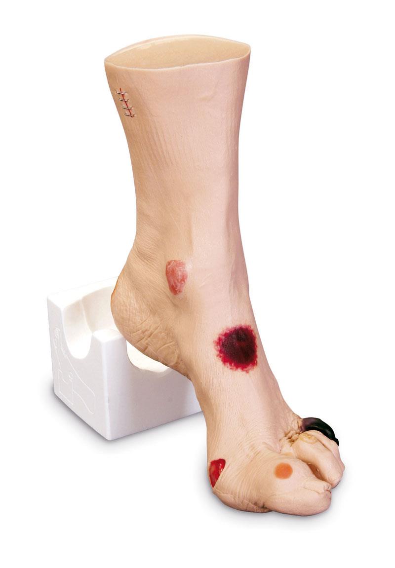
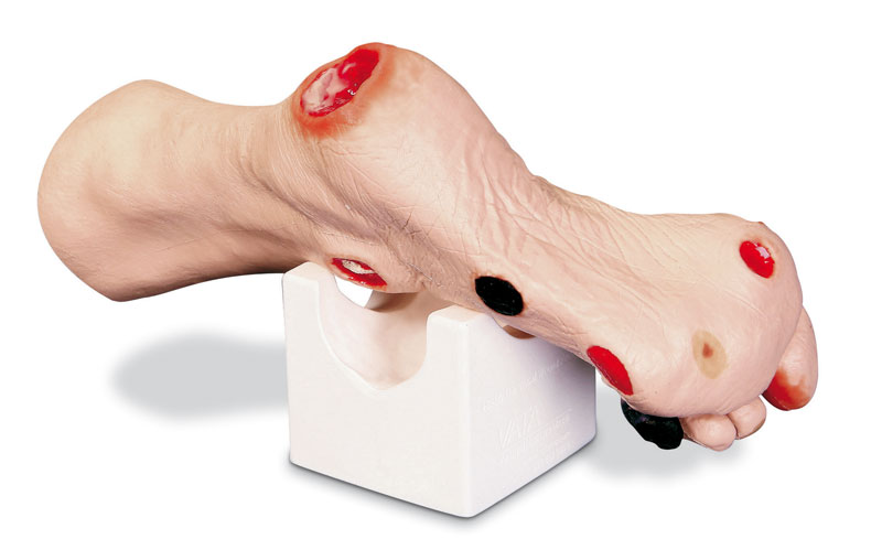
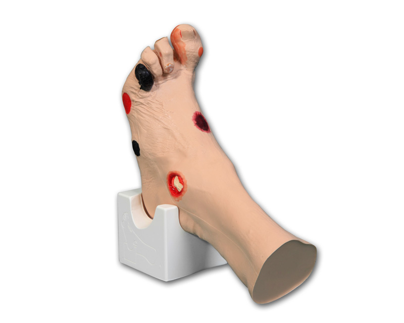
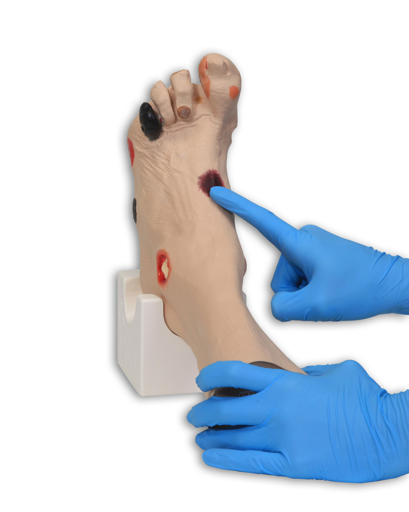
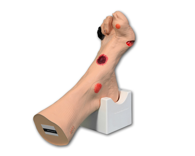
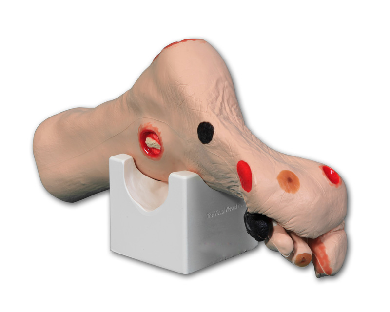
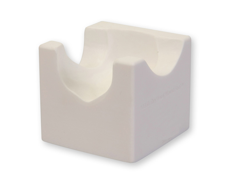
Moulded from a real foot for a true-life experience when assessing the various wounds. Twenty different conditions are presented so you can see and understand how they are different. Great care has been taken to colour each wound just as you would see it on a patient. Once the different etiologies are understood, you can discuss and devise treatment plans that will deliver optimized patient care. The following wounds and abnormalities are included: pressure ulcers, deep tissue injury, callus, amputated toe, gangrene, maceration, partial thickness wounds, corn, ingrown toenail, blister, hammer toes, and skin stapled wound. Comes with positioning base for "hands free" access to all the sites when applying dressings or teaching.
Size: 32 x 20 x 9 cm

0 of 0 reviews

Leave a review!

Share your experiences with other customers.


VATA

Vascular Access Teaching Aids, Inc.
Anatomical Healthcare Models
308 South Sequoia Parkway
97013 Canby, OR
United States of America
info@vatainc.com

Verantwortlich/Responsible:

Erler-Zimmer GmbH & Co. KG
Hauptstrasse 27
77886 Lauf
Germany
info@erler-zimmer.de


Achtung! Medizinisches Ausbildungsmaterial, kein Spielzeug. Nicht geeignet für Personen unter 14 Jahren.

Attention! Medical training material, not a toy. Not suitable for persons under 14 years of age.



Other customers also bought

Elderly Pressure Ulcer Foot
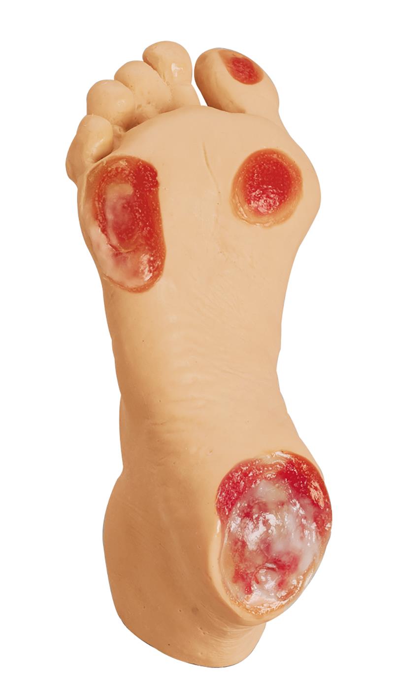
Chronic foot ulcers affect elderly the most, we have designed the Elderly Pressure Ulcer Foot for instruction on care and cleaning of pressure ulcers in various stages. Pressure ulcers are more likely to appear over pressure points such as heels, tips of toes, between toes, or anywhere the bones may protrude and rub against socks, shoes, or bed sheets. This replica contains all four severity stages:- Stage I: Located on the bottom of the toe. Surface of the skin is red.- Stage II: Located on the bottom of the foot just under the toe. Surface of the skin is red and deeper into the skin layers.- Stage III: Located on the side of the foot. Surface of the skin is red, looks more like a crater, and reaches the bottom layer of the skin.- Stage IV: Located on the heel of the foot. Surface of the skin is red. A great amount of tissue has been damaged, including muscle, bone, joints, and tendons.

€408.17*

Continuous innovation

Social responsibility

Active customer orientation

Understanding quality

Sustainable actions

ISO 9001 certification

Your last viewed products