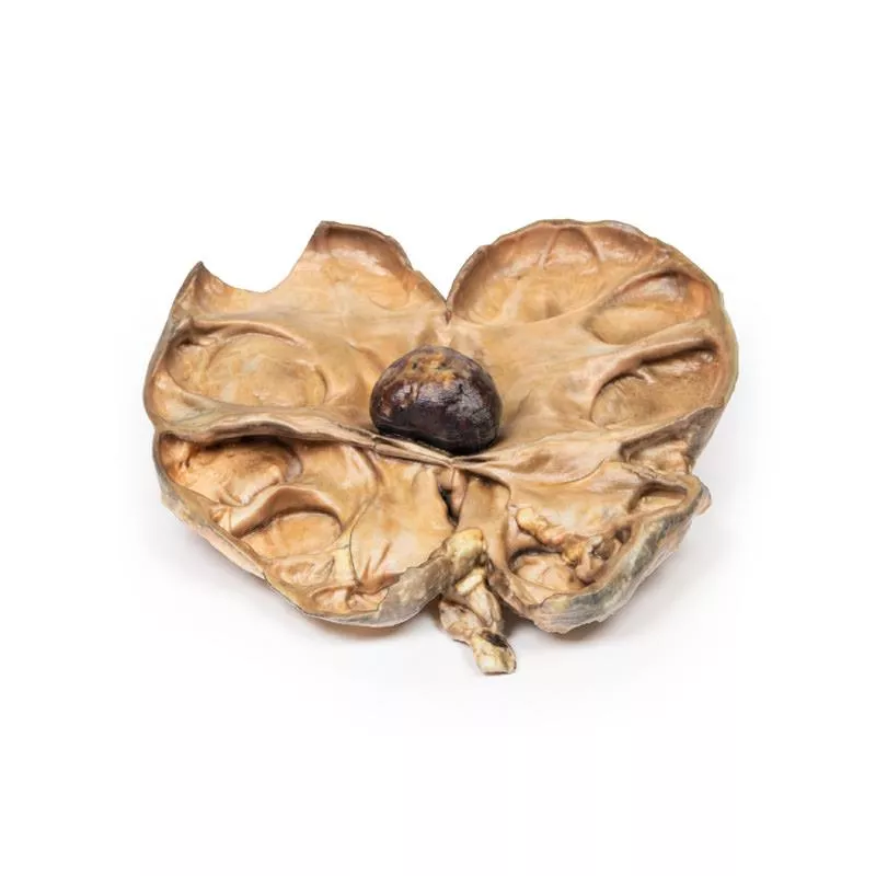
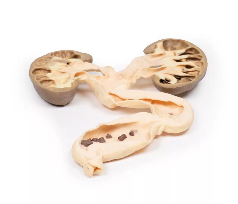
Product information "Horseshoe Kidney"
Clinical History
This specimen was found during a routine post-mortem of a 56-year-old male who died of rheumatic heart disease.
Pathology
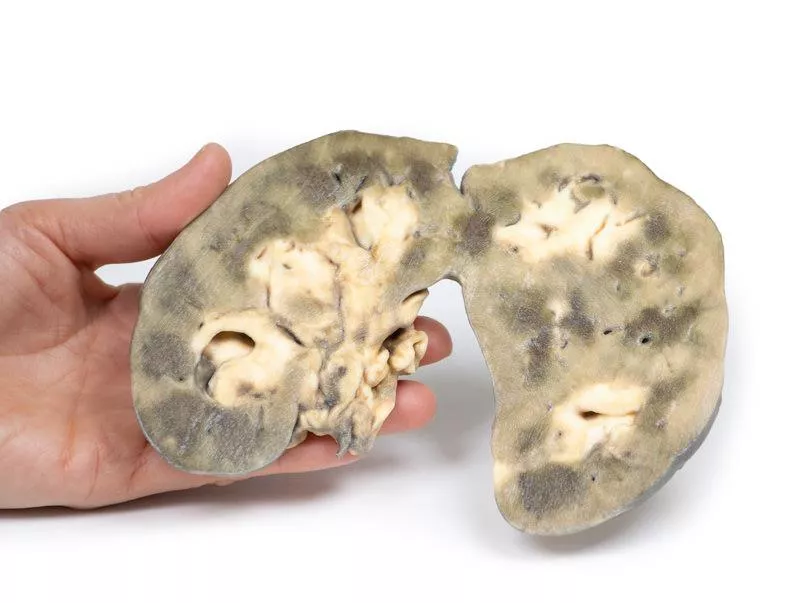
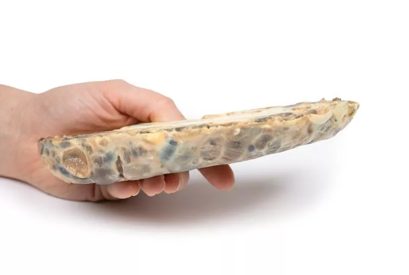
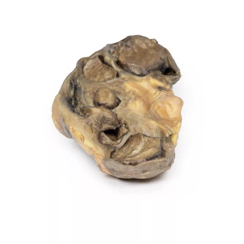
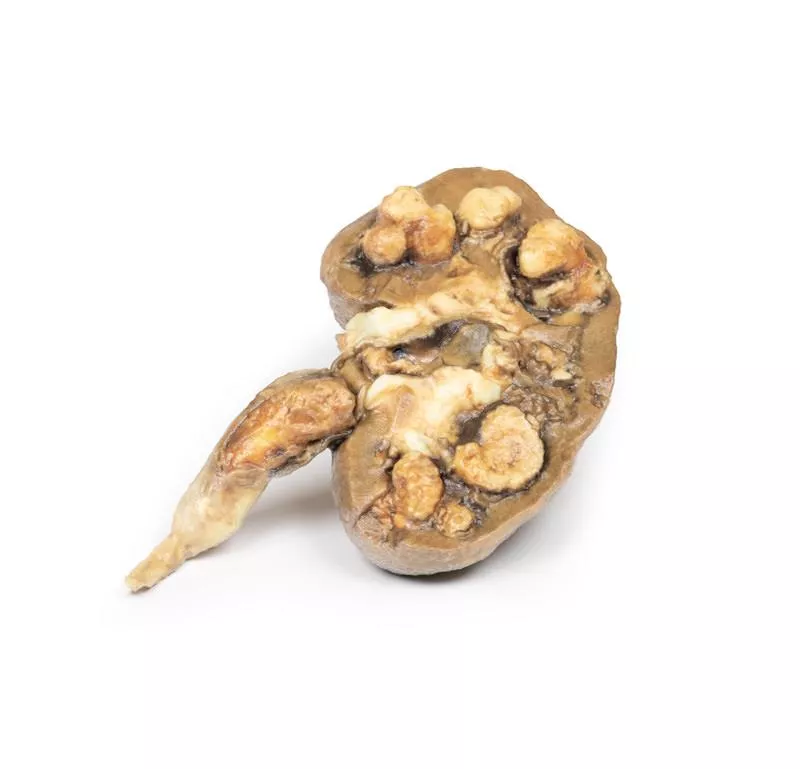
The kidney measures 12 cm in length and shows fusion of the two parts at the lower pole, forming a characteristic horseshoe shape. The ureters emerge from the hilum on the anterior side. The kidney is bisected horizontally, visible on the posterior surface, and shows persistent fetal lobulation. The renal pelvis is positioned antero-medially, with the ureters passing anterior to the fused lower poles or isthmus.
Further Information
Horseshoe kidney is the most common renal developmental anomaly, occurring twice as often in males than females, found in about 1 in 500 to 1000 autopsies. Most cases are sporadic but can be linked to chromosomal anomalies such as Down and Edwards syndromes or non-aneuploidic conditions like VACTERL association. In 90% of cases, the lower poles are connected by an isthmus of renal tissue; fusion of upper poles is rare. The renal pelvises are directed more anteriorly, and ureters angulate as they cross the isthmus.
This condition is usually asymptomatic and discovered incidentally on ultrasound or CT scans. Kidney function is typically normal, but there is a higher incidence of urinary stones, likely due to ureteral angulation and stasis. Patients also have an increased risk of hydronephrosis, mainly from pelvi-ureteric junction obstruction, and urinary tract infections often caused by vesico-ureteric reflux. Some renal malignancies, such as transitional cell carcinoma and Wilms tumour, occur more frequently.
Erler-Zimmer
Erler-Zimmer GmbH & Co.KG
Hauptstrasse 27
77886 Lauf
Germany
info@erler-zimmer.de
Achtung! Medizinisches Ausbildungsmaterial, kein Spielzeug. Nicht geeignet für Personen unter 14 Jahren.
Attention! Medical training material, not a toy. Not suitable for persons under 14 years of age.


