


Product information "Hydronephrosis Hydroureter"
Clinical History
A 49-year old male presents with a 6-week history of malaise, urinary frequency and haematuria for 6 weeks. Further questioning revealed intermittent left flank pain. Abdominal ultrasound showed severe hydronephrosis and hydroureter, secondary to multiple obstructing ureteric calculi at the uretero-vesical junction. He underwent a left nephrectomy and ureterectomy, and made a successful recovery.
Pathology
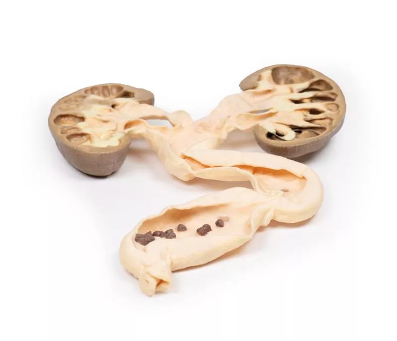
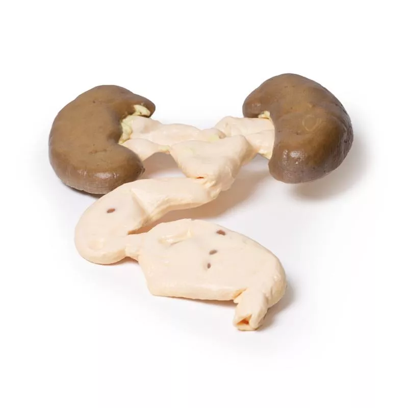
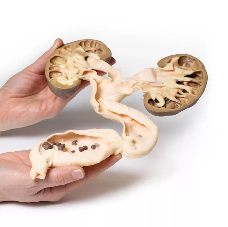
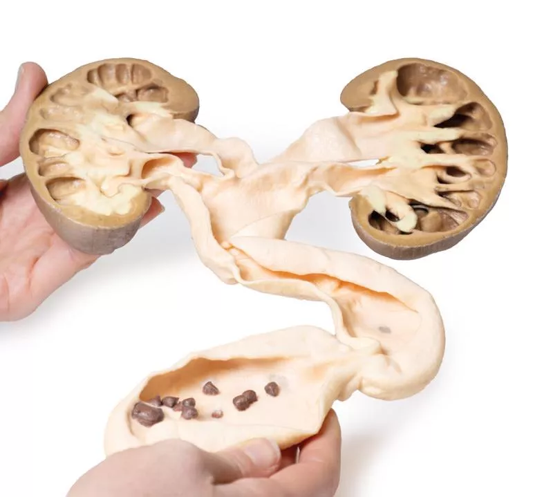
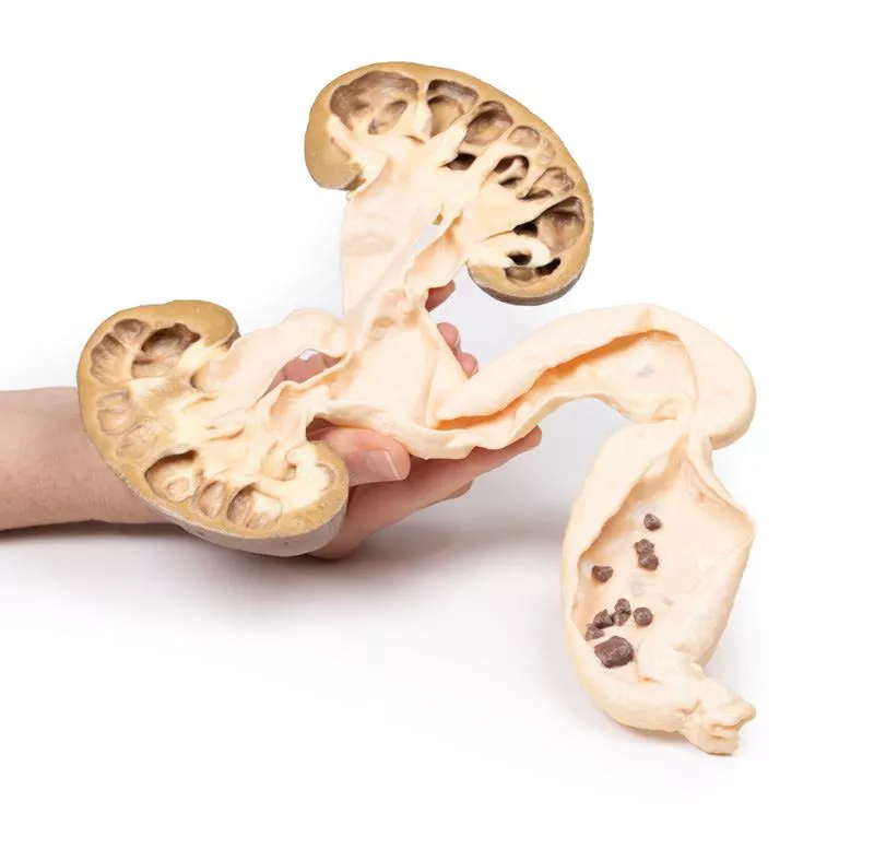
This is the patient’s left nephrectomy and ureterectomy specimen. The kidney has been bisected and the cut surface of both halves is displayed, mounted in continuity with the ureter, which has been opened. The kidney is grossly hydronephrotic, and there is considerable atrophic thinning and loss of renal parenchymal tissue. The ureter is extremely dilated and distally contains a number of small brown-black calculi with irregular sharp surface projections. These are calcium oxalate stones.
This is an example of hydronephrosis and hydroureter due to calculi obstructing the lower end of the ureter.
Further Information
Hydronephrosis, or obstructive uropathy, is the dilation of the renal pelvis and calyces caused by an obstruction in the urine outflow. Obstruction can occur at any point in the urinary tract. Any lesion-, intrinsic (within the outflow system) or extrinsic (outwith the ureter)-, that impedes the flow of urine can lead to hydronephrosis. Common causes include: congenital anomalies, urinary calculi, urinary tract tumours, urinary tract inflammation, prostatic hypertrophy, and prostate tumours. Symptoms of the hydronephrosis relate to the pathology causing the obstruction (e.g. renal colic pain with calculi), the time period of the obstruction (acute or chronic), the site (unilateral or bilateral) and whether it is complete or partial.
If the obstruction is not relieved it will ultimately cause pressure to build up proximal to the obstruction. This pressure is transmitted in a retrograde manner through the collecting ducts to the cortex causing progressive atrophy of the kidney with dilatation of the renal calyces and pelvis. The pressure also compresses the vasculature in the medulla leading to ischaemic medullary damage. Glomerular filtration persists in the affected kidney until late in the disease process when the filtration gradually diminishes or ceases. Obstruction triggers an interstitial inflammatory process leading to fibrosis. Ultrasound is the key diagnostic tool for diagnosis followed by CT or urogram. Most obstructing lesions require surgical intervention to relieve the blockage. Surgical interventions depend on each individual cause, but include nephrostomy or stenting for upper urinary tract obstruction and urinary catheter or suprapubic catheter insertions for lower urinary tract obstructions.
A 49-year old male presents with a 6-week history of malaise, urinary frequency and haematuria for 6 weeks. Further questioning revealed intermittent left flank pain. Abdominal ultrasound showed severe hydronephrosis and hydroureter, secondary to multiple obstructing ureteric calculi at the uretero-vesical junction. He underwent a left nephrectomy and ureterectomy, and made a successful recovery.
Pathology
This is the patient’s left nephrectomy and ureterectomy specimen. The kidney has been bisected and the cut surface of both halves is displayed, mounted in continuity with the ureter, which has been opened. The kidney is grossly hydronephrotic, and there is considerable atrophic thinning and loss of renal parenchymal tissue. The ureter is extremely dilated and distally contains a number of small brown-black calculi with irregular sharp surface projections. These are calcium oxalate stones.
This is an example of hydronephrosis and hydroureter due to calculi obstructing the lower end of the ureter.
Further Information
Hydronephrosis, or obstructive uropathy, is the dilation of the renal pelvis and calyces caused by an obstruction in the urine outflow. Obstruction can occur at any point in the urinary tract. Any lesion-, intrinsic (within the outflow system) or extrinsic (outwith the ureter)-, that impedes the flow of urine can lead to hydronephrosis. Common causes include: congenital anomalies, urinary calculi, urinary tract tumours, urinary tract inflammation, prostatic hypertrophy, and prostate tumours. Symptoms of the hydronephrosis relate to the pathology causing the obstruction (e.g. renal colic pain with calculi), the time period of the obstruction (acute or chronic), the site (unilateral or bilateral) and whether it is complete or partial.
If the obstruction is not relieved it will ultimately cause pressure to build up proximal to the obstruction. This pressure is transmitted in a retrograde manner through the collecting ducts to the cortex causing progressive atrophy of the kidney with dilatation of the renal calyces and pelvis. The pressure also compresses the vasculature in the medulla leading to ischaemic medullary damage. Glomerular filtration persists in the affected kidney until late in the disease process when the filtration gradually diminishes or ceases. Obstruction triggers an interstitial inflammatory process leading to fibrosis. Ultrasound is the key diagnostic tool for diagnosis followed by CT or urogram. Most obstructing lesions require surgical intervention to relieve the blockage. Surgical interventions depend on each individual cause, but include nephrostomy or stenting for upper urinary tract obstruction and urinary catheter or suprapubic catheter insertions for lower urinary tract obstructions.
Login
Erler-Zimmer
Erler-Zimmer GmbH & Co.KG
Hauptstrasse 27
77886 Lauf
Germany
info@erler-zimmer.de
Achtung! Medizinisches Ausbildungsmaterial, kein Spielzeug. Nicht geeignet für Personen unter 14 Jahren.
Attention! Medical training material, not a toy. Not suitable for persons under 14 years of age.
Other customers also bought

Continuous innovation

Social responsibility

Active customer orientation

Understanding quality

Sustainable actions

ISO 9001 certification