Produktinformationen "Bicuspid Aortic Valve"
Klinische Vorgeschichte
Eine 64-jährige Frau klagte über Brustschmerzen seit 5 Monaten, begleitet von Atemnot und Keuchen seit 4 Monaten. Bei der Untersuchung zeigte sie Dyspnoe, exspiratorisches Keuchen, linksseitige Rasselgeräusche und Zeichen eines rechtsseitigen Pleuraergusses. Puls und Blutdruck waren normal. Ein präkordialer systolischer Herzton und ein kräftiger Herzspitzenstoß im 5. linken Zwischenrippenraum wurden festgestellt. Periphere Ödeme lagen nicht vor. Die Patientin verstarb 4 Tage nach Aufnahme.
Pathologie
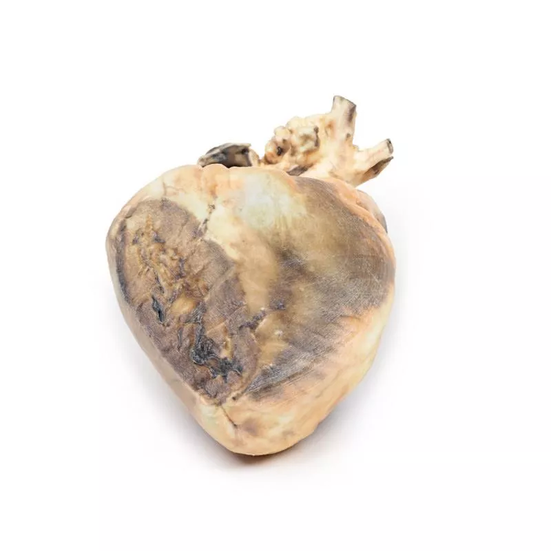
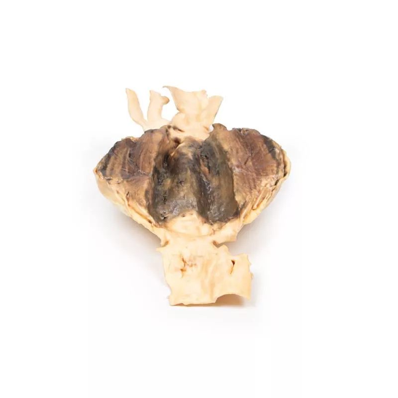
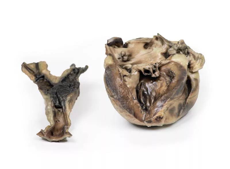
Das Herz wurde geöffnet und zeigte eine zweisegelige Aortenklappe mit zwei statt drei Klappensegeln, die nur geringfügig verdickt waren. Die Koronararterien, einschließlich der linken Zirkumflexarterie, waren weit offen. Am Herzen fanden sich dichte perikardiale Verwachsungen und Fibrose, Hinweis auf eine konstringierende Perikarditis. Die Autopsie ergab außerdem Aszites, eine kleine zirrhotische Leber, beidseitige Pleuraergüsse und einen Kollaps der rechten Lunge. Todesursache war Leberzirrhose mit Versagen, möglicherweise durch die Perikarditis bedingt. Die zweisegelige Aortenklappe war ein Zufallsbefund.
Weitere Informationen
Die zweisegelige Aortenklappe ist eine häufige angeborene Anomalie, die oft erst im höheren Alter Symptome zeigt. Sie begünstigt die Entwicklung einer verkalkten Aortenstenose, meist zwischen dem 50. und 70. Lebensjahr. Die Klappe kann isoliert oder Teil eines Syndroms wie der Fallot-Tetralogie sein. Die Fusion von zwei Klappensegeln führt zu ungleich großen Segeln, was abnorme Bewegungen und Turbulenzen verursacht. Dadurch steigt das Risiko für Aortendilatation, Dissektion und Verkalkung. Im Verlauf kann es zu Aortenstenose oder -insuffizienz kommen, die sich durch Atemnot und verminderte Belastbarkeit bemerkbar machen. Die Diagnose wird durch transthorakale Echokardiografie bestätigt.
Erler-Zimmer
Erler-Zimmer GmbH & Co.KG
Hauptstrasse 27
77886 Lauf
Germany
info@erler-zimmer.de
Achtung! Medizinisches Ausbildungsmaterial, kein Spielzeug. Nicht geeignet für Personen unter 14 Jahren.
Attention! Medical training material, not a toy. Not suitable for persons under 14 years of age.


