


Product information "Bronchopneumonia"
Clinical History
There is no clinical history available for this specimen.
Pathology
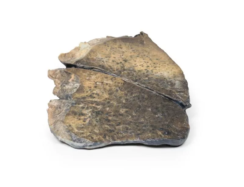
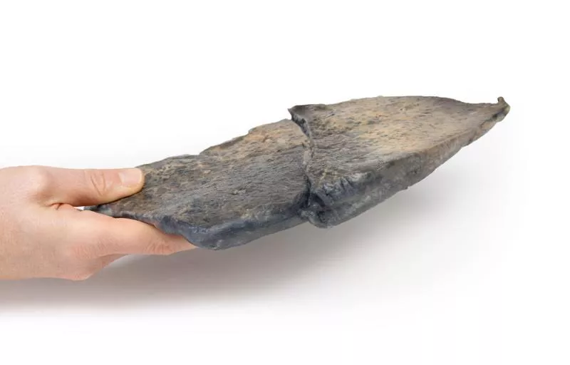
This parasagittal section of the left lung shows patchy consolidations and discolouration, especially in the upper lobe, where the pleural surface appears notably affected. The consolidations are centred around ectatic bronchioles, with signs of congestion and hyperaemia in both lobes.
Further Information
Bronchopneumonia is a form of pneumonia marked by inflammatory exudate within the alveolar spaces, leading to focal consolidation. These areas show red hepatization caused by vascular congestion, infiltration of neutrophils, red blood cells, and fibrin. The resulting consolidation gives the lung a solid appearance.
It involves acute inflammation of the bronchi with affected peribronchial and peribronchiolar lobules. Though traditionally distinguished from lobar pneumonia, patterns often overlap in clinical practice. Bronchopneumonia may progress into lobar pneumonia and is usually caused by bacterial infection. It is more often a hospital-acquired than a community-acquired pneumonia.
There is no clinical history available for this specimen.
Pathology
This parasagittal section of the left lung shows patchy consolidations and discolouration, especially in the upper lobe, where the pleural surface appears notably affected. The consolidations are centred around ectatic bronchioles, with signs of congestion and hyperaemia in both lobes.
Further Information
Bronchopneumonia is a form of pneumonia marked by inflammatory exudate within the alveolar spaces, leading to focal consolidation. These areas show red hepatization caused by vascular congestion, infiltration of neutrophils, red blood cells, and fibrin. The resulting consolidation gives the lung a solid appearance.
It involves acute inflammation of the bronchi with affected peribronchial and peribronchiolar lobules. Though traditionally distinguished from lobar pneumonia, patterns often overlap in clinical practice. Bronchopneumonia may progress into lobar pneumonia and is usually caused by bacterial infection. It is more often a hospital-acquired than a community-acquired pneumonia.
Login
🔬 3D Anatomy Series - Human Body Replicas to Enhance Teaching!
August 26, 2025
Discover exclusive 3D-printed models of the human body – created directly from radiological data or real specimens.
Erler-Zimmer
Erler-Zimmer Medical GmbH
Hauptstrasse 27
77886 Lauf
Germany
info@erler-zimmer.de
Achtung! Medizinisches Ausbildungsmaterial, kein Spielzeug. Nicht geeignet für Personen unter 14 Jahren.
Attention! Medical training material, not a toy. Not suitable for persons under 14 years of age.
Other customers also bought

Carcinoma of Larynx
Clinical History A 47-year-old man presented with a 13-month history of dysphonia and odynophagia around the thyroid cartilage. He had a significant history of smoking. Imaging revealed a laryngeal tumour. He underwent radiotherapy followed by laryngectomy. Unfortunately, pulmonary metastases were detected six months later, and he passed away shortly after.Pathology The specimen shows a posterior view of the larynx opened longitudinally. A large ulcerating tumour distorts the right vocal cord, with visible mucosal congestion. Histology confirmed a well-differentiated squamous cell carcinoma (SCC).Further InformationMore than 95% of laryngeal cancers are SCCs. These tumours typically originate on the vocal cords but can also affect the epiglottis, aryepiglottic folds, or pyriform sinuses. The disease often begins as carcinoma in situ and may progress to an invasive form if exposure to carcinogens such as tobacco and alcohol continues. Other risk factors include HPV infection, asbestos exposure, and radiation. The condition occurs more often in men, mostly in their 60s.Laryngeal cancer can spread locally, to cervical lymph nodes, or via the bloodstream—most commonly to the lungs. Symptoms include dysphonia, dysphagia, odynophagia, globus sensation, and cough. Less frequent signs are haemoptysis, dyspnoea, or halitosis.Treatment depends on staging. Early-stage therapy may focus on organ-preserving techniques like laser treatment, microsurgery, or radiotherapy. Advanced stages often require a combination of laryngectomy, radiotherapy, and chemotherapy. Smoking and alcohol cessation remain essential across all stages.

Carcinoma of Pyriform Fossa
Clinical History A 60-year-old man presented with a 6-week history of globus sensation and dysphonia. Clinical examination revealed enlarged cervical lymph nodes. A laryngeal tumour was diagnosed, and he underwent a laryngectomy with cervical lymph node dissection. The patient made a full recovery.Pathology The specimen shows the larynx from the posterior view. An irregular, fungating tumour originates from the left pyriform fossa, accompanied by distortion and oedema of surrounding tissues. Histological analysis confirmed a squamous cell carcinoma.Further InformationOver 95% of laryngeal cancers are squamous cell carcinomas (SCC), typically arising on the vocal cords, epiglottis, aryepiglottic folds, or pyriform sinuses. The disease often begins as carcinoma in situ and can become invasive if carcinogenic exposure continues. Main risk factors include tobacco, alcohol, HPV infection, asbestos, and radiation. Men are more commonly affected, usually in their 60s.Laryngeal SCC can invade surrounding structures and spread via lymphatic or haematogenous routes, most often to cervical nodes or the lungs. Symptoms include hoarseness, dysphagia, throat discomfort (globus), and cough; less commonly, haemoptysis or breathing difficulty.Treatment depends on the stage and may include laser therapy, microsurgery, or radiotherapy for early cases. Advanced disease may require laryngectomy and chemotherapy. HPV-positive tumours have better outcomes, and vaccination programmes aim to reduce HPV-related head and neck cancers.

Tracheoesophageal Fistula and Oesophagus Atresia
Clinical History A 32-year-old woman presented in preterm labor at 25 weeks and delivered a live male infant. The baby showed polydactyly, imperforate anus, excessive drooling, and a loud pan-systolic murmur. A single umbilical artery was noted. The infant developed feeding difficulties and respiratory distress, and died two days later from aspiration pneumonia.Pathology This specimen includes the tongue, larynx, trachea, bronchi, both lungs, and oesophagus. A Type C tracheoesophageal fistula is visible just above the tracheal bifurcation, connecting the distal oesophagus to the trachea. Oesophageal atresia may be present but is not clearly visible.Further Information Tracheoesophageal fistula (TEF) occurs in about 1 in 4000 live births. Type C – oesophageal atresia with distal TEF – is the most common (86%). TEF results from faulty foregut separation during development and is associated with syndromes such as VACTERL and CHARGE. Prenatally, signs include polyhydramnios and an absent stomach bubble. Postnatally, symptoms include drooling, choking, feeding difficulties, and respiratory distress. Diagnosis is confirmed by failed nasogastric tube placement and imaging. Surgical repair is the main treatment. Prognosis is good unless associated with other major anomalies or prematurity.

Metastatic Tumour in Lung from Primary Testicular Cancer
Clinical History A 37-year-old male presented with lethargy, cough, and weight loss over 1 month. He had an orchiectomy 18 months earlier for a testicular tumour and received neck radiotherapy for metastasis 12 months later. He became acutely dyspnoeic and hypoxic on admission and died.PathologyThe right lung specimen shows multiple tumour nodules (5–30mm), some extending along bronchi and pleural surfaces. The nodules have mixed pale yellow and dark brown areas with necrosis and hemorrhage, typical of pulmonary metastases from a mixed germ cell testicular tumour, likely choriocarcinoma in a malignant teratoma.Further Information Germ cell testicular tumours (GCT) are the most common tumours in men, mainly diagnosed around age 30. Risk factors include cryptorchidism and family history. GCTs are seminomatous or non-seminomatous; over one-third are mixed types combining seminoma, teratoma, embryonal carcinoma, yolk sac tumour, and choriocarcinoma. Choriocarcinoma produces elevated AFP and beta-hCG. Metastases first spread to retroperitoneal lymph nodes and later to mediastinal, supraclavicular nodes, lungs, liver, brain, and bones. Symptoms include painless testicular mass, haematospermia, cough, dyspnoea, and haemoptysis. Treatment usually involves orchiectomy, chemotherapy, and sometimes radiotherapy, with >95% cure rate in early stages.

Inhaled Foreign Body—trachea
Clinical History A 57-year-old man presented with a 3-week history of cough and pleuritic left chest pain. Chest X-ray showed left upper lobe collapse and a large left pleural effusion. Pleurodesis yielded frank pus from the pleural cavity. Despite drainage and antibiotics, he died.PathologyThe specimen includes the lower trachea and main bronchi with the left upper lobe sliced open. At the origin of the left upper lobe bronchus was an impacted foreign body — an inhaled rabbit vertebra. The obstruction caused the lobe to collapse, pneumonia to develop, and fibrinous exudate to cover the pleura. This represents foreign body aspiration with collapse, pneumonia, and empyema of the left upper lobe.Further InformationForeign body aspiration (FBA) occurs when an object blocks the airway partially or completely, posing a serious risk of death, especially in children under 1 year and adults over 75. Adult risk factors include decreased consciousness, intoxication, anesthesia, medication impairing cough/swallowing, stroke, and neurodegenerative diseases like Alzheimer’s or Parkinson’s. Common aspirated items include inorganic objects (nails, pins) and organic materials (bones, poorly chewed meat). Symptoms depend on obstruction size, ranging from sudden choking to gradual cough, dyspnea, fever, chest pain, and haemoptysis. Distal airway collapse can cause infection. Treatment requires bronchoscopic removal or emergency tracheostomy.

Fibrocaseous Tuberculosis
Clinical History An 89-year-old man presented with massive haemoptysis. He had diabetes and was immunosuppressed due to steroids for rheumatoid arthritis. Symptoms included a long history of cough, haemoptysis, fever, and weight loss. He was cachexic and hypoxic. Chest X-ray showed multiple cavitary lesions in the left lung. He died after another massive haemoptysis.PathologyThe left lung’s upper lobe was almost completely replaced by large necrotic cavities with fibrosis and haemorrhage. The lower lobe showed smaller caseous necrotic areas with scarring. The pleura was thickened. This is fibrocaseous tuberculosis with cavitation.Further InformationTuberculosis (TB) is a chronic infectious disease caused by Mycobacterium tuberculosis, transmitted via inhalation. Risk factors include immunosuppression, diabetes, chronic lung disease, alcoholism, and malnutrition. Most (90%) develop latent TB which may reactivate as secondary TB with symptoms like cough, haemoptysis, fever, night sweats, and weight loss. TB causes granulomas with caseous necrosis. Progressive TB leads to cavitation and haemoptysis. Diagnosis uses clinical history, X-ray, sputum culture, and skin or blood tests. Treatment requires prolonged multi-drug antibiotics.

Lobar pneumonia
Clinical History There is no clinical history available for this specimen.Pathology This is a parasagittal section of the right lung, clearly showing the boundaries between the three lobes. The upper and middle lobes are notably congested and hyperaemic, giving them a darker appearance. Smaller areas of similar changes are also visible in the left lung.Further InformationLobar pneumonia is characterized by inflammatory exudate filling the alveolar spaces, causing consolidation over a large, continuous area of a lobe. In this case, the affected lobe shows classic red hepatization, resulting from vascular congestion, leakage of red blood cells, and accumulation of neutrophils and fibrin. This leads to a solidified, liver-like appearance of the lung.Common causative organisms include Streptococcus pneumoniae (pneumococcus), Haemophilus influenzae, Moraxella catarrhalis, and occasionally Mycobacterium tuberculosis or Klebsiella pneumoniae. Legionella pneumophila is another possible cause. Lobar pneumonia can occur as a community-acquired infection, in immunosuppressed patients, or in a hospital setting, though most are community-acquired.On a chest X-ray, the affected lobe appears radiopaque, with no visible air, clearly indicating lobar pneumonia.

Carcinoma of Larynx
Clinical History A 74-year-old man presented with dysphagia, dysphonia and weight loss. He had a history of heavy alcohol use and smoked 40 cigarettes daily for 40 years. A laryngeal tumour was diagnosed and treated with radiotherapy. The tumour recurred, and he died 9 months after initial presentation.Pathology The specimen includes tongue, pharynx, larynx, oesophagus and trachea. A 5 x 4 x 2 cm fungating carcinoma, with necrotic surface, extends from the larynx into both vocal cords, the left aryepiglottic fold, and both pyriform fossae.Further Information Over 95% of laryngeal cancers are squamous cell carcinomas. Risk factors include smoking, alcohol, HPV, asbestos, and radiation. It typically affects older men and can spread locally or metastasize to the lungs. Symptoms include hoarseness, difficulty swallowing, and pain.Early-stage disease may be treated with laser therapy or radiotherapy; advanced cases may require laryngectomy, chemotherapy or combined treatment. HPV-positive tumours have better outcomes. Vaccination programmes aim to reduce HPV-related head and neck cancers.

Metastatic carcinoma
Clinical History This 47-year-old woman was admitted in a terminal stage of carcinomatosis. Examination revealed a hard liver and a palpable right pelvic mass. After months of constitutional symptoms, she sought medical care only late in her illness. She was admitted for palliative treatment and passed away shortly thereafter.Pathology The specimen is a longitudinally sliced left lung, showing multiple pale tumour nodules of varying size dispersed throughout the lung tissue. Near the hilum, some nodules are confluent. Hilar lymph nodes are infiltrated with tumour tissue, and small nodules are also visible beneath a thickened pleura. Histology confirmed these were metastatic adenocarcinoma. At autopsy, the primary tumour was found in the ovary, with metastases in the lungs, heart, liver, and pericardium.Further Information Metastatic tumours in the lung are more common than primary lung cancers. Malignancies from various sites in the body may spread to the lungs. Sarcomas typically metastasize via the bloodstream, while carcinomas may spread through the bloodstream, lymphatic system, or both.

Continuous innovation

Social responsibility

Active customer orientation

Understanding quality

Sustainable actions

ISO 9001 certification