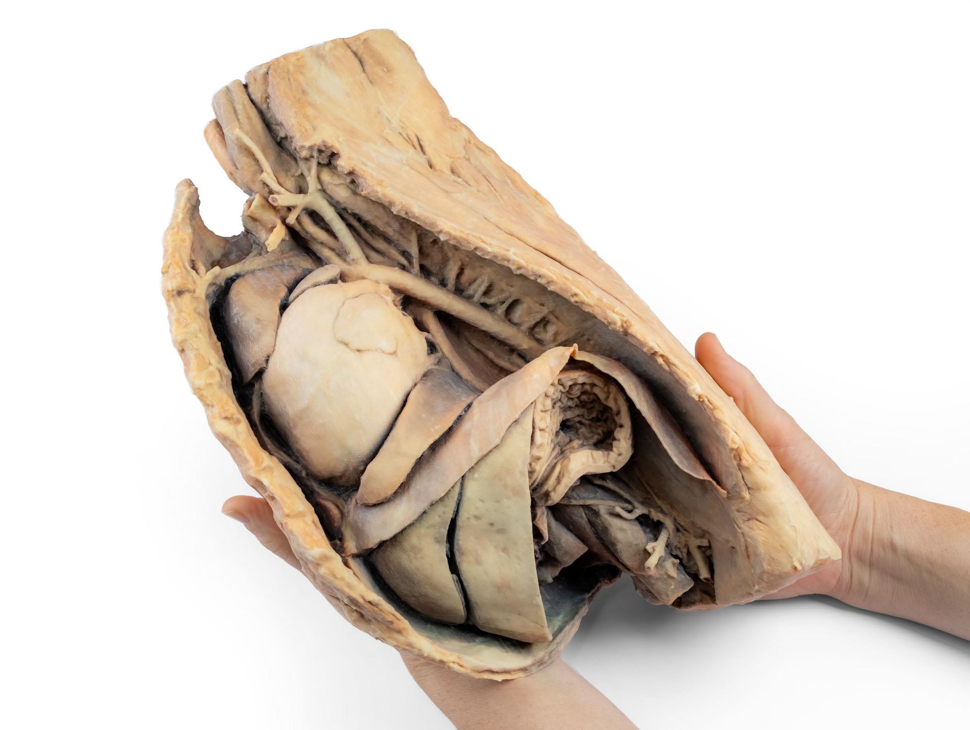
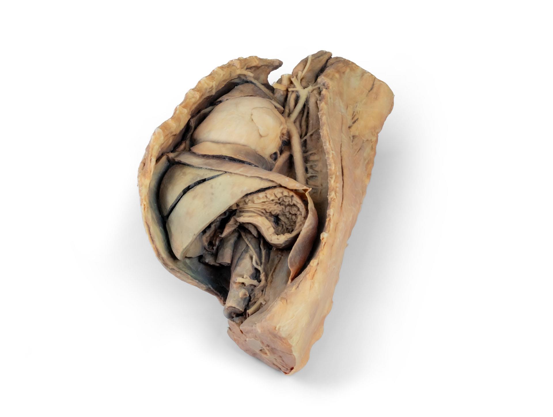
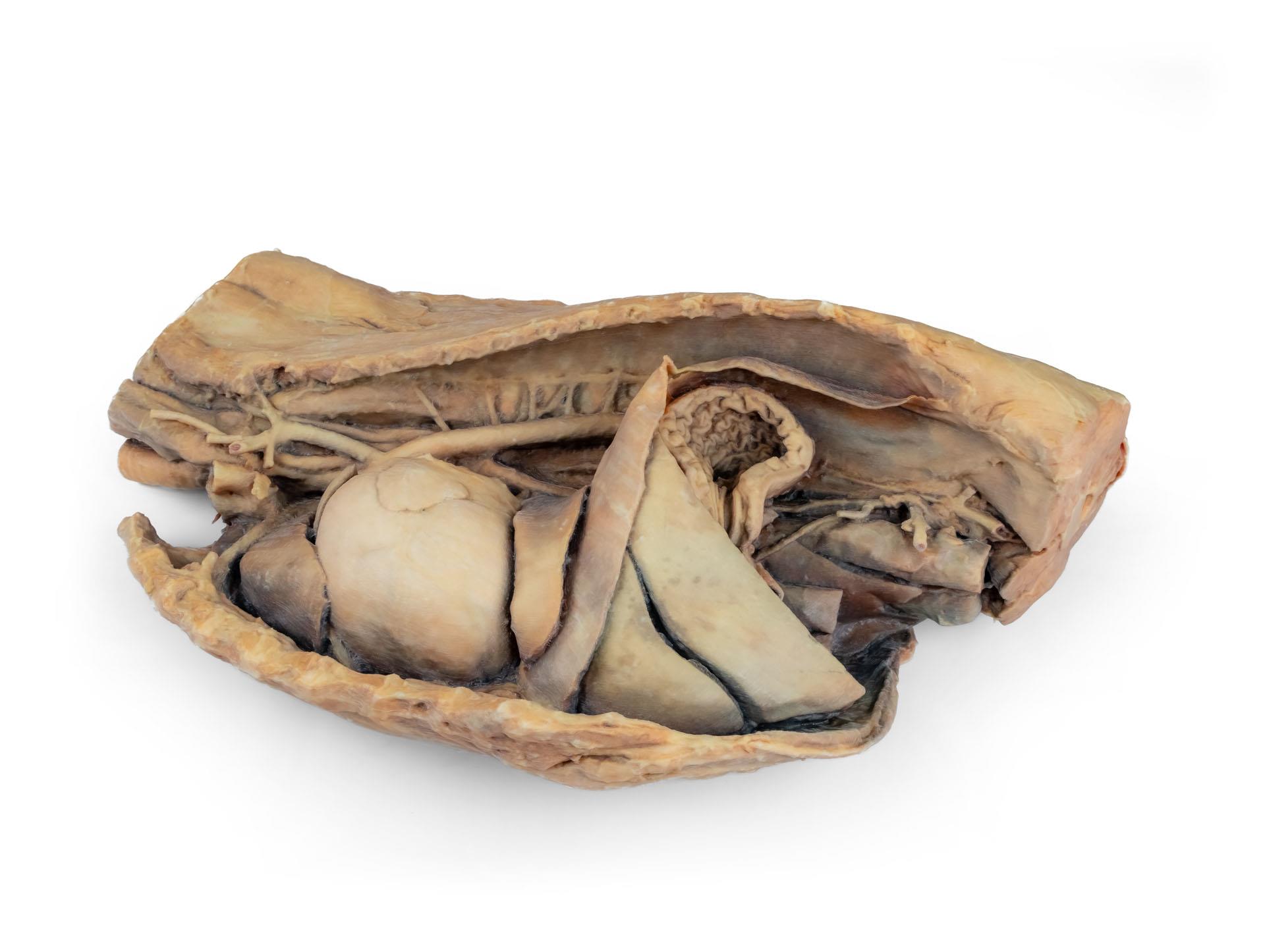
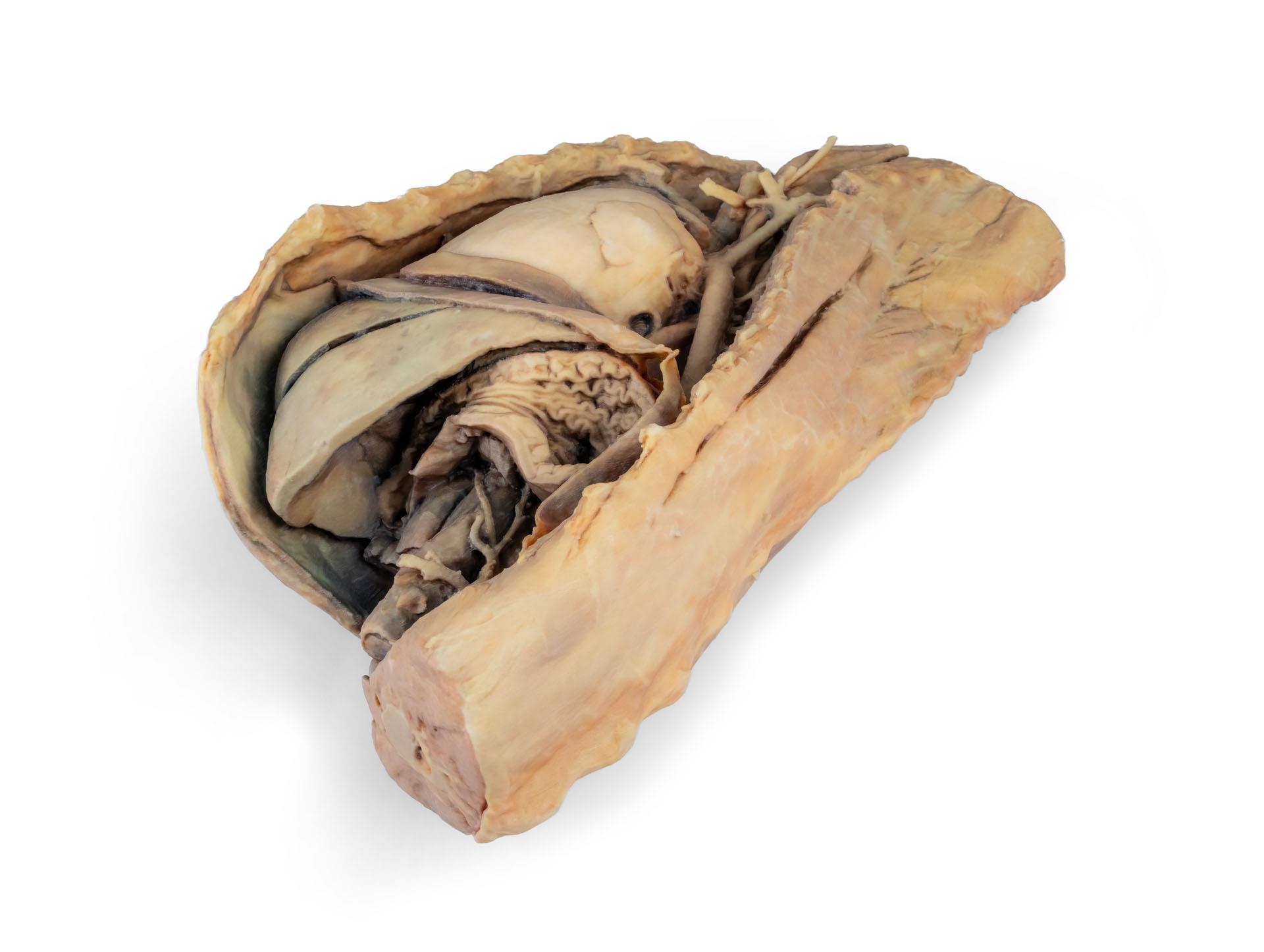
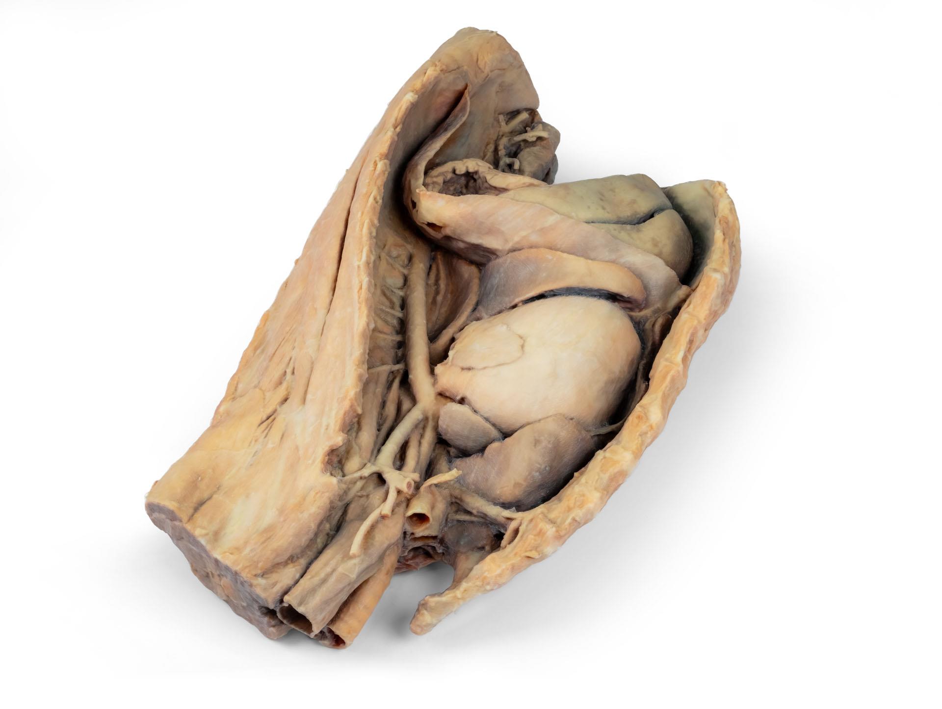
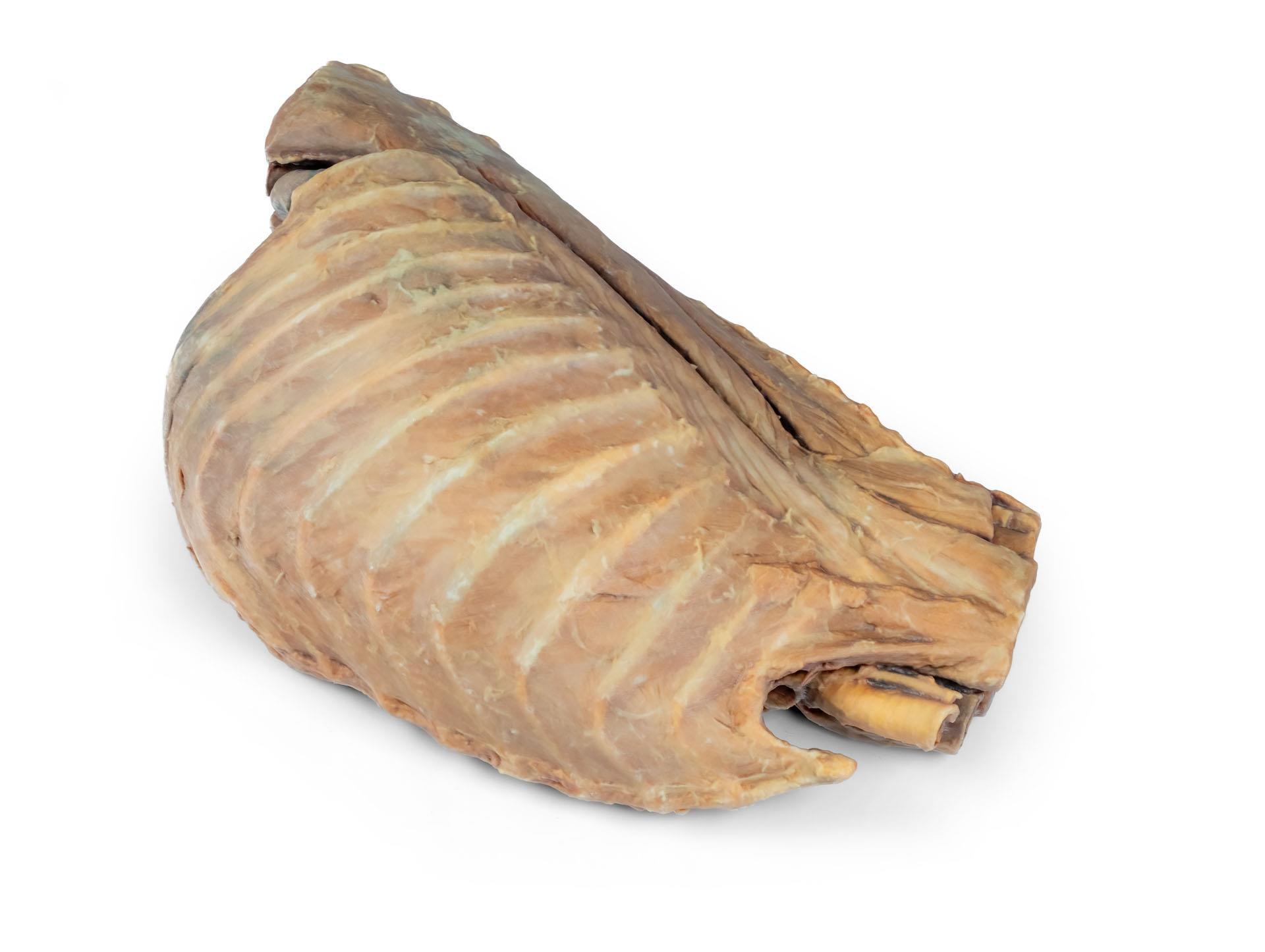
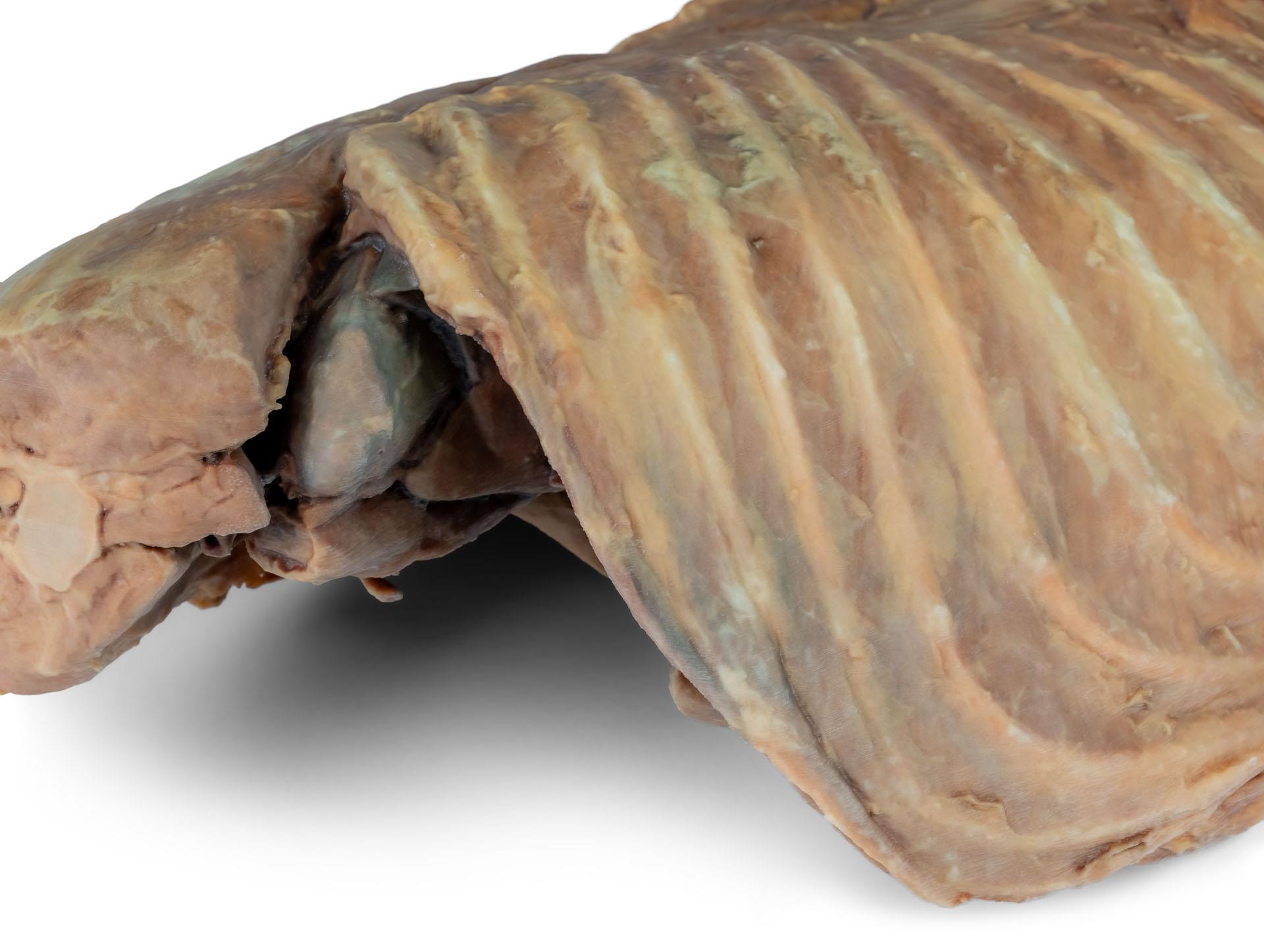
Canine thoracic cavity left side
€3,973.41*
Article in production, available in about 2-3 weeks
Product number:
VP9050
Item number: VP9050
Product information "Canine thoracic cavity left side"
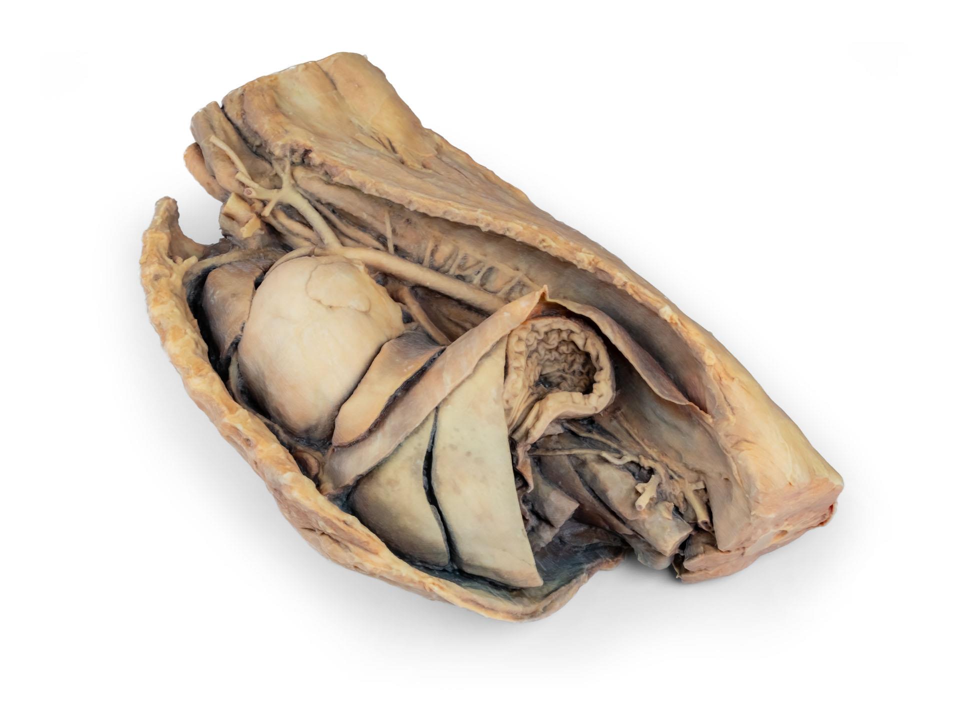
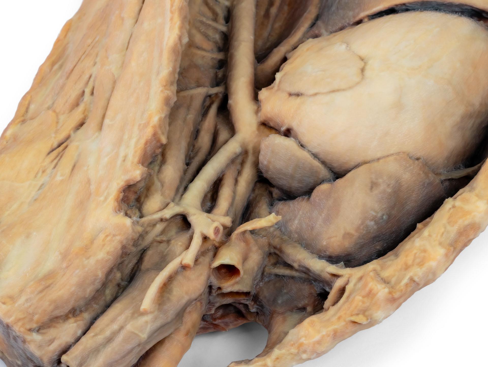
This specimen depicts the topography of the heart following theremoval of the left lung during a dissection of the thoracic cavityand cranial abdomen of a dog approached from the left side. The cranial and accessory lobes of the right lung remain in situ, servingas anatomical reference points. Within the cranial mediastinum, the brachiocephalic trunk and the left subclavian artery areidentifiable as major branches arising from the aortic arch.
Thethoracic portion of the esophagus runs through the mediastinumin a craniocaudal direction. The diaphragm has been preserved to serve as a landmark forretrodiaphragmatic organs such as the liver and the stomach.
The diaphragmatic crura and their relation to the aortic hiatus aremaintained, as are the initial visceral branches of the abdominalaorta—specifically, the celiac trunk and the cranial mesentericartery.
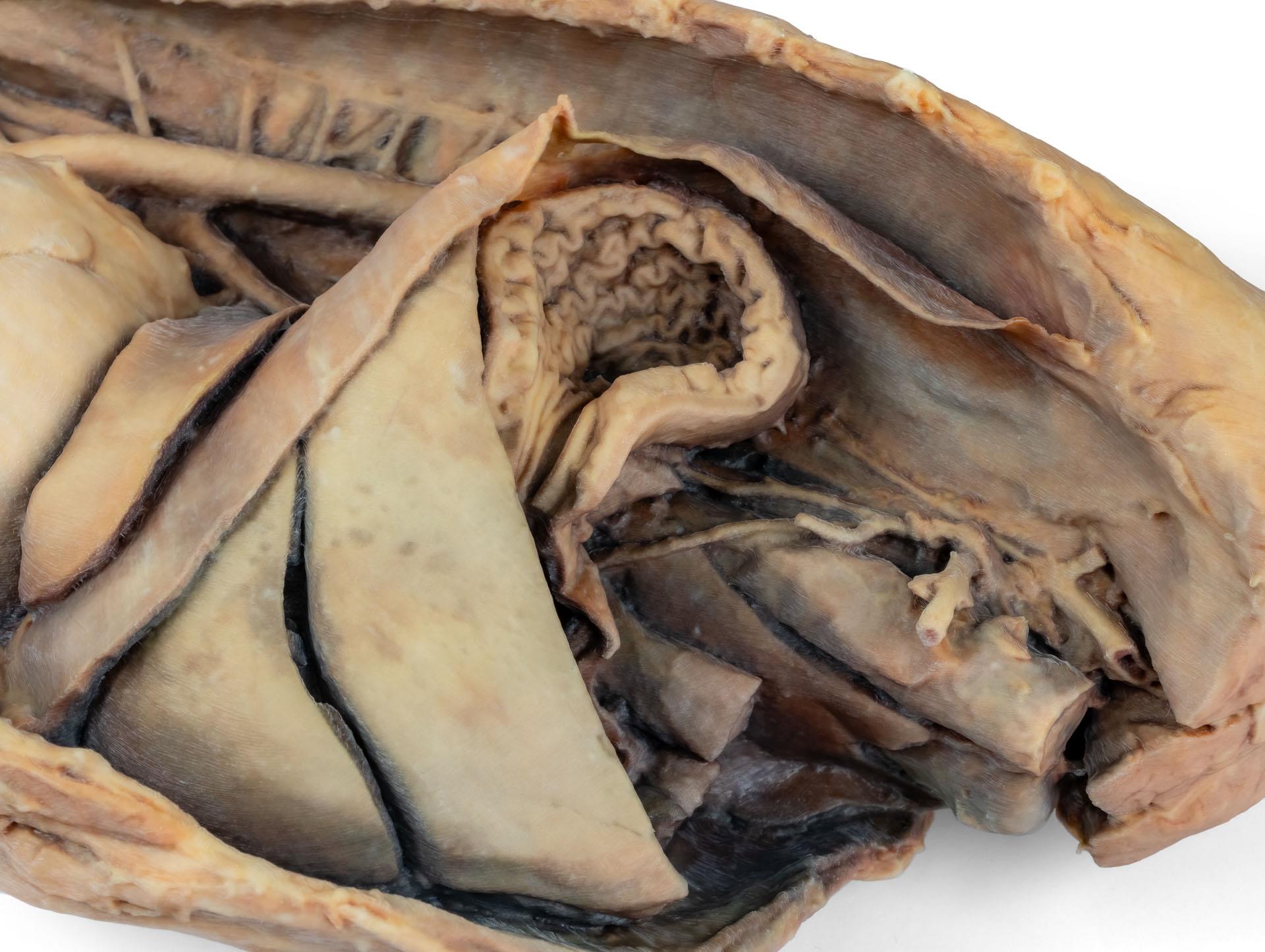
Both the interventricular and coronary grooves are visible. A portion of the right atrial wall has been removed to exposethe right atrioventricular (tricuspid) valve, including the chordaetendineae and papillary muscles.
Additionally, the septomarginaltrabecula (moderator band) is visible within the right ventricle, as well as the trajectory from the ventricle to the pulmonary trunk.The left atrial wall is also open to display its lumen.
Erler-Zimmer
Erler-Zimmer GmbH & Co.KG
Hauptstrasse 27
77886 Lauf
Germany
info@erler-zimmer.de
Achtung! Medizinisches Ausbildungsmaterial, kein Spielzeug. Nicht geeignet für Personen unter 14 Jahren.
Attention! Medical training material, not a toy. Not suitable for persons under 14 years of age.


