


Hydronephrosis and Hydroureter Caused by Obstruction by a Renal Calculus
Question regarding article:
Product information "Hydronephrosis and Hydroureter Caused by Obstruction by a Renal Calculus"
Clinical History
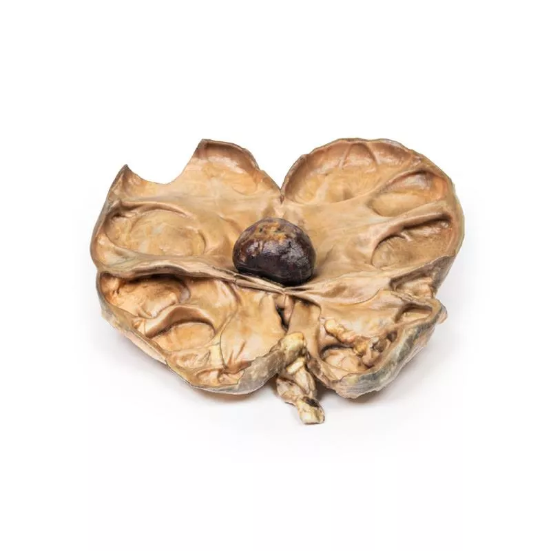
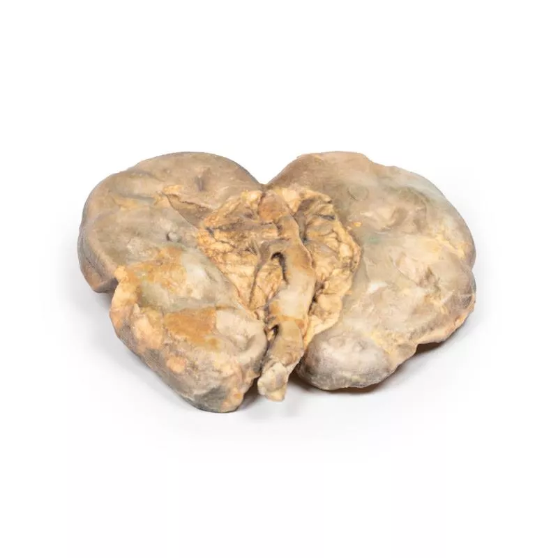
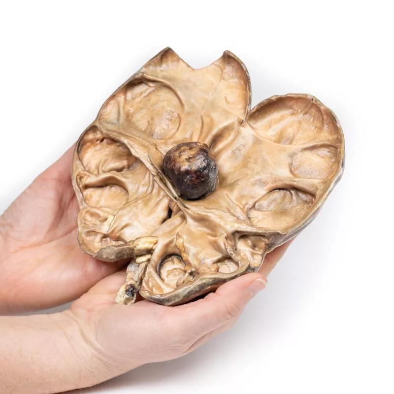
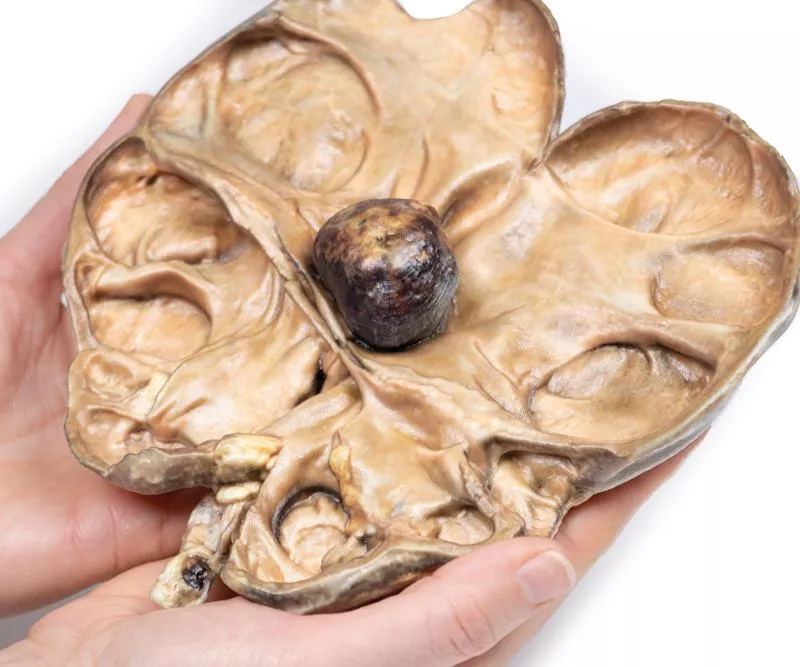
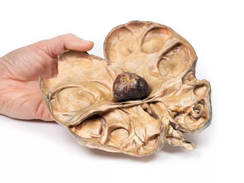
A 72-year-old female presented with colicky flank pain, malaise, and intermittent haematuria. Blood tests showed significantly impaired renal function. CT scan revealed congenital absence of the left kidney and severe hydronephrosis and hydroureter on the right side, caused by obstruction from a small calculus. Percutaneous lithotomy was attempted to remove the stone, but the patient died from a cardiac event during the procedure.
Pathology
The right kidney specimen is grossly enlarged and partially bisected. There is marked dilation of the pelvi-calyceal system and significant cortical atrophy. A large brown calculus is visible in the renal pelvis at the ureteropelvic junction.
Further Information
Urolithiasis (kidney stones) affects up to 10% of people. Stones can form anywhere in the urinary tract, most commonly in the kidneys. Risk factors include male sex, urine composition changes (e.g., hypercalciuria), metabolic disorders (cystinuria, gout), diet (high oxalate, animal protein), low fluid intake, and hot climates. Most stones are unilateral. Symptoms include severe colicky pain, haematuria, nausea, vomiting, and urinary urgency. Pain occurs when stones move into the ureter, often resolving once the stone passes.
Diagnosis is based on history and imaging such as non-contrast CT or ultrasound. Untreated obstruction causes pressure buildup leading to hydronephrosis, cortical atrophy, ischemic damage, and fibrosis, ultimately risking renal failure. Stones also predispose to infection due to obstruction and urothelial trauma.
Treatment includes analgesics (NSAIDs, opioids) and medications to facilitate stone passage (alpha blockers, calcium channel blockers). If conservative treatment fails or complications occur, surgical options include lithotripsy, laparoscopic or percutaneous stone removal. Open surgery is rarely needed.
Login
Erler-Zimmer
Erler-Zimmer Medical GmbH
Hauptstrasse 27
77886 Lauf
Germany
info@erler-zimmer.de
Achtung! Medizinisches Ausbildungsmaterial, kein Spielzeug. Nicht geeignet für Personen unter 14 Jahren.
Attention! Medical training material, not a toy. Not suitable for persons under 14 years of age.
Other customers also bought

Adrenal haemorrhage / Waterhouse-Friderichsen Syndrome
Clinical HistoryA 77-year-old man presented with abdominal and flank pain, fever, and rigors three days in duration. He was two weeks post-operative from duodenal ulcer repair. On admission, he was hypotensive, hyperkalemic, hyponatremic, and had a purpuric rash. Blood cultures grew Escherichia coli. Despite treatment, he rapidly deteriorated and died from septic shock.PathologyThe kidney and adrenal gland specimen shows extensive haemorrhage within the adrenal medulla, with blood extravasation into the surrounding fat. This is characteristic of adrenal haemorrhage in severe septic shock, known as Waterhouse-Friderichsen syndrome.Further InformationWaterhouse-Friderichsen syndrome is adrenal haemorrhage caused by overwhelming sepsis, leading to hypotensive shock, disseminated intravascular coagulation (DIC), and adrenal insufficiency. It mainly affects children but can occur in adults. Over 80% of cases are due to Neisseria meningitidis, but other bacteria like Escherichia coli, Streptococcus pneumoniae, and Pseudomonas aeruginosa can also cause it. The haemorrhage likely results from bacterial invasion, DIC, or endothelial damage from toxins. It usually affects both adrenal glands, starting in the medulla and extending outward, leading to adrenal failure. Patients present with rapid septic shock, purpuric rash, and adrenal crisis. Treatment includes supportive care, intravenous antibiotics, and steroids. Mortality exceeds 50%.

Pyonephrosis
Clinical HistoryA 38-year-old female presented with severe nausea, vomiting, fevers, and rigors. She had recurrent urinary tract infections over 6 months, treated with multiple antibiotics including IV therapy. Blood tests showed raised inflammatory markers, and urinalysis revealed white blood cells. CT scan detected unilateral left hydronephrosis and pyelonephritis. After failing conservative treatment, she underwent nephrectomy and fully recovered.PathologyA 38-year-old female presented with severe nausea, vomiting, fevers, and rigors. She had recurrent urinary tract infections over 6 months, treated with multiple antibiotics including IV therapy. Blood tests showed raised inflammatory markers, and urinalysis revealed white blood cells. CT scan detected unilateral left hydronephrosis and pyelonephritis. After failing conservative treatment, she underwent nephrectomy and fully recovered.Further InformationPyonephrosis occurs when an obstruction in the upper urinary tract combines with pyelonephritis, causing pus accumulation in a hydronephrotic kidney. It is rare and linked to risk factors like immunosuppression, diabetes, or urinary tract abnormalities (strictures, stones, tumours). Symptoms may include sepsis signs, flank pain, haematuria, dysuria, and pyuria. Diagnosis is mainly by CT, ultrasound, or MRI.Treatment focuses on urgent drainage of pus via percutaneous or ureteral stents plus antibiotics. Further surgery depends on obstruction cause. Untreated cases risk severe sepsis, abscess formation, and fistulae.

Hydronephrosis Hydroureter
Clinical HistoryA 49-year-old male presented with 6 weeks of malaise, urinary frequency, and haematuria. He also reported intermittent left flank pain. Ultrasound revealed severe hydronephrosis and hydroureter caused by multiple ureteric calculi blocking the uretero-vesical junction. He underwent left nephrectomy and ureterectomy with successful recovery.PathologyThe left nephrectomy and ureterectomy specimen shows a grossly hydronephrotic kidney with marked thinning and loss of renal parenchyma. The ureter is extremely dilated and contains several small brown-black stones with sharp edges, identified as calcium oxalate calculi. This illustrates hydronephrosis and hydroureter due to obstruction by ureteric stones.Further InformationHydronephrosis is dilation of the renal pelvis and calyces caused by obstruction of urine flow anywhere in the urinary tract. Causes include congenital anomalies, stones, tumors, inflammation, and prostatic diseases. Symptoms depend on cause, duration, site, and severity of obstruction. If untreated, pressure buildup leads to kidney atrophy, ischemic damage, and fibrosis. Diagnosis is primarily by ultrasound, supported by CT or urogram. Treatment usually involves surgery to relieve obstruction, such as nephrostomy, stenting, or catheterization depending on the site.

Renal Cell Carcinoma
Clinical History A 64-year-old male presented with 5 months of malaise, weight loss, and dull right flank pain. Examination revealed a palpable right abdominal mass and hypertension. Urinalysis showed microscopic haematuria. He underwent right nephrectomy.Pathology The kidney specimen shows a 5 cm irregular mass replacing the lower pole, compressing the renal parenchyma. The tumour surface shows haemorrhage and necrosis. Several small tumour nodules represent intrarenal metastases. The renal pelvis is slightly dilated, indicating mild hydronephrosis. Histology confirmed renal cell carcinoma (RCC).Further InformationRCC accounts for 85% of primary kidney cancers, mostly arising in the renal cortex. It affects men twice as often, typically in their 60s. Risk factors include smoking, obesity, hypertension, certain toxins, and familial syndromes (e.g., Von Hippel Lindau).Major RCC types are clear cell (70–80%), papillary (10–15%), chromophobe (5–10%), oncocytic (3–7%), and rare collecting duct carcinoma (less than 1%). Clear cell carcinoma is linked to chromosome 3p deletion and may be sporadic or hereditary. Papillary carcinoma often shows genetic trisomies and is multifocal. Chromophobe and oncocytic variants usually have better prognosis. Collecting duct carcinoma is aggressive with poor response to treatment.Clinical signs include flank pain, palpable mass, and haematuria, with possible systemic symptoms. RCC metastasizes early, mainly to lungs and bones. Tumours can invade the renal vein and inferior vena cava.Diagnosis uses ultrasound and CT; biopsy may be needed. Many RCC cases are incidental findings.The 5-year survival is ~70%. Treatment involves radical nephrectomy, and for metastases, chemotherapy, VEGF inhibitors, and tyrosine kinase inhibitors.

Septic Renal Infarct
Clinical History A 54-year-old male active intravenous drug user presented with flank pain, intermittent haematuria, fevers, malaise, and vomiting. On exam, he was hypertensive, pyrexic, and had Janeway lesions and IV drug track marks. A systolic murmur was heard. Blood tests showed raised inflammatory markers, impaired renal function, elevated LDH, and positive blood cultures. Echocardiogram revealed a large mobile tricuspid valve vegetation. Despite treatment for infective endocarditis, he died from sudden cardiac arrest.Pathology The post-mortem kidney specimen shows multiple well-demarcated wedge-shaped pale yellow-white infarcts in the cortex, with areas of hemorrhage. The largest infarct is at the lateral upper pole. These findings correspond to renal infarction.Further InformationRenal infarction occurs due to interrupted blood flow to the kidney, which has limited collateral circulation. The cortex is most vulnerable. Common causes include cardioembolic events (e.g., septic emboli from endocarditis), renal artery injury, hypercoagulable states, and idiopathic origins. Cardioembolic causes are the most frequent. Bilateral infarcts occur in ~15% of cases.Symptoms vary but often include flank pain, haematuria, hypertension, nausea, vomiting, and sometimes fever. Urinalysis and serum creatinine help in diagnosis. CT with contrast is preferred, showing a characteristic wedge-shaped perfusion defect.Treatment focuses on supportive care and managing the underlying cause.

Papillary Transitional Cell Carcinoma of the Renal Pelvis
Clinical History A 60-year-old man, after 40 years working in a paint factory, developed painless haematuria lasting one month. A CT scan revealed a suspected tumour in the left renal pelvis. He underwent nephrectomy.Pathology The kidney specimen shows preserved foetal lobulation and a friable papillary tumour, 35mm in diameter, obstructing and dilating the renal pelvis. Histology confirmed a papillary transitional cell carcinoma of the renal pelvis.Further Information5-10% of primary renal cancers arise from the urothelium lining the renal pelvis and calyces, similar to tumours in the ureter and bladder. They range from rare benign papillomas to common well-differentiated papillary carcinomas and aggressive poorly differentiated forms.Early symptoms include haematuria due to tumour friability, with later signs like hydronephrosis and flank pain from obstruction. Tumours may be multiple, affecting pelvis, ureter, and bladder.Risk factors include Lynch syndrome, analgesic nephropathy, smoking, and exposure to aromatic amines used in dye industries, such as benzidine and beta-naphthylamine.Wall infiltration is common and worsens prognosis. Five-year survival ranges from 50-100% for low-grade non-invasive lesions to 10% for high-grade infiltrating tumours.

Horseshoe Kidney
Clinical HistoryThis specimen was found during a routine post-mortem of a 56-year-old male who died of rheumatic heart disease.PathologyThe kidney measures 12 cm in length and shows fusion of the two parts at the lower pole, forming a characteristic horseshoe shape. The ureters emerge from the hilum on the anterior side. The kidney is bisected horizontally, visible on the posterior surface, and shows persistent fetal lobulation. The renal pelvis is positioned antero-medially, with the ureters passing anterior to the fused lower poles or isthmus.Further InformationHorseshoe kidney is the most common renal developmental anomaly, occurring twice as often in males than females, found in about 1 in 500 to 1000 autopsies. Most cases are sporadic but can be linked to chromosomal anomalies such as Down and Edwards syndromes or non-aneuploidic conditions like VACTERL association. In 90% of cases, the lower poles are connected by an isthmus of renal tissue; fusion of upper poles is rare. The renal pelvises are directed more anteriorly, and ureters angulate as they cross the isthmus.This condition is usually asymptomatic and discovered incidentally on ultrasound or CT scans. Kidney function is typically normal, but there is a higher incidence of urinary stones, likely due to ureteral angulation and stasis. Patients also have an increased risk of hydronephrosis, mainly from pelvi-ureteric junction obstruction, and urinary tract infections often caused by vesico-ureteric reflux. Some renal malignancies, such as transitional cell carcinoma and Wilms tumour, occur more frequently.

Multiple Renal Calculi
Clinical HistoryA 68-year-old male presented with fevers, rigors, and a 6-month history of intermittent bilateral flank pain and haematuria. Biochemical tests showed impaired renal function with normal serum calcium. CT revealed bilateral hydronephrosis, multiple renal calculi, and perinephric and subphrenic abscesses. The patient died from progressive renal failure.PathologyThe bisected kidney specimen shows gross dilatation of the pelvi-calyceal system and severe renal atrophy, sometimes reduced to a thin rim. A large mottled brown-white calculus lies in the pelvis, with smaller stones in the calyces and one occluding the ureter, causing proximal dilation.Further InformationUrolithiasis (renal stones) affects up to 1 in 10 people, most commonly in the kidneys. Risk factors include male gender, urine composition changes (hypercalciuria, hyperoxaluria), metabolic disorders (cystinuria, gout), diet, and environment. Symptoms range from severe colicky pain and haematuria to asymptomatic cases if stones remain in the pelvis or bladder.There are four main types of stones: - Calcium stones (70%), mainly calcium oxalate/phosphate, linked to hypercalciuria and hyperoxaluria. - Struvite stones (5–10%), magnesium ammonium phosphate, associated with infections, forming large “staghorn” calculi. - Uric acid stones (5–10%), in hyperuricemia (gout, leukemia). - Cysteine stones, due to impaired amino acid reabsorption.

Adult polycystic kidney disease
Histoire cliniqueUn homme de 40 ans s’est présenté avec deux semaines d’hématurie, de nouveaux maux de tête et une vision floue. Son médecin a relevé une tension artérielle très élevée de 260/110 et l’a adressé à l’hôpital. À son arrivée, il s’est effondré. Le scanner cérébral a montré une hémorragie sous-arachnoïdienne importante due à la rupture d’un anévrisme en sac. Le patient est décédé peu après.PathologieLe rein est hypertrophié et presque entièrement remplacé par de multiples kystes allant jusqu’à 3 cm de diamètre. Ces kystes ont des parois fines et translucides et contiennent des matériaux de couleurs variées, donnant un aspect « marbré » à la coupe. La surface externe est lobulée à cause des nombreux kystes, et le parenchyme restant est sévèrement atrophié par la pression. Ceci correspond à la maladie polykystique rénale adulte (APKD). Informations complémentairesL’APKD est une maladie autosomique dominante caractérisée par la formation de multiples kystes dans le rein, due à une altération de l’épithélium tubulaire. L’expansion des kystes détruit les glomérules, provoquant ischémie, atrophie et insuffisance rénale. Elle touche environ 1 naissance sur 40 à 1000. Les mutations des gènes PKD1 (chromosome 16) et PKD2 (chromosome 4) sont responsables, la mutation PKD1 étant plus fréquente et sévère. Les symptômes comprennent hématurie, douleur liée à l’expansion des kystes, hypertension et signes d’insuffisance rénale comme protéinurie et urémie. Des manifestations extra-rénales telles que les anévrismes intracrâniens en sac, kystes hépatiques et pancréatiques, ainsi que des maladies des valves cardiaques peuvent survenir. Le diagnostic se fait par échographie, scanner ou IRM, avec tests génétiques possibles. Le traitement repose sur la dialyse et la transplantation. Plus d’un tiers des patients décèdent d’insuffisance rénale, un tiers de maladies cardiaques, et environ 1?% d’une hémorragie sous-arachnoïdienne due à la rupture d’anévrisme.

Continuous innovation

Social responsibility

Active customer orientation

Understanding quality

Sustainable actions

ISO 9001 certification