Product information "Septic Renal Infarct"
Clinical History
A 54-year-old male active intravenous drug user presented with flank pain, intermittent haematuria, fevers, malaise, and vomiting. On exam, he was hypertensive, pyrexic, and had Janeway lesions and IV drug track marks. A systolic murmur was heard. Blood tests showed raised inflammatory markers, impaired renal function, elevated LDH, and positive blood cultures. Echocardiogram revealed a large mobile tricuspid valve vegetation. Despite treatment for infective endocarditis, he died from sudden cardiac arrest.
Pathology
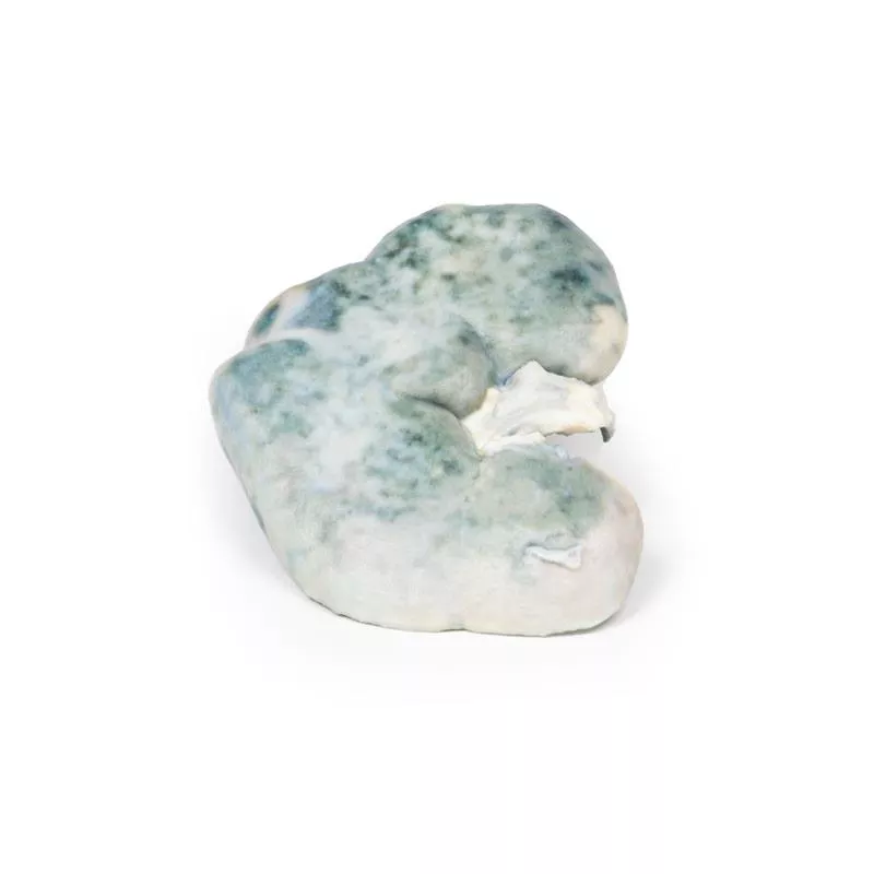
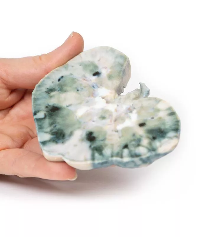
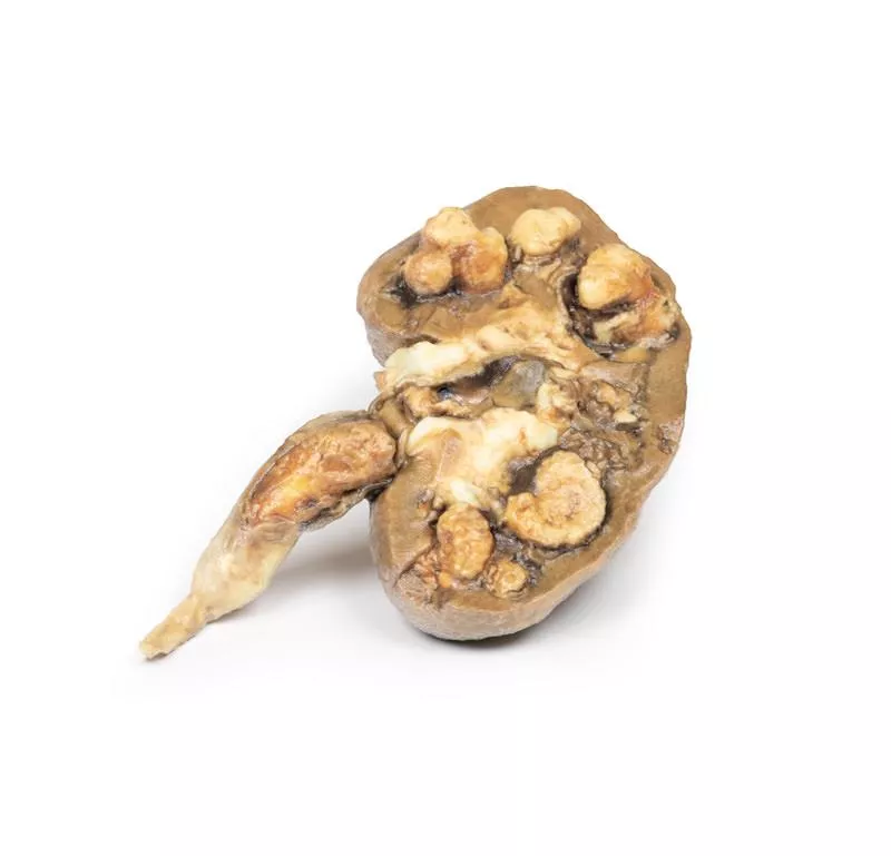
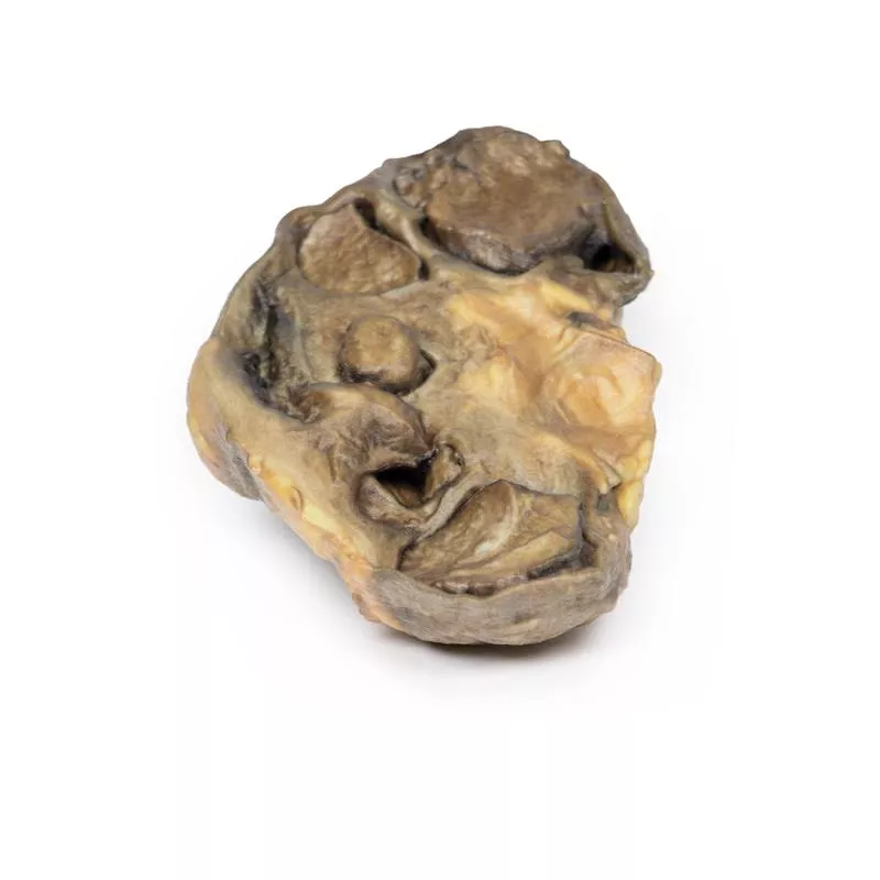
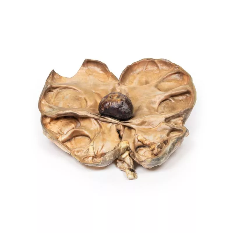
The post-mortem kidney specimen shows multiple well-demarcated wedge-shaped pale yellow-white infarcts in the cortex, with areas of hemorrhage. The largest infarct is at the lateral upper pole. These findings correspond to renal infarction.
Further Information
Renal infarction occurs due to interrupted blood flow to the kidney, which has limited collateral circulation. The cortex is most vulnerable. Common causes include cardioembolic events (e.g., septic emboli from endocarditis), renal artery injury, hypercoagulable states, and idiopathic origins. Cardioembolic causes are the most frequent. Bilateral infarcts occur in ~15% of cases.
Symptoms vary but often include flank pain, haematuria, hypertension, nausea, vomiting, and sometimes fever. Urinalysis and serum creatinine help in diagnosis. CT with contrast is preferred, showing a characteristic wedge-shaped perfusion defect.
Treatment focuses on supportive care and managing the underlying cause.
Erler-Zimmer
Erler-Zimmer GmbH & Co.KG
Hauptstrasse 27
77886 Lauf
Germany
info@erler-zimmer.de
Achtung! Medizinisches Ausbildungsmaterial, kein Spielzeug. Nicht geeignet für Personen unter 14 Jahren.
Attention! Medical training material, not a toy. Not suitable for persons under 14 years of age.


