Product information "NG & Tracheostomy Torso"
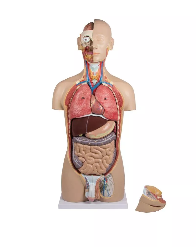
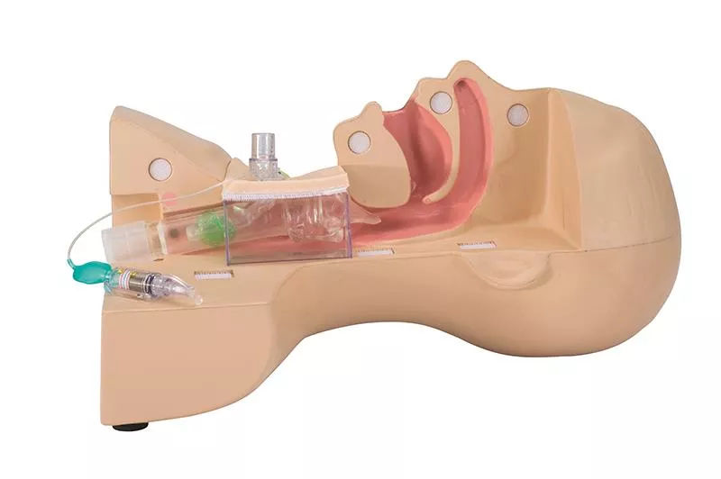
This model shows a cross-section of an upper body in the sagittal plane. The cross-section runs through the nose, mouth, pharynx, trachea, oesophagus and stomach. A feeding tube or catheter can be inserted through the nose or mouth into the oesophagus and stomach. A tracheostoma is available to demonstrate endotracheal suctioning.

0 of 0 reviews

Leave a review!

Share your experiences with other customers.


Erler-Zimmer

Erler-Zimmer GmbH & Co.KG
Hauptstrasse 27
77886 Lauf
Germany
info@erler-zimmer.de


Achtung! Medizinisches Ausbildungsmaterial, kein Spielzeug. Nicht geeignet für Personen unter 14 Jahren.

Attention! Medical training material, not a toy. Not suitable for persons under 14 years of age.


Other customers also bought

Torso with open back, 27-part, dual sex
This life-size torso can be dismantled into 27 individual parts and shows the most important organs and anatomical details of the human body. The right half of the head is open and shows the skull, brain, mouth and throat as well as the eye. The front of the neck is open and allows the larynx, thyroid gland and neck vessels to be studied. The chest and abdomen are completely open and all internal organs can be removed and partially dissected. Male and female genitalia can be exchanged. The back is open and shows parts of the back muscles, the spinal column, the spinal nerves and a removable vertebra with spinal cord.The torso can be disassembled into the following individual parts:- torso- half of the brain- eye- Left lung, 2 parts- Right lung, 2 parts- Heart, 2-part- Trachea- Oesophagus- Liver- Stomach, 2-part- Pancreas, spleen and duodenum- Kidney half- Intestinal package- Appendix valve- Female genital insert, 4-part- Male genital insert, 4-part- Thoracic vertebraeOn base plate.Size: 88 x 40 x 23 cmWeight: approx. 10.4 kgSupplied with torso guide on CD-ROM.

€903.21*
Torso with open back, 28-part, dual sex
This life-size torso with open back consists of 28 parts and offers an exceptionally realistic reproduction of the anatomical structures in their finest details.All body systems are represented with full accessibility. The left side shows all major superficial and deep muscles with vessels and bones. The open back shows the muscle layers as well as the spinal column and the associated nerve branches. A thoracic vertebra, including a section of the spinal cord, can be removed to examine its anatomical details in detail. The head is open and the brain is completely exposed on one side. The neck is opened to expose muscle, nerve, vascular and glandular structures. Significant features are numbered and noted on the enclosed key card. The male and female urogenital systems that can be disassembled are interchangeable.Detachable structures include the following:- Eye with optic nerve and extraocular muscles- Cerebral hemisphere, divided along the longitudinal fissure, with visualisation of the internal structures and blood vessels- Trachea- Descending thoracic aorta and oesophagus- Right and left lung (2 parts each), showing the bronchial tree and the vascular system- Chest lid- mammary gland- Liver with gall bladder- One half of the kidney in frontal section showing the internal structure and vessels- 2-part heart with clear depiction of atria, ventricles, valves and section through the heart wall- Stomach divided into 2 parts- Pancreas, duodenum and spleen with vessels and ducts- Small and large intestine with removable lid over part of the appendix- Male urogenital system (4 parts)- Female urogenital system (3 parts) including embryo- Thoracic vertebrae (T12) with spinal cord

€1,879.01*
Miniaturtorso, 16-teilig, zweigeschlechtig

€213.01*
Tracheostomy management simulator
This model can be used to practice the procedures carried out for tracheostomypatients in the fields of nursing and caregiving when replacing cannulas or performing suction through a cannula. The model can be divided into two halves and the trachea section is transparent, allowing confirmation of the state of cannula insertion and the position of the suction catheter in the cannula during suction. In addition, with the specially provided cannula attached, a ventilator can be operated by connection a test lung (simulated lung) to the model.Features- Allows the user to practice the procedures for replacing cannulas.- As the trachea section is transparent, the state of inflation of the cuff can beobserved, and by using the specially provided cannula, the approximate optimum pressure can be confirmed. (Because the posterior wall of the trachea is soft, the user can feel the compression of the trachea that results when the cuff is overinflated.)- The transparent trachea facilitates explanation of how the suction cathetershould be positioned, and of how to perform suction using the upper part of the cuff. (The above mentioned practicing of suction procedures can be performed using the simulated sputum.)- The soft neck surface skin allows the user to feel the thyroid cartilage throughthe surface skin.- The detachable trachea section facilitates explanation of the areas where granulation tends to develop.- A ventilator can be operated by attaching the specially provided cannula andconnecting a test lung, allowing confirmation of the alarm tone generated whenair leakage occurs.

€950.81*
Sticker set "Medical gas connections" with 5pcs 3D stickers
Make your simulation environment even more realistic with this 3D sticker set. This turns every cable duct into a wall supply unit.

€35.70*

Continuous innovation

Social responsibility

Active customer orientation

Understanding quality

Sustainable actions

ISO 9001 certification

Your last viewed products