Product information "Tracheostomy management simulator"
This model can be used to practice the procedures carried out for tracheostomy
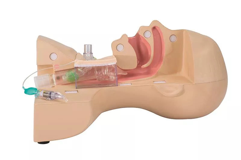
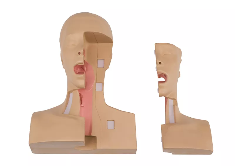
patients in the fields of nursing and caregiving when replacing cannulas or performing suction through a cannula. The model can be divided into two halves and the trachea section is transparent, allowing confirmation of the state of cannula insertion and the position of the suction catheter in the cannula during suction. In addition, with the specially provided cannula attached, a ventilator can be operated by connection a test lung (simulated lung) to the model.


Features

- Allows the user to practice the procedures for replacing cannulas.

- As the trachea section is transparent, the state of inflation of the cuff can be
observed, and by using the specially provided cannula, the approximate optimum pressure can be confirmed. (Because the posterior wall of the trachea is soft, the user can feel the compression of the trachea that results when the cuff is overinflated.)

- The transparent trachea facilitates explanation of how the suction catheter
should be positioned, and of how to perform suction using the upper part of the cuff. (The above mentioned practicing of suction procedures can be performed using the simulated sputum.)

- The soft neck surface skin allows the user to feel the thyroid cartilage through
the surface skin.

- The detachable trachea section facilitates explanation of the areas where granulation tends to develop.

- A ventilator can be operated by attaching the specially provided cannula and
connecting a test lung, allowing confirmation of the alarm tone generated when
air leakage occurs.

0 of 0 reviews

Leave a review!

Share your experiences with other customers.


Koken

Koken Co., Ltd.
1-4-14 Koraku, Bunkyo-Ku
112-0004 Tokyo
Japan
narumi.kida@kokenmpc.co.jp

Verantwortlich/Responsible:

Erler-Zimmer GmbH & Co. KG
Hauptstrasse 27
77886 Lauf
Germany
info@erler-zimmer.de


Achtung! Medizinisches Ausbildungsmaterial, kein Spielzeug. Nicht geeignet für Personen unter 14 Jahren.

Attention! Medical training material, not a toy. Not suitable for persons under 14 years of age.


Other customers also bought

NG & Tracheostomy Torso
This model shows a cross-section of an upper body in the sagittal plane. The cross-section runs through the nose, mouth, pharynx, trachea, oesophagus and stomach. A feeding tube or catheter can be inserted through the nose or mouth into the oesophagus and stomach. A tracheostoma is available to demonstrate endotracheal suctioning.

€529.55*
Sticker set "Medical gas connections" with 5pcs 3D stickers
Make your simulation environment even more realistic with this 3D sticker set. This turns every cable duct into a wall supply unit.

€35.70*
Training port
This training port is designed for use in suitable puncture trainers. The port has a fluid supply that can be connected with a blood reservoir (not included). The port can be punctured with real needles. Not sterile, not for human use.

€151.13*
CVC Insertion Simulator III
The CVC Insertion Simulator III provides training in a sequence of procedural skills from the needle insertion to catheter placement, including Seldinger technique. It provides training opportunities in an array of procedural skills from determining the insertion site to placement of the catheterwithout interruption. Both landmark and ultrasound-guided catheterization are possible in one pad.Features:- Repeated insertionThe improved frictionless tissue of the pad allows Seldinger technique and repeated insertion and removing of the catheter with less needle marks left on the surface of the pad.- Both landmark and ultrasound-guided CVC:The anatomically correct structure facilitate training in both landmark and ultrasound-guided CVC.- Mechanical complications, such as arterial puncture and pneumothorax can be simulated for training.- New materialThe close-to-human tissue material of the pad provides true-to-life sensation inserting the catheter.- Realistic venous collapseThe vein collapses under the pressure of the ultrasound probe.The pad shows the following anatomical structures:- Internal jugular vein & carotid artery- Subclavian vein & artery- Superior vena cava- Ribs- Sternum- LungTraining Skills:- Ultrasound-guided CVC- Landmark guided CVC- Ultrasound-guided venous access- Prevention of mechanical complicationsThe new and innovative material does provide an incredible realism and tactile excellence as well as ultrasound imaging quality. However, due to its special material, the injection pad has a limited shelf life once it is unpacked.

€2,773.89*

Continuous innovation

Social responsibility

Active customer orientation

Understanding quality

Sustainable actions

ISO 9001 certification

Your last viewed products