


Product information "Popliteal Fossa distal thigh and proximal leg"
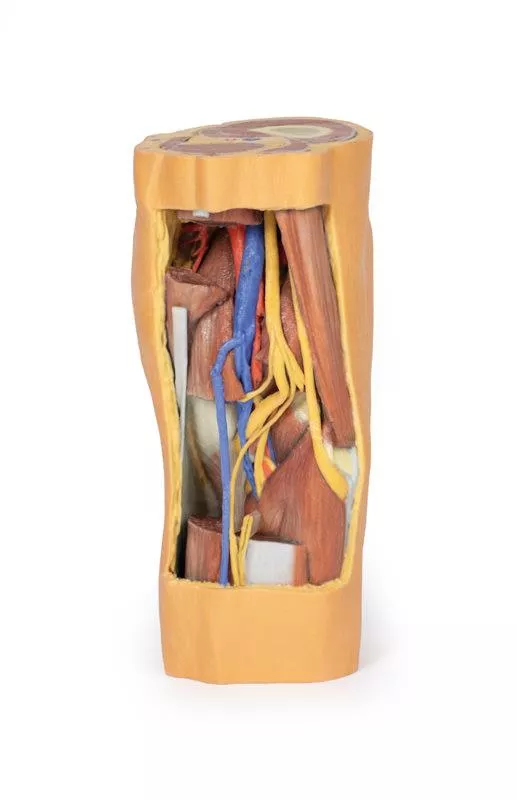
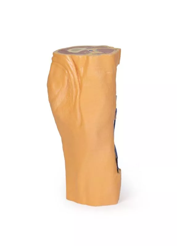
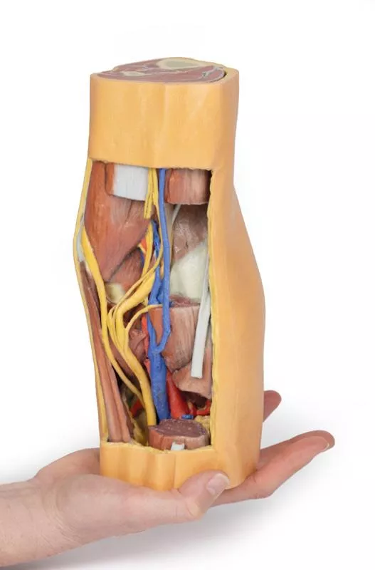
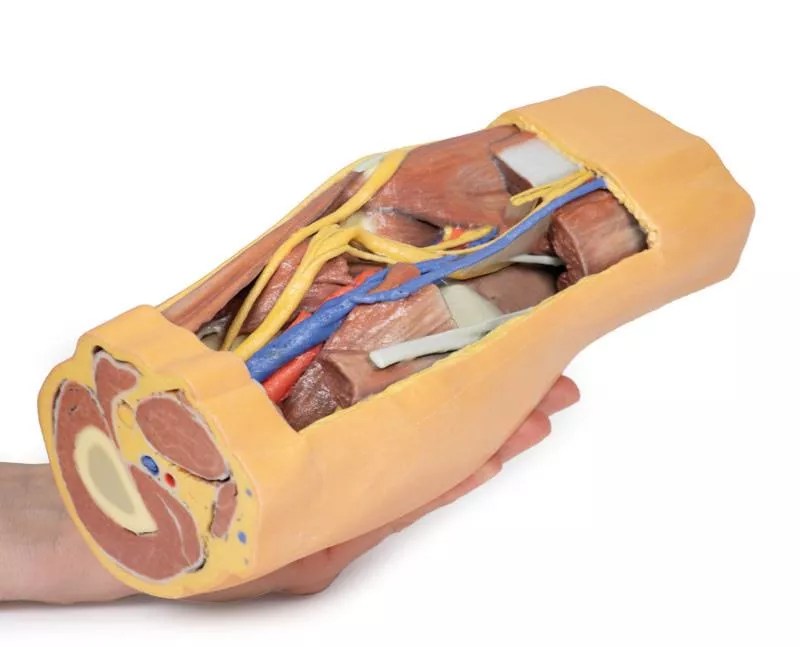
This 3D printed specimen preserves the distal thigh and proximal leg, dissected posteriorly to highlight the popliteal fossa and surrounding structures.
Proximal Cross-Section: Compartments and Vessels
The proximal cross-section displays the anterior, posterior, and medial compartment muscles, with the popliteal artery and vein entering the fossa via the adductor hiatus. The sciatic nerve and great saphenous vein are also visible. Posteriorly, the skin, superficial fascia, fascia lata, and crural fascia have been removed to clearly demonstrate the course of the popliteal vessels, tibial nerve, and common peroneal nerve. Medial sectioning of the semitendinosus and semimembranosus muscles reveals the superior medial genicular artery and the medial head of the gastrocnemius, while distal sectioning exposes the popliteus muscle and the tendon of the plantaris.
Popliteal Vessels and Nerves
The popliteal artery and vein can be traced through the fossa to their passage deep to the soleus muscle, accompanied by the tibial nerve. With the lateral head of the gastrocnemius removed, several tibial nerve branches and the medial sural cutaneous nerve are visible. The common peroneal nerve runs parallel, descending laterally over the exposed soleus to the fibular neck, just distal to the biceps femoris. Deep to the biceps femoris, the superior lateral genicular branch passes toward the anterior compartment.
Distal Cross-Section: Leg Neurovascular Structures
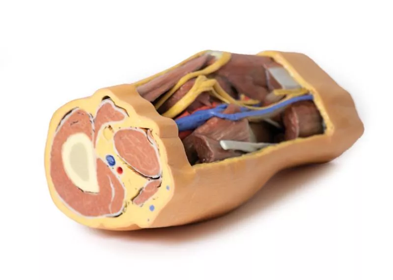
The distal cross-section shows the continuation of the popliteal contents and branches. The great and small saphenous veins and sural nerves are visible within the superficial fascia. Between the posterior, lateral, and anterior compartment muscles, the neurovascular bundles of the leg can be observed, including the posterior tibial artery and veins with tibial nerve, the peroneal artery and veins, and the anterior tibial artery, veins, and deep peroneal nerve.
Proximal Cross-Section: Compartments and Vessels
The proximal cross-section displays the anterior, posterior, and medial compartment muscles, with the popliteal artery and vein entering the fossa via the adductor hiatus. The sciatic nerve and great saphenous vein are also visible. Posteriorly, the skin, superficial fascia, fascia lata, and crural fascia have been removed to clearly demonstrate the course of the popliteal vessels, tibial nerve, and common peroneal nerve. Medial sectioning of the semitendinosus and semimembranosus muscles reveals the superior medial genicular artery and the medial head of the gastrocnemius, while distal sectioning exposes the popliteus muscle and the tendon of the plantaris.
Popliteal Vessels and Nerves
The popliteal artery and vein can be traced through the fossa to their passage deep to the soleus muscle, accompanied by the tibial nerve. With the lateral head of the gastrocnemius removed, several tibial nerve branches and the medial sural cutaneous nerve are visible. The common peroneal nerve runs parallel, descending laterally over the exposed soleus to the fibular neck, just distal to the biceps femoris. Deep to the biceps femoris, the superior lateral genicular branch passes toward the anterior compartment.
Distal Cross-Section: Leg Neurovascular Structures
The distal cross-section shows the continuation of the popliteal contents and branches. The great and small saphenous veins and sural nerves are visible within the superficial fascia. Between the posterior, lateral, and anterior compartment muscles, the neurovascular bundles of the leg can be observed, including the posterior tibial artery and veins with tibial nerve, the peroneal artery and veins, and the anterior tibial artery, veins, and deep peroneal nerve.
Login
Erler-Zimmer
Erler-Zimmer Medical GmbH
Hauptstrasse 27
77886 Lauf
Germany
info@erler-zimmer.de
Achtung! Medizinisches Ausbildungsmaterial, kein Spielzeug. Nicht geeignet für Personen unter 14 Jahren.
Attention! Medical training material, not a toy. Not suitable for persons under 14 years of age.
Other customers also bought

Flexed knee joint deep dissection
This 3D printed specimen shows a left knee joint in flexion, highlighting the internal joint capsule and surrounding muscles, ligaments, and neurovascular structures.Proximal Thigh & Popliteal FossaThe proximal section includes parts of the quadriceps femoris and sartorius, with the iliotibial tract visible. The popliteal fossa displays the popliteal vessels, tibial nerve, and common peroneal nerve. Posteriorly, insertions of the adductor magnus, gracilis, and posterior thigh muscles (biceps femoris, semitendinosus, semimembranosus) are preserved.Distal Leg & Neurovascular StructuresThe distal section shows muscles of the anterior, lateral, and posterior compartments. Key vessels and nerves include the anterior tibial artery and deep peroneal nerve, posterior tibial artery and tibial nerve, and the fibular vessels. Ligaments & MenisciAnteriorly, remnants of the quadriceps tendon and patellar ligament remain. The anterior and posterior cruciate ligaments and menisci are visible between the femur and tibia. Medially, the tibial collateral ligament and semitendinosus insertion are preserved; laterally, the fibular collateral ligament, biceps femoris tendon, and common peroneal nerve are exposed.

Lower Limb - deep dissection
This 3D printed specimen presents a right lower limb, sectioned just above the knee and including a partially dissected foot, showing detailed structures on the dorsum.Proximal Section & Popliteal FossaThe proximal cross-section shows the patella articulating with the distal femur. Posteriorly, the superior popliteal fossa is preserved, revealing the popliteal artery, vein, and terminal sciatic nerve.Distal Leg & Neurovascular StructuresDistal to the knee, most posterior muscles are removed to highlight neurovascular structures, including the common peroneal and tibial nerves as well as the posterior and anterior tibial arteries. The popliteus muscle and the interosseous membrane between tibia and fibula are visible. Medially, the pes anserinus inserts onto the proximal tibia; laterally, the biceps femoris inserts into the fibular head near the common peroneal nerve. Muscle Compartments & TendonsMost of the posterior and lateral compartment muscles are removed, leaving the anterior compartment intact and visible under the crural fascia. The anterior tibial artery and vein pass distally deep to the exposed interosseous membrane. Distal tendons of the anterior muscles pass beneath the extensor and peroneal retinaculae to their insertions. Foot & DorsumOn the dorsum, the dorsalis pedis artery and the terminal deep peroneal nerve are visible between the extensor hallucis longus and extensor hallucis brevis tendons. The extensor digitorum brevis is clearly seen beneath the extensor digitorum longus and peroneus tertius tendons.

Lower Limb Musculature
This 3D printed specimen presents a superficial dissection of the lower limb from mid-thigh to mid-leg, highlighting muscles, nerves, and vessels of the popliteal fossa.Thigh Musculature & Knee JointThe insertions of anterior, medial, and posterior thigh muscles are visible, including the pes anserinus medially and iliotibial tract laterally. The knee joint capsule is opened anteriorly, revealing the menisci and the tibial and fibular collateral ligaments.Leg Muscles & Compartment StructuresSuperficial leg muscles are preserved, with anterior and lateral compartment muscles shown beneath the crural fascia. The proximal cross-section displays distal thigh muscles, the femoral artery and vein, and the saphenous nerve in the adductor canal. The sciatic nerve and perforating branches of the profunda femoris artery are also visible in the posterior compartment. Popliteal Fossa & Neurovascular StructuresIn the popliteal fossa, the popliteal artery and vein descend from the adductor hiatus. The sciatic nerve bifurcates into the common fibular, tibial, and sural nerves. Distal cross-sections highlight the anterior, lateral, and posterior compartment muscles, with the deep fibular nerve alongside the anterior tibial artery and veins, and posteriorly, the posterior tibial and fibular arteries and veins near the tibial nerve.

Knee Joint, flexed
This 3D printed specimen showcases the ligaments of the knee joint with the leg in flexion, providing a detailed anatomical view.Anterior View DetailsWith the patella and part of the patellar ligament removed, the medial and lateral menisci as well as the anterior and posterior cruciate ligaments are clearly visible. Both the tibial and fibular collateral ligaments remain intact, offering insight into the knee’s stabilizing structures.Medial and Lateral StructuresMedially, the insertions of the adductor magnus and semimembranosus muscles are preserved, along with the oblique popliteal ligament reflected over the posterior joint capsule. Laterally, the specimen retains the insertion of the biceps femoris and the origins of the popliteus and soleus muscles.

Lower limb – superficial dissection
This 3D printed model represents the remainder of the lower limb from our male abdominopelvic and proximal thigh specimen (MP1765). Sectioned proximally near the mid-thigh, it continues to the partially dissected foot, providing a detailed view of lower limb anatomy.Thigh Neurovascular StructuresA transverse section through the thigh exposes the neurovascular structures of the anterior, medial, and posterior compartments. Visible structures include the great saphenous vein superficial to the terminal branches of the femoral nerve, the femoral artery and vein in the anterior compartment, and perforating branches of the deep femoral artery in the medial and posterior compartments.Superficial Structures and MusculatureThe remaining thigh, leg, and dorsum of the foot are partially dissected to demonstrate superficial structures and compartmental musculature, while the posterior aspect remains undissected for comparative study. The course of the great saphenous vein is visible from the medial thigh to the medial malleolus and the medial aspect of the dorsal venous plexus. The small saphenous vein can be traced from lateral branches of the dorsal venous plexus to the margin of the dissected superficial fascia near the lateral malleolus. Deep StructuresDeeper branches of the femoral artery, vein, and nerve are visible beneath the anterior compartment muscles (including a sectioned sartorius) as they enter the adductor canal. Nerve PathwaysNear the medial knee, the saphenous nerve runs superficially alongside the great saphenous vein on the posterior crural fascia, terminating as the medial cutaneous nerve of the leg branches. On the lateral leg, the medial and intermediate dorsal cutaneous branches of the superficial fibular nerve extend onto the dorsum of the foot adjacent to the dorsal venous plexus tributaries.

Lower Limb superficial veins
This high-quality 3D printed model presents a detailed superficial dissection of the left lower limb, spanning from just above the knee to the complete foot.Superficial Venous StructuresThe skin and superficial fascia have been removed to clearly display the leg’s venous anatomy. Visible structures include the dorsal venous plexus, the great saphenous vein with its numerous tributaries, and the small saphenous vein with its tributaries along the crural fascia.Cutaneous NervesSeveral cutaneous nerves accompany the veins: the sural nerve posteriorly, the saphenous nerve medially, and the superficial peroneal nerve anteriorly, providing a comprehensive view of nerve pathways in the leg. Foot AnatomyOn the dorsum of the foot, lateral to the extensor hallucis longus tendon, the dorsal digital branch of the deep peroneal nerve emerges, supplying the skin between the first two toes.

Knee Joint, extended
This 3D printed specimen demonstrates the ligaments of the knee joint with the leg in extension. It represents the same specimen as LW29B, which is imaged in a flexed position, allowing comparison of joint anatomy under different conditions.Anterior ViewWith the patella and part of the patellar ligament removed, the specimen clearly shows the tibial and fibular collateral ligaments, which remain intact for full structural reference.Medial and Lateral StructuresMedially, the insertions of the adductor magnus and semimembranosus muscles are preserved, with the oblique popliteal ligament reflected onto the posterior joint capsule. Laterally, the insertion of the biceps femoris and the origins of the popliteus (covered by the arcuate popliteal ligament) and soleus muscles are maintained, providing a detailed view of lateral knee anatomy.

Popliteal Fossa
This 3D printed specimen preserves the distal thigh and proximal leg, highlighting the popliteal fossa and surrounding structures.Proximal Thigh and Popliteal FossaThe proximal cross-section shows the anterior, posterior, and medial compartment muscles, with the femoral artery and vein in the adductor canal, as well as the sciatic nerve and great saphenous vein. Posteriorly, the skin and fascia are removed to expose the popliteal space. A window in the semimembranosus reveals the popliteal vessels, while the great saphenous vein runs over the sartorius, which joins the semitendinosus and semimembranosus to form the pes anserinus. The superior lateral genicular artery and posterior iliotibial tract are also visible.Distal Leg and Neurovascular StructuresThe distal cross-section displays the continuation of popliteal contents. The great and small saphenous veins and medial and lateral sural cutaneous nerves are visible in the superficial fascia. Between muscles of the posterior, lateral, and anterior compartments, the neurovascular bundles of the leg are preserved, including the posterior tibial, anterior tibial, peroneal vessels and nerves, and superficial and deep peroneal nerves.

Lower limb – superficial dissection with male left pelvis
This 3D printed specimen combines the male left pelvis (MP1765) with the lower limb – superficial dissection (MP1816), offering a comprehensive view from the pelvis to the foot.Lower Limb: Thigh, Leg, and FootThe lower limb is sectioned proximally near mid-thigh and continues to the partially dissected foot. A transverse section through the thigh reveals the neurovascular structures of the anterior, medial, and posterior compartments, including the great saphenous vein, femoral artery and vein, and perforating branches of the deep femoral artery. The remainder of the thigh, leg, and dorsum of the foot show superficial structures and compartmental musculature, while the posterior aspect remains undissected. The great and small saphenous veins are traced from the thigh to the medial and lateral malleoli. Deeper femoral vessels and nerves are visible beneath the anterior compartment muscles entering the adductor canal. Saphenous and superficial fibular nerves are preserved along their course to the foot.Male Pelvis: Superficial and Deep StructuresThe male left pelvis displays superficial and deep structures of the true and false pelvis, including the inguinal and femoral regions. Transverse sections reveal epaxial and abdominal wall musculature (rectus abdominis, external/internal obliques, transversus abdominis), psoas major, and quadratus lumborum, separated by fascial layers such as the rectus sheath and thoracolumbar fascia. The external iliac artery, left testicular vessels, ilioinguinal, lateral cutaneous, and femoral nerves, as well as the internal iliac branches are visible, tracking to their respective regions. Pelvic Viscera and Urogenital AnatomyMidline sections display the pubic symphysis and pelvic viscera, including the bladder, left seminal vesicle, vas deferens, and rectum with surrounding musculature. The urethra is visible through the prostate, pelvic diaphragm, and penis, while the scrotum shows the parietal tunica vaginalis. Pelvic-to-Thigh TransitionThe proximal thigh highlights the fascia lata removal, revealing the transition of neurovasculature and musculature from the pelvis. The femoral artery, vein, and nerve, the great saphenous vein, and anterior thigh muscles (sartorius, rectus femoris, vastus muscles) are displayed. Lateral structures include the tensor fasciae latae inserting onto the iliotibial tract and a window exposing the gluteus medius to the greater trochanter.

Foot - Parasagittal cross-section
This 3D printed specimen provides a parasagittal cross-section through the medial aspect of the right distal tibia and foot, showcasing the skeletal structures of the medial longitudinal arch and surrounding soft tissues.Proximal Structures: Tendons and MusclesProximally, the tendocalcaneus (Achilles tendon) is visible, lying superficial to the deep posterior compartment muscles and inserting into the posterior calcaneus.Plantar Surface: Musculature and AponeurosisOn the plantar surface of the medial arch, the plantar aponeurosis extends from the calcaneus toward the toes. A sectioned lateral sesamoid is positioned at the head of the hallux. Portions of the lateral head of the flexor hallucis brevis, flexor digitorum brevis, and quadratus plantae are preserved, while the lateral plantar neurovascular bundle is sectioned. Deep Structures: Tendons and InsertionsDeep to these muscles, the flexor digitorum longus tendon passes obliquely near the calcaneus and talar neck. The tibialis posterior tendon is visible inserting at the navicular and medial cuneiform articulation.

Lower Limb - deep dissection of a left pelvis and thigh
This 3D printed specimen shows a deep dissection of the left pelvis and thigh, highlighting the femoral artery and sciatic nerve from their proximal origins to mid-femur.Pelvic StructuresThe pelvis is sectioned mid-sagittally with pelvic organs removed. The coccygeus muscle, obturator artery and nerve, and lumbosacral trunk forming the sciatic nerve are clearly visible.Sciatic Nerve & ThighThe sciatic nerve exits via the greater sciatic foramen, passing over gluteal muscles. Posterior thigh muscles are removed to show the nerve’s tibial and common peroneal components. Femoral Artery & MusclesThe femoral artery crosses the femoral triangle, giving off circumflex femoral and profunda femoris arteries, while anterior and posterior thigh muscles are removed to reveal deep neurovascular structures, including the obturator externus.

Continuous innovation

Social responsibility

Active customer orientation

Understanding quality

Sustainable actions

ISO 9001 certification