
Product information "Skeleton of foot with tibia and fibula insertion, flexible"
True to life casting of a human foot with tibia and fibular insertion. The foot is movable mounted on rubber and the rolling motion can therefore be demonstrated admirably.
Length: 25 cm

2 of 2 reviews

5 out of 5 stars


100%

0%

0%

0%

0%


Leave a review!

Share your experiences with other customers.


5 October 2021 09:43

äußerst flexibel

Das Fußskelett ist sehr flexibel montiert, Knochen sind auch i.O.

29 September 2021 08:02

knöcherne Strukturen sind gut erkennbar

Die knöcheren Strukturen sind dank der befestigung auf einem Gummi sehr gut erkennbar, da laut Hersteller das Fußskelett in Deutschland produziert wird ist auch die Qualität der Montage sehr sauber!

Erler-Zimmer

Erler-Zimmer Medical GmbH
Hauptstrasse 27
77886 Lauf
Germany
info@erler-zimmer.de


Achtung! Medizinisches Ausbildungsmaterial, kein Spielzeug. Nicht geeignet für Personen unter 14 Jahren.

Attention! Medical training material, not a toy. Not suitable for persons under 14 years of age.


Other customers also bought

Chart Foot and Ankle, 70x100cm
Size: 70 cm x 100 cm
High Quality anatomical chart, made of 200µ plastic foil, size appr. 70x100cm, incl. metal edging with hanger cord. Nomenclature in German and English.

Variants from €19.04*
€26.18*
Skeleton of foot with tibia and fibula insertion, flexible and numbered
True to life casting of a human foot with tibia and fibular insertion. The foot is movable mounted on rubber and the rolling motion can therefore be demonstrated admirably. With bone numbering.

€153.51*
Skeleton of arm with shoulder girdle
True to life casting of a skeleton of the human arm. The rolling movements of the bones of the lower arm (pronation and supination) and movements of the hand joint can be demonstrated. The hand is mobile-mounted on wire.

€124.95*
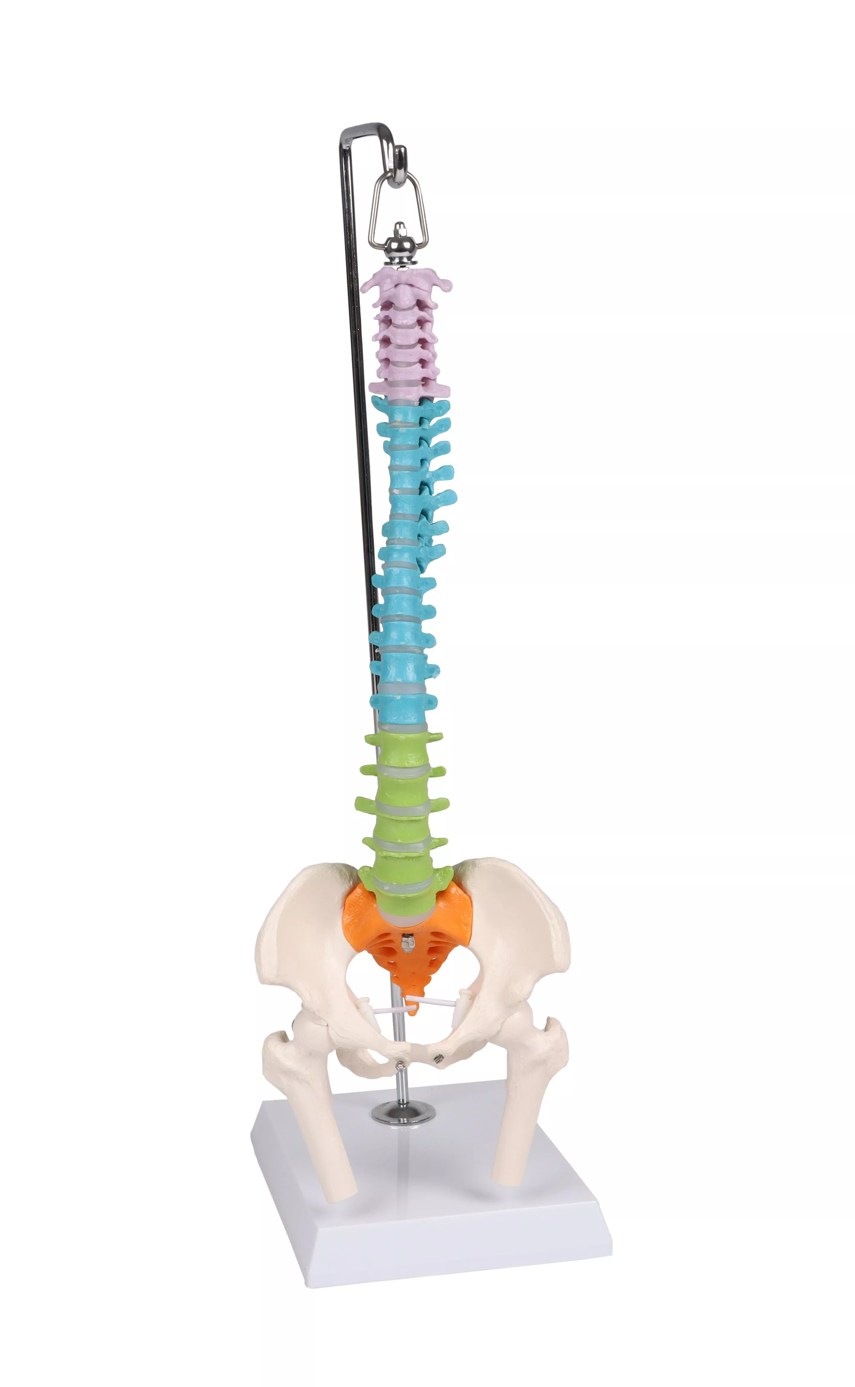
Flexible miniature spine, didactic
This spine model is an advanced tool designed for medical education and patient education, aiming to help users gain a deeper understanding of the structure and function of the human spine.The spine model is divided into five different sections, each distinguished by different colors, including 7 cervical vertebrae, 12 thoracic vertebrae, 5 lumbar vertebrae, the sacrum, and the coccyx. This color differentiation aids in quickly and accurately locating and explaining key points about the human spine during the teaching process, providing convenience for learning.The model also includes simulations of L3-L4 disc prolapse, spinal nerve exits, and the cervical vertebral artery, facilitating a deeper understanding of spine-related diseases and issues.To enhance the practicality of the model, we've added a male pelvis and movable femur heads. These features comprehensively demonstrate various aspects of the human skeletal system, improving the learning experience.Lastly, our educational spine model is made from high-quality PVC plastic, ensuring durability and resistance to damage. Its texture is clear, and the mold accuracy is high, guaranteeing the accuracy of anatomical structures.In summary, this interactive educational human spine model is an ideal tool for medical professionals, educators, and students. It will contribute to a better understanding and teaching of the anatomy of the human spine, ultimately enhancing the quality of medical education and patient education.

€130.90*
Female pelvis with sacrum, 2 lumbar vertebrae and femoral stumps, flexible
Natural casting of an adult female pelvis The pelvis can be disassembled fast and easily. It is mounted with rubber bands completey to allow an exceptional range of movement. With flexibly mounted L5 and L4. The femoral stumps are moveable and removable.

€152.32*
Reduced
Osteopathic Skull Model, 22 part, didactical version - EZ Augmented Anatomy
This fascinating model of an average European adult skull can be disassembled into 22 single bones. During development of this model one of the main targets was to make the model easy to assemble and dismantle. Stable parts with convenient magnet connections make handling of the product a child‘s play. The detailed bones do not need any complicated pins to be stuck into holes, they almost slide into position, guided by realistic bone sutures and held by strong magnets. Due to the very good anatomy and the easy handling this skull is the ideal training aid for osteopathy.This version is perfect for visual distinction of the single bones. The bones are produced in different colours, commonly used in anatomy, geminate bones do have identical colour. The following individual bones are shown:•Parietal bone left and right•Occipital bone•Temporal bone left and right•Sphenoid bone•Frontal bone•Ethmoid bone•Vomer•Palatine bone, left and right•Inferior nasal concha left and right•Maxilla with teeth, left and right•Lacrimal bone left and right•Nasal bone left and right•Zygomatic bone left and right•Mandible with teethScope of delivery:•User manual in English and German•Detailed description on CD in Latin, English, French, German, Spanish, Portuguese, Italian, Polish, Russian, Arabic, Korean and Japanese.Augmented- Anatomy- AppThe new Augmented Anatomy App in combination with this high quality anatomical model makes learning now even easier and more efficient!This app automatically recognises our anatomical models and displays the nomenclature in augmented reality. As an Erler Zimmer customer, you can use it completely free of charge and for an unlimited period of time. •High-quality Augmented Reality learning app•Free of charge and without registration•Nomenclature always and everywhere available•Further online links in the learning lexiconOur augmented anatomy app works on all common smartphones or tablets. Click here to Download the App.

€234.37* €360.57* (35% saved)
Chart Foot and Ankle, 70x100cm
High Quality anatomical chart, made of 200µ plastic foil, size appr. 70x100cm, incl. metal edging with hanger cord. Nomenclature in German and English.

Variants from €19.04*
€26.18*
Reduced
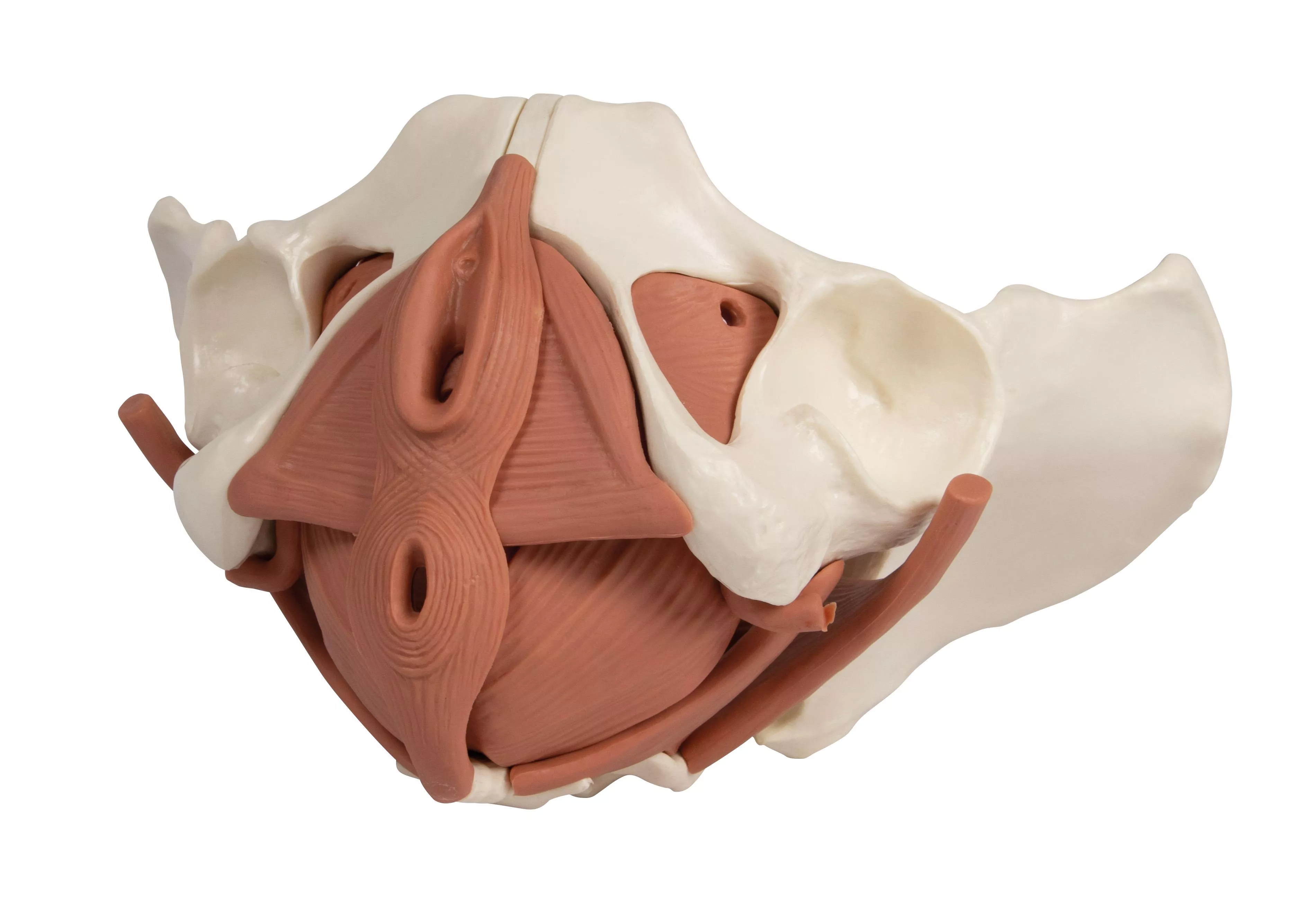
Female Pelvic Floor Model, 12 part, magnetic
This improved model of a female pelvis with pelvic floor muscles depicts the pelvic floor in its layers. The following muscles are represented and magnetically detachable: - M. obturatorius internus (left and right), - M. piriformis (left and right), - M. coccygeus (left and right), - Diaphragma pelvis (M. levator ani consisting of M. puborectalis, M. pubococcygeus, and M. iliococcygeus), - Diaphragma urogenitale (consisting of M. transversus perinei profundus, M. transversus perinei superficialis, and M. ischiocavernosus), and the - sphincter muscles of the urogenital and digestive tracts (consisting of M. sphincter ani externus, M. sphincter urethrae, and M. bulbospongiosus). The model consists of a total of 12 parts, including the two halves of the pelvis and the sacrum. The model is held together by magnets, allowing individual parts to be easily removed for demonstration of the different layers. This model is highly suitable for courses in pelvic floor exercises, childbirth preparation, midwifery training, or simply for studying the pelvic floor muscles. Size: 27 x 18 x 17 cm Weight: 960 g

€212.41* €283.22* (25% saved)

Continuous innovation

Social responsibility

Active customer orientation

Understanding quality

Sustainable actions

ISO 9001 certification

Your last viewed products