


Product information "Uterus Bicornuate Unicollis"
Clinical History
A 36-year-old woman experienced a severe postpartum hemorrhage following the breech delivery of her fourth child. All her previous three deliveries were also breech, and there was no history of miscarriage. She had a background of intermittent mild abdominal pain. As the bleeding could not be controlled, the obstetric team performed an emergency radical hysterectomy with bilateral salpingo-oophorectomy. Both mother and child made a full recovery.
Pathology
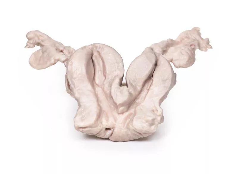
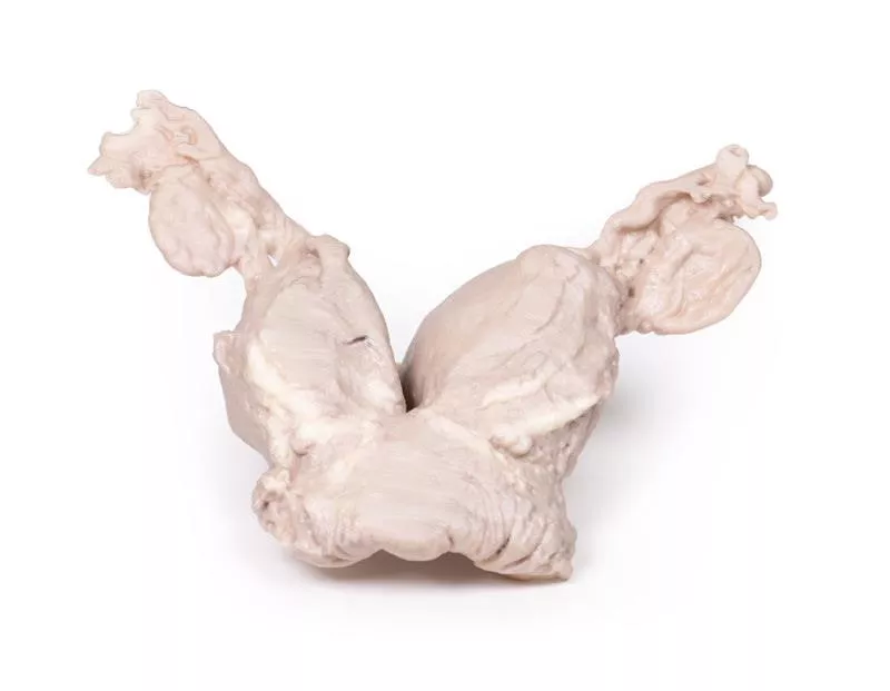
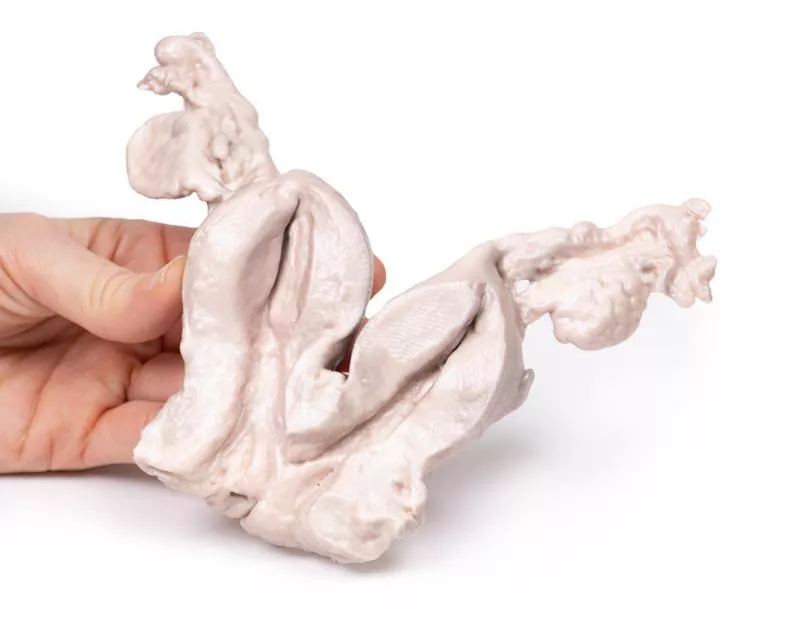
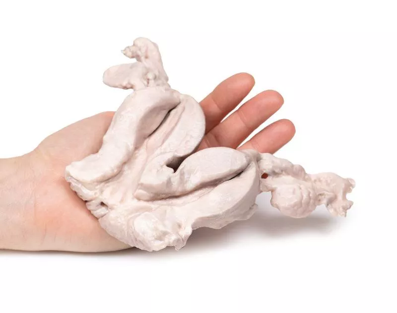
The specimen includes a bicornuate uterus, fallopian tubes, and ovaries, sliced coronally to show both internal and external surfaces. The two uterine bodies are of equal size and share a single cervical canal. A few small cervical cysts are visible.
Further Information
A bicornuate uterus is a congenital malformation in which the uterine fundus has an indentation greater than 1 cm. While the cervix and vagina are usually normal, the uterus typically contains two partially separated endometrial cavities due to incomplete fusion of the Müllerian ducts during embryonic development.
This condition affects approximately 0.5% of women, though the actual number may be higher, as many are asymptomatic. Symptoms, when present, may include pelvic pain (cyclic or non-cyclic), abnormal bleeding, discharge, or urinary tract infections.
In pregnancy, a bicornuate uterus is associated with increased risk of recurrent miscarriage, preterm labour, fetal malpresentation, growth restriction, and placenta previa. Malpresentation often necessitates a caesarean section. After birth, there is a higher risk of placental retention and postpartum hemorrhage.
Diagnosis is usually made via pelvic ultrasound; MRI may be used in select cases to confirm the diagnosis. Most cases do not require treatment.
A 36-year-old woman experienced a severe postpartum hemorrhage following the breech delivery of her fourth child. All her previous three deliveries were also breech, and there was no history of miscarriage. She had a background of intermittent mild abdominal pain. As the bleeding could not be controlled, the obstetric team performed an emergency radical hysterectomy with bilateral salpingo-oophorectomy. Both mother and child made a full recovery.
Pathology
The specimen includes a bicornuate uterus, fallopian tubes, and ovaries, sliced coronally to show both internal and external surfaces. The two uterine bodies are of equal size and share a single cervical canal. A few small cervical cysts are visible.
Further Information
A bicornuate uterus is a congenital malformation in which the uterine fundus has an indentation greater than 1 cm. While the cervix and vagina are usually normal, the uterus typically contains two partially separated endometrial cavities due to incomplete fusion of the Müllerian ducts during embryonic development.
This condition affects approximately 0.5% of women, though the actual number may be higher, as many are asymptomatic. Symptoms, when present, may include pelvic pain (cyclic or non-cyclic), abnormal bleeding, discharge, or urinary tract infections.
In pregnancy, a bicornuate uterus is associated with increased risk of recurrent miscarriage, preterm labour, fetal malpresentation, growth restriction, and placenta previa. Malpresentation often necessitates a caesarean section. After birth, there is a higher risk of placental retention and postpartum hemorrhage.
Diagnosis is usually made via pelvic ultrasound; MRI may be used in select cases to confirm the diagnosis. Most cases do not require treatment.
Login
Erler-Zimmer
Erler-Zimmer Medical GmbH
Hauptstrasse 27
77886 Lauf
Germany
info@erler-zimmer.de
Achtung! Medizinisches Ausbildungsmaterial, kein Spielzeug. Nicht geeignet für Personen unter 14 Jahren.
Attention! Medical training material, not a toy. Not suitable for persons under 14 years of age.
Other customers also bought

Retrosternal Goiter
Clinical HistoryA 60-year-old woman presented with an abnormal swelling in her neck, a persistent cough, and difficulty swallowing. Over the previous years, she had experienced weight gain. She later died of an unrelated cardiovascular condition, and the specimen was collected during the post-mortem examination.PathologyThe post-mortem specimen includes the larynx, trachea, and a large, multilobular thyroid gland. The thyroid is significantly enlarged, especially the right lobe, which features two prominent lobes extending 7–8 mm superiorly and inferiorly—well beyond normal boundaries from the front view. Posteriorly, the oesophagus has been opened to show the rear wall of the trachea. The right lobe appears even larger from this angle, with the abnormal growth concentrated at its inferior pole. No major pigmentation changes are observed on the surfaces, but prominent veins are visible on the right lobe.Further InformationGoitre often presents as a visible neck swelling. Depending on its size and location, it may cause pressure symptoms such as difficulty breathing, swallowing issues (dysphagia), hoarseness, and persistent cough. In rare cases, an expanding goitre can paralyse the recurrent laryngeal nerve. Symptoms of tracheal obstruction, including stridor and shortness of breath, may occur. Sudden tenderness and rapid enlargement may result from cystic expansion or bleeding into a thyroid nodule.Common causes of goitre include autoimmune diseases (such as Hashimoto’s thyroiditis and Grave’s disease), thyroid nodules, and iodine deficiency. Goitre usually results from reduced thyroid hormone synthesis due to biosynthetic defects or iodine deficiency. This leads to increased TSH (thyroid-stimulating hormone), which stimulates thyroid growth in compensation. In Hashimoto’s thyroiditis, elevated TSH and autoimmune-related fibrosis both contribute to gland enlargement. In Grave’s disease, thyroid growth is primarily driven by TSH receptor antibodies.Reference: Hughes et al. (2012). Goitre: Causes, investigation and management. Aust Family Physician, 41, 572–576.

Carcinoma of Breast
Clinical HistoryA 76-year-old woman was admitted to the emergency department with a sudden loss of consciousness and signs of a left-sided stroke. She was intubated and treated in the ICU, where a fixed mass was noted in her left breast along with palpable axillary lymphadenopathy. She later died from pneumonia related to mechanical ventilation.PathologyThe specimen shows the left breast with a large, oval tumour mass (11?cm), located beneath and attached to the skin and adherent to the muscle. The cut surface is heterogeneous, showing areas of necrosis, haemorrhage, and cyst formation. Histology confirmed a breast adenocarcinoma with regional lymph node involvement.Further InformationBreast cancer is the second most common cancer in women and mainly affects those over 30, peaking between 70 and 80 years. Risk factors include estrogen exposure, family history, nulliparity, obesity, and mutations in genes such as BRCA1, BRCA2, and others.Most cases are adenocarcinomas originating in the duct or lobular system, often first appearing as DCIS. Subtypes are defined by hormone receptor status (ER) and HER2 expression, which guide treatment. Common metastatic sites are bone, liver, lung, and brain.In countries with screening programs, most cases are found by mammography. Symptomatic signs include a hard, irregular, immobile mass, skin changes like dimpling (peau d’orange), and nipple retraction.Treatment varies by stage and tumour profile and may include mastectomy, lumpectomy, radiotherapy, targeted therapies (e.g. trastuzumab for HER2+), anti-estrogen therapy (e.g. tamoxifen for ER+), and systemic chemotherapy.

Multinodular goitre
Clinical HistoryA 53-year-old female presented with an abnormal neck swelling and persistent cough. She also experienced lethargy and weight gain over several years. During investigations, she died months later from unrelated cardiovascular disease.Pathology The post-mortem specimen includes the base of the tongue, larynx, and trachea. The thyroid gland is grossly enlarged, especially the right lobe, extending beyond its normal boundaries. The cut surfaces show multiple hyper- and hypopigmented nodules and cystic areas in both lobes. The tongue base, larynx, and trachea appear normal.Further InformationNodular goitre is usually detected as a neck swelling. Depending on size and location, it can cause pressure symptoms like difficulty breathing, dysphagia, cough, or hoarseness. Rarely, recurrent laryngeal nerve paralysis occurs. Sudden growth or tenderness can result from cystic expansion or hemorrhage within nodules.Causes include autoimmune diseases (Hashimoto’s thyroiditis, Grave’s disease), thyroid nodules, and iodine deficiency. Goitre develops when thyroid hormone synthesis is reduced—due to biosynthetic defects or iodine shortage—leading to increased TSH stimulation and thyroid growth. In Hashimoto’s, elevated TSH combined with fibrosis enlarges the thyroid. In Grave’s disease, stimulation by TSH receptor antibodies causes the goitre.

Nodular hyperplasia of the Prostate
Clinical History A 63-year-old man presented with acute abdominal pain and had been unable to urinate for five days. He reported a two-year history of urinary frequency, hesitancy, double voiding, nocturia, and poor stream. Examination revealed a distended bladder and an enlarged prostate. Bladder scan showed >1?L urine retention; labs confirmed acute renal failure. Catheterisation attempts failed, leading to a total prostatectomy. He recovered well post-op.Pathology The enlarged prostate, sliced transversely, shows multiple nodules (2–10?mm) consistent with benign prostatic hyperplasia (BPH).Further Information BPH is a common condition in older men, caused by nodular overgrowth of stromal and epithelial cells in the periurethral zone, stimulated by dihydrotestosterone. The median lobe may enlarge disproportionately and obstruct the urethra.Prevalence increases with age: 20% by 40, 70% by 60, 90% by 80. Risk factors include family history, obesity, and androgenic steroids.Typical symptoms: urinary frequency, nocturia, hesitancy, double voiding, dribbling. Acute urinary retention can lead to UTIs and kidney damage.Diagnosis includes history, digital rectal exam (DRE), PSA test, and imaging. Treatments: alpha-blockers, 5-alpha-reductase inhibitors, or in severe cases transurethral resection of the prostate (TURP). Total prostatectomy is rarely used due to complications.

Lymphoma of the thyroid
Clinical HistoryA 68-year-old woman presented with a small, hard thyroid lump. Over six weeks, the mass rapidly enlarged, causing laryngeal stridor and oesophageal obstruction. No lymphadenopathy or splenomegaly was noted.Pathology The specimen includes the larynx, thyroid, upper trachea, and oesophagus. The enlarged left thyroid lobe, and to a lesser extent the right, is replaced by homogeneous pale tumour tissue. The tumour compresses the larynx and oesophagus. Histology confirmed lymphoblastic lymphoma of the thyroid. Due to its rarity, anaplastic carcinoma and secondary lymphoma spread must be excluded.Further InformationPrimary thyroid lymphoma is rare but important to consider in thyroid masses. Most are non-Hodgkin lymphomas; lymphoblastic lymphoma is aggressive and usually seen in children. The main known risk factor is chronic autoimmune (Hashimoto’s) thyroiditis, present in about 50% of cases.Over 90% of patients present with a rapidly enlarging goitre causing compression of trachea, oesophagus, and neck vessels, resulting in symptoms like stridor, hoarseness, dysphagia, and neck pain. Systemic ‘B-symptoms’ may include night sweats, fever, and weight loss.Diagnosis requires ultrasound with fine needle aspiration or biopsy. Cytology and immunohistochemistry are essential to differentiate lymphoma from Hashimoto’s thyroiditis or carcinoma.

Aorta & para-aortic lymph nodes
Clinical HistoryThis case involves a 75-year-old woman who presented with recurrent symptoms five years after initial treatment for stage IIIc serous adenocarcinoma of the ovary. She was diagnosed with chemo-resistant retroperitoneal lymph node metastases, involving both para-aortic and pelvic nodes, as shown on PET/CT imaging. Unfortunately, she died of liver complications before options like radical lymphadenectomy could be explored.PathologyThe specimen includes the abdominal aorta and common iliac arteries, surrounded by a large number of enlarged para-aortic and iliac lymph nodes. Histopathological analysis confirmed metastatic high-grade adenocarcinoma in several of these nodes.Further InformationIn some cases of recurrent ovarian cancer, lymph node metastases may be the only sign of disease. In this patient, PET/CT findings accurately predicted all metastatic nodes. While ultrasound (US) could have also detected the enlarged nodes, both PET/CT and US may miss microscopic disease, making it difficult to fully exclude lymph node involvement during follow-up.In younger women with recurrent ovarian cancer but no other spread, systematic dissection of aortic and pelvic nodes may offer symptom relief and open the door for novel therapies, although it is rarely curative.More typically, pelvic and aortic lymph node sampling forms part of initial surgical staging in epithelial ovarian cancer. For women with advanced-stage disease, surgical removal of enlarged retroperitoneal lymph nodes may be appropriate if it enables complete tumour debulking.

Endometrial Carcinoma
Clinical HistoryA 63-year-old woman presented with dull lower abdominal pain lasting two months and persistent heavy vaginal bleeding for one week, 13 years after menopause. Following a biopsy-confirmed diagnosis of endometrial carcinoma, she underwent radical abdominal hysterectomy and bilateral salpingo-oophorectomy as part of her treatment.PathologyThe specimen includes uterus, fallopian tubes, and ovaries. The endometrial lining shows significant abnormalities, especially on the right side, where a brown polypoid tumour has invaded the myometrium and extended into the cervical canal. Histology confirmed a well-differentiated endometrial adenocarcinoma. The left ovary is enlarged and contains multiple large follicular cysts.Further InformationEndometrial carcinoma is the most common gynecologic cancer in developed countries. It exists mainly in two types: endometrioid carcinoma (around 80% of cases), which often arises from atypical hyperplasia and presents early with better prognosis, and serous carcinoma, which is less common and more aggressive.Common mutations include PTEN, PIK3Ca, ARID1A in endometrioid tumours and TP53 in serous types. Risk factors for endometrioid carcinoma include obesity, glucose intolerance, infertility, and unopposed estrogen exposure. Serous carcinoma typically affects older women with lower BMI and atrophic uterus. Women with Lynch Syndrome also have a significantly increased risk.Post-menopausal bleeding is the most frequent symptom, allowing early detection. Some cases are asymptomatic or found incidentally. Imaging shows thickened endometrium on ultrasound or CT. Diagnosis is confirmed via biopsy, curettage or hysterectomy. Treatment depends on stage and includes surgery, radiotherapy and chemotherapy.

Uterine Leiomyoma
Clinical History A 30-year-old woman presented with infertility, pelvic pain, heavy bleeding and dysmenorrhea. A pelvic mass was palpable and ultrasound showed a hypoechoic lesion in the myometrium. A planned myomectomy was converted to an emergency hysterectomy due to complications. She recovered well.Pathology The uterus (with cervix and fundus) is sectioned sagittally. A 4?×?2?cm ovoid mass arises from the posterior uterine wall, protruding into the cavity and reaching the cervical canal.Further Information Uterine leiomyomas (fibroids) are benign smooth muscle tumors, common in women of reproductive age, especially among Black women. They may cause bleeding, pelvic pressure, infertility, and complications in pregnancy. Diagnosis is by ultrasound. Treatment options include hormonal therapy, myomectomy, hysterectomy and uterine artery embolisation.

Hydrocoele
Clinical History A patient with diabetes and previous myocardial infarctions presented with bilateral pleural effusion, peripheral oedema, and a swollen scrotum showing transillumination. Chest x-ray revealed congestive heart failure. Despite treatment, the patient died during admission.Pathology The testis and coverings show a distended cavity between the visceral and parietal layers of the tunica vaginalis due to fluid accumulation, representing a hydrocele secondary to generalized oedema from heart failure.Further Information A hydrocele is serous fluid between the layers of the tunica vaginalis. It can be communicating (linked to the peritoneal cavity) or non-communicating. Communicating hydroceles arise from failure of processus vaginalis closure and may be congenital or develop later, often due to increased abdominal pressure, like heart failure. Non-communicating types result from fluid imbalances due to infections, trauma, tumors, or lymphatic issues.Patients notice a scrotal swelling, uni- or bilateral. Communicating hydroceles may vary in size with pressure; non-communicating are usually stable. Swellings are generally painless unless complicated by infection or torsion. Larger hydroceles can cause skin problems.Diagnosis is clinical, aided by transillumination and ultrasound to rule out other causes. Tumor markers like AFP and B-HCG may exclude cancer. Many congenital hydroceles resolve by age two; persistent or symptomatic cases may require surgical repair. Treating the underlying cause can also resolve reactive hydroceles.

Continuous innovation

Social responsibility

Active customer orientation

Understanding quality

Sustainable actions

ISO 9001 certification