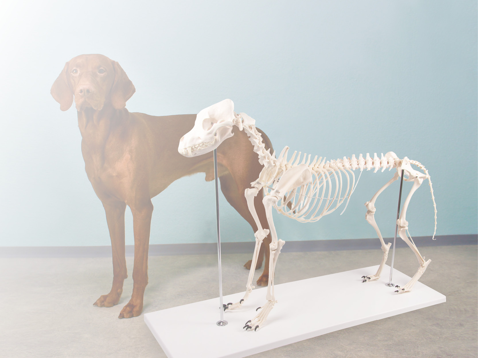
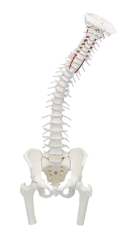
This fascinating model of an average European adult skull can be disassembled into 22 single bones. During development of this model one of the main targets was to make the model easy to assemble and dismantle. Stable parts with convenient magnet connections make handling of the product a child‘s play. The detailed bones do not need any complicated pins to be stuck into holes, they almost slide into position, guided by realistic bone sutures and held by strong magnets. Due to the very good anatomy and the easy handling this skull is the ideal training aid for osteopathy.This version is perfect for visual distinction of the single bones. The bones are produced in different colours, commonly used in anatomy, geminate bones do have identical colour. The following individual bones are shown:•Parietal bone left and right •Occipital bone •Temporal bone left and right •Sphenoid bone •Frontal bone •Ethmoid bone •Vomer •Palatine bone, left and right •Inferior nasal concha left and right •Maxilla with teeth, left and right •Lacrimal bone left and right •Nasal bone left and right •Zygomatic bone left and right •Mandible with teeth Scope of delivery:•User manual in English and German •Detailed description on CD in Latin, English, French, German, Spanish, Portuguese, Italian, Polish, Russian, Arabic, Korean and Japanese. Augmented- Anatomy- AppThe new Augmented Anatomy App in combination with this high quality anatomical model makes learning now even easier and more efficient!This app automatically recognises our anatomical models and displays the nomenclature in augmented reality. As an Erler Zimmer customer, you can use it completely free of charge and for an unlimited period of time. •High-quality Augmented Reality learning app •Free of charge and without registration •Nomenclature always and everywhere available •Further online links in the learning lexicon Our augmented anatomy app works on all common smartphones or tablets. Click here to Download the App.
High Quality anatomical chart, made of 200g/sqm art paper, size 50x70cm, incl. attachable plastic rods with hanger. Nomenclature in German, Latin and English.
This model, which is unique world-wide, consists of cervical vertebra C3, axis C2, atlas C1 and a segment of the occipital bone. All components are represented in 2 x enlargement for improved demonstration. With this model, the movements in the upper cervical area can be understood in a true to life and anatomically correct manner. All possible movements in the atlanto-occipital and atlantoaxial articulation can be demonstrated admirably. With removable stand.