Product information "
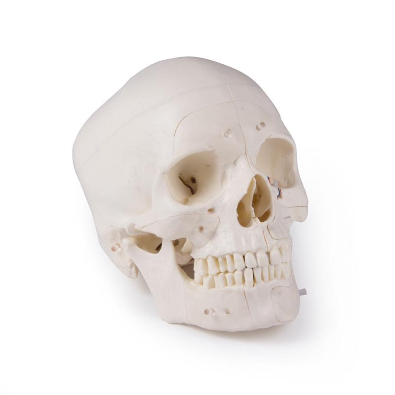
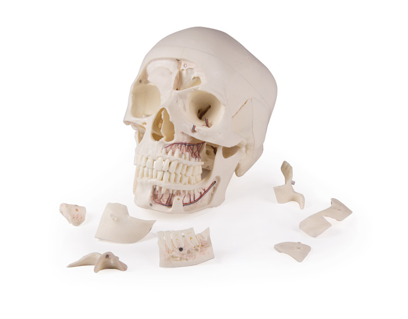
Deluxe demonstration skull; 14-part; for advanced studies"
This skull model is an actual cast of a real human specimen and shows all anatomical structures in highest detail. It is made for students in anatomy, medicine, surgery, otolaryngology, ophthalmology and dentistry. The Skull is intricately sectioned and reassembled with metal and magnet connections.
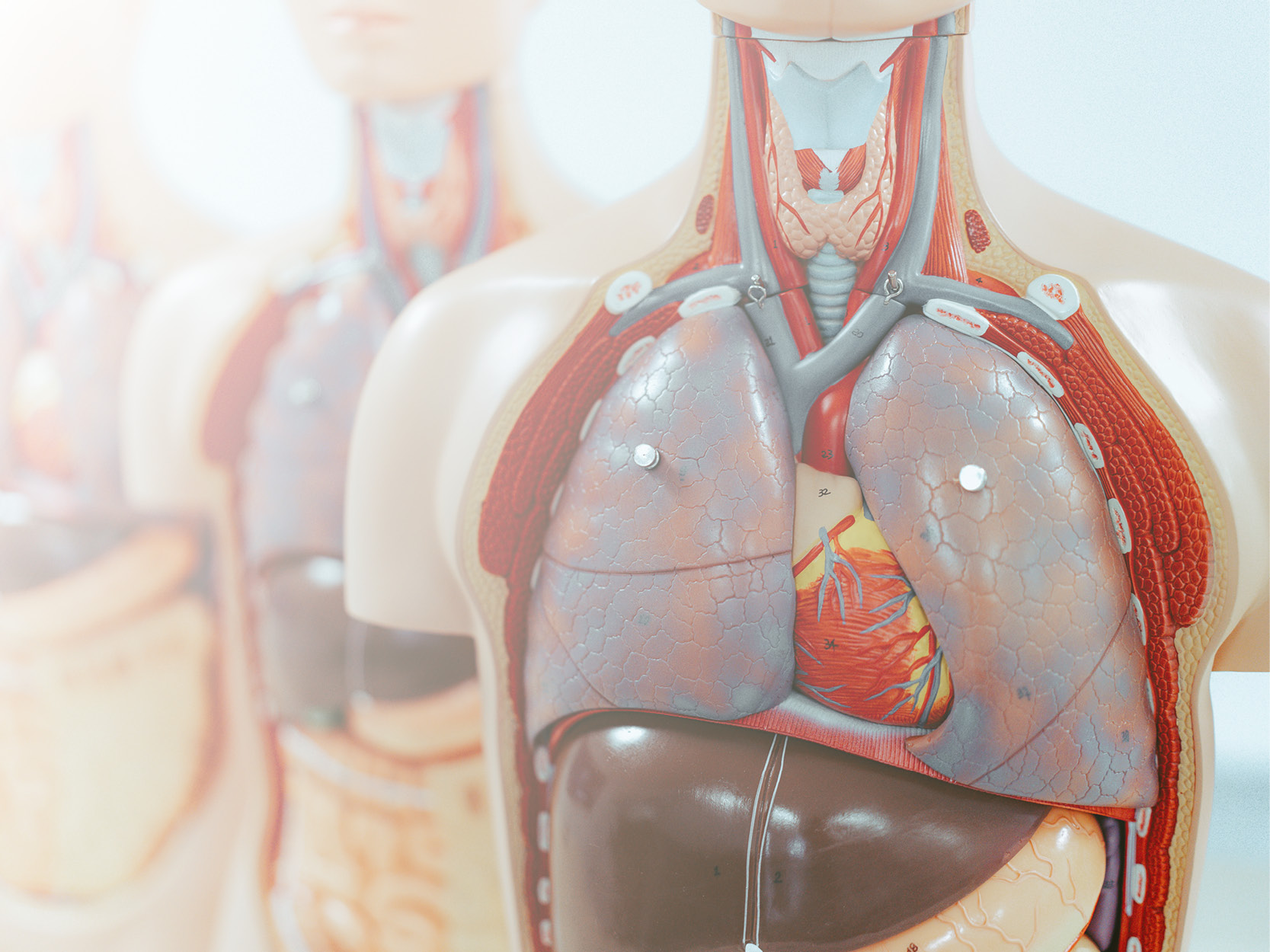
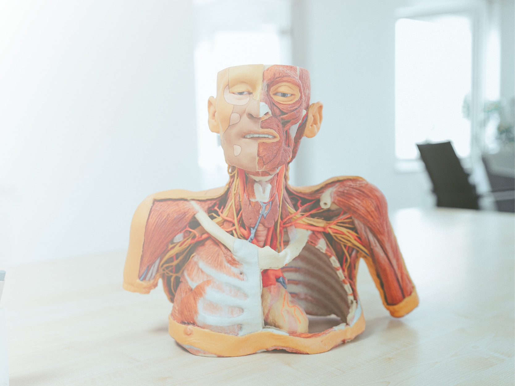
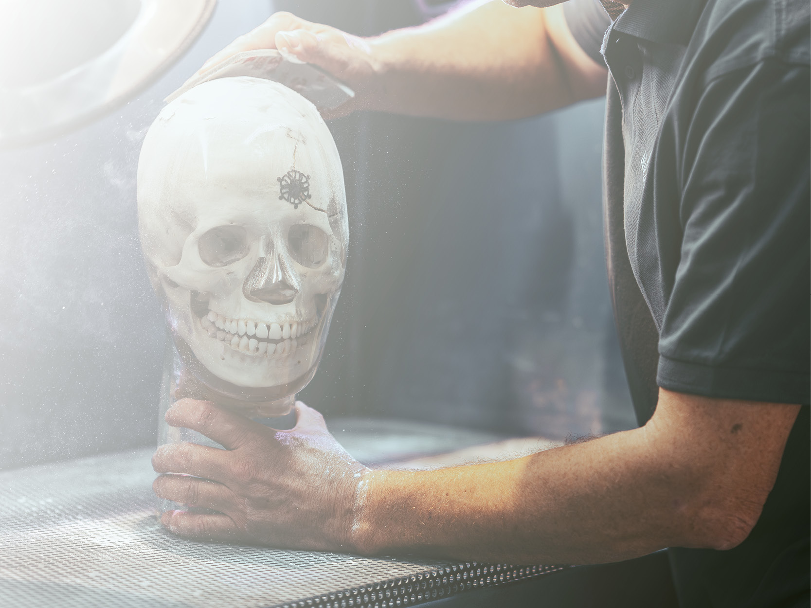
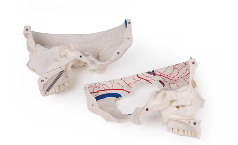
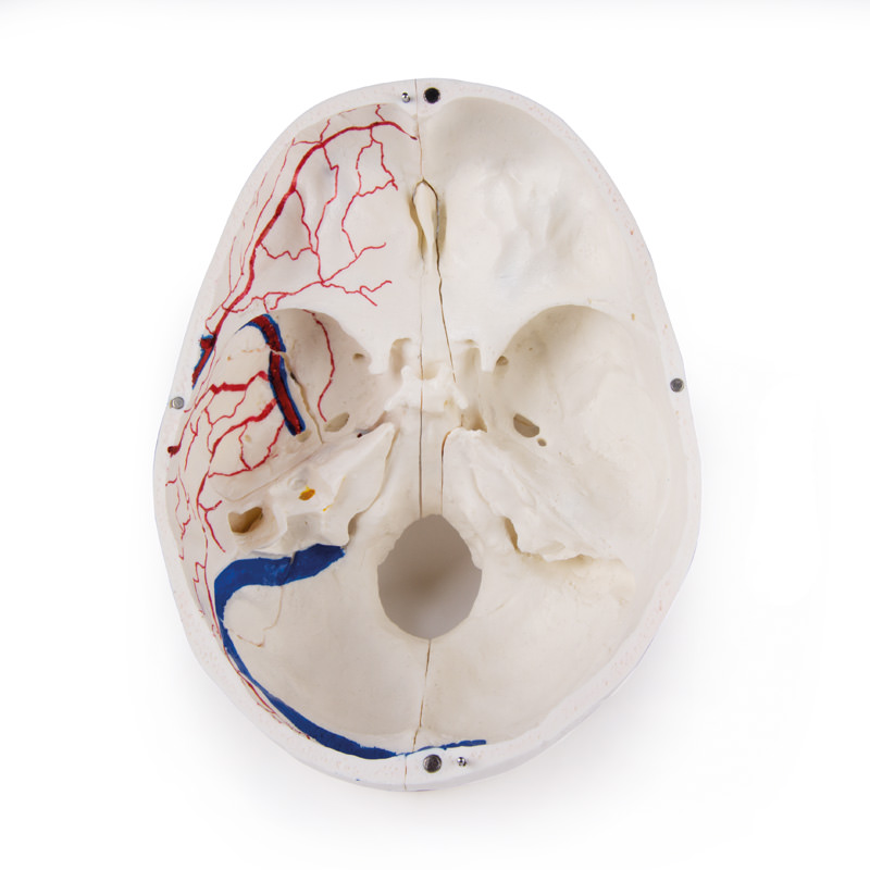
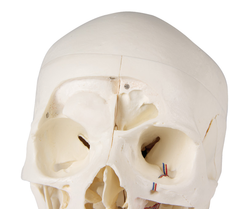
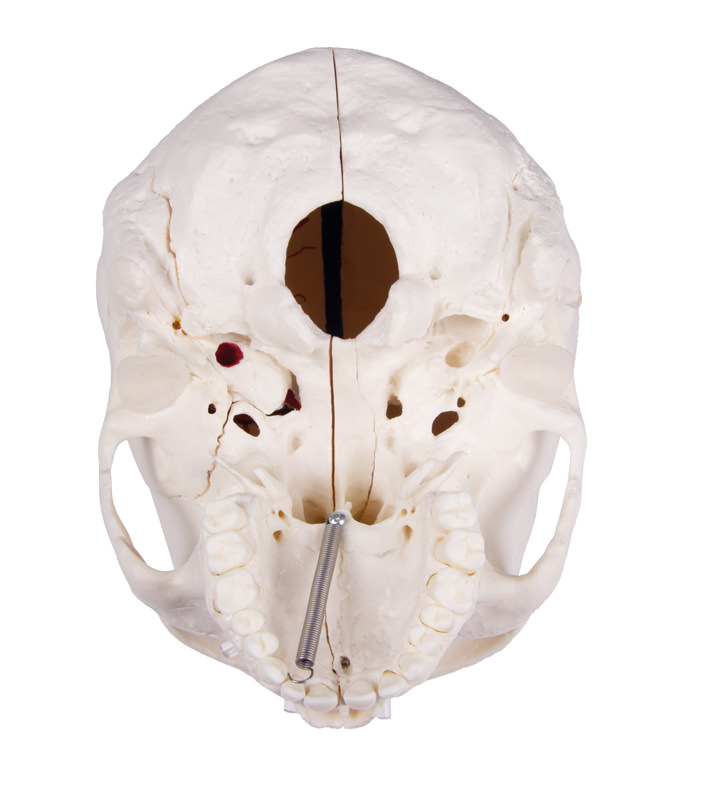
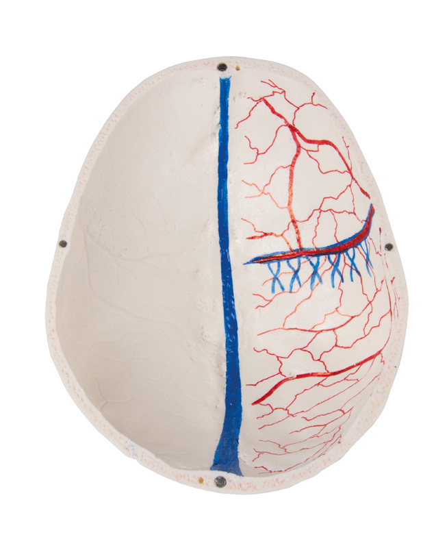
The calvarium is sectioned horizontally leaving the temporal bone and its sutures intact. Bony impressions of the superior sagittal sinus, transverse sinus and sigmoid sinus as well as the meningeal vessels have been painted. The base portion of the skull has been sagittaly sectioned in the way that it passes through the one cribriform plate on one side and another section in the same plane passes through the other cribriform plate of the ethmoid leaving the crista galli perpendicular plate of the ethmoid intact as also the whole nasal septum. The structures of anterior, middle and posterior cranial fossae are easily accessible. One can directly visualize the nasal cavity, the concha, the nasal septum, the bony pharyngeal and naso-pharyngeal spaces. The nasal septum is separable from surrounding bones. The frontal sinuses have been dissected on one side to show the sinus as whole and on the other side chiselled out for full access to the sinus. The relation of this sinus to the nasal cavity is clearly shown and is especially valuable for otolaryngologists.
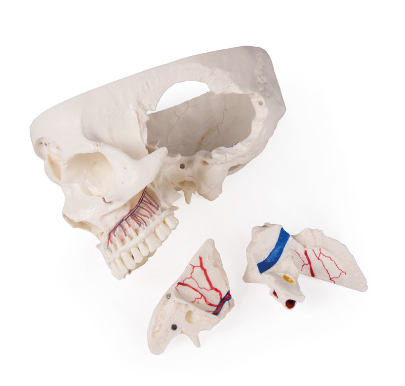
On one side of the skull the temporal bone has been left in situ. The other temporal bone is removable from the skull. A portion of the mastoid and squama can be removed along with the tympanic antrum, baring internal ear in full view. All three semicircular canals are visible along with the course of the facial nerve coursing backwards and then downwards emerging finally through the stylo-mastoid foramen. The removable temporal bone has the external auditory meatus intact. An almost vertical section through the squama mastoid process and carried inwards along the petro-squamosal junction has been made and when apart, one sees the position of tympanic membrane. The carotid canal has been opened as also the cochlea , showing the internal canal, and the course of the facial nerve has been depicted. Oval window, the semi-circular canals, and aditus of the tympanic antrum are visible.
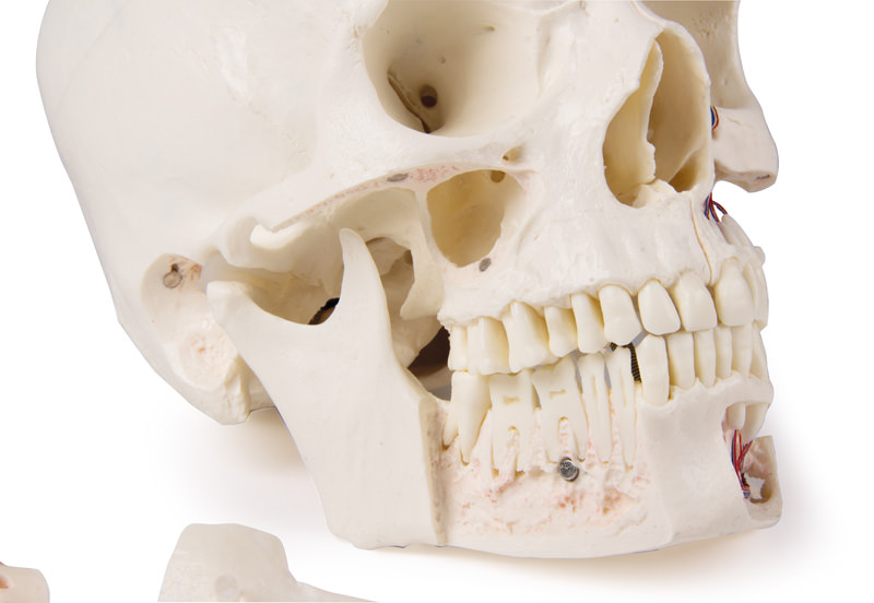
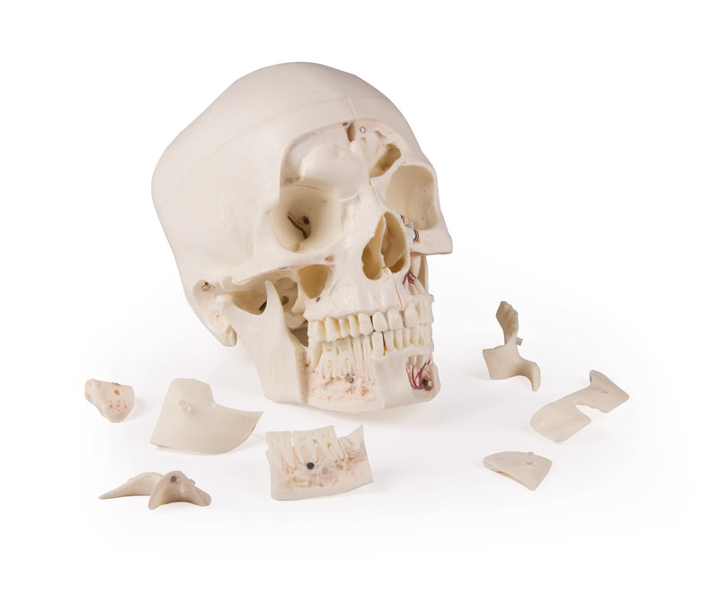
The maxilla and mandible expose the structures of dentition, the roots, the bony margin of the alveolar process, dental vessels and nerves are visible. The maxillary sinus can by opened by removing a bone flap. Teeth of the right mandible are sectioned to show the inner tooth structure.

4 of 4 reviews

5 out of 5 stars


100%

0%

0%

0%

0%


Leave a review!

Share your experiences with other customers.


October 4, 2021 08:39

einfaches zusammenbauen

Ich bin positiv von der Qualität des Schädels überrascht! Das zusammenbauen ist Dank der Magnete ein Kinderspiel.

November 20, 2021 11:53

awesome details / dental skull

great teaching tool for my dental studies :)The skull has very accurate anatomical structures and is a relief when learning. Through the openings in the jaw, all the important nerve tracts can be shown.

February 5, 2021 13:50

sehr schöne Strukturen, Schädel hält gut zusammen

sehr schöne Strukturen, Schädel hält gut zusammen

October 4, 2021 09:45

great structures

very detailed structures are visible, especially in the dental area.

Other customers also bought

Skull model, 3 parts - EZ Augmented Anatomy
Developing this lifelike reproduction of a human skull we have used the latest technology to digitalize a real human skull and idealized it under the aspects of medical education. This means the skull was adjusted to be anatomically ideal, all anatomical details and structures are present and correspond to anatomical teaching. The three part model consists of skull base, skull cap and lower jaw. The teeth correspond with real dentition concerning position and embrasure. The lower jaw is movably mounted and can be removed. The skull cap is aligned with the skull base using metal pins and held by strong magnets. Due to this there is no need for disturbing hooks, no plastic pins that could break and no risk of having a gap between skull cap and skull base. This distinguishes the model from competitor’s skulls with plastic pins and disturbing metal hooks. The model corresponds with an average European adult concerning size and proportions. Due to modern production technologies and high production volume this very successful model can be offered at a very reasonable price, so everyone can afford this extraordinary model.Size: 18 x 19 x 12 cm, weight: 0.7 kgAugmented- Anatomy- AppThe new Augmented Anatomy App in combination with this high quality anatomical model makes learning now even easier and more efficient!This app automatically recognises our anatomical models and displays the nomenclature in augmented reality. As an Erler Zimmer customer, you can use it completely free of charge and for an unlimited period of time. - High-quality Augmented Reality learning app- Free of charge and without registration- Nomenclature always and everywhere available- Further online links in the learning lexiconOur augmented anatomy app works on all common smartphones or tablets. Click here to Download the App

€86.87*
Osteopathic Skull Model, 22 part, didactical version - EZ Augmented Anatomy
This fascinating model of an average European adult skull can be disassembled into 22 single bones. During development of this model one of the main targets was to make the model easy to assemble and dismantle. Stable parts with convenient magnet connections make handling of the product a child‘s play. The detailed bones do not need any complicated pins to be stuck into holes, they almost slide into position, guided by realistic bone sutures and held by strong magnets. Due to the very good anatomy and the easy handling this skull is the ideal training aid for osteopathy.This version is perfect for visual distinction of the single bones. The bones are produced in different colours, commonly used in anatomy, geminate bones do have identical colour.The following individual bones are shown:- Parietal bone left and right- Occipital bone- Temporal bone left and right- Sphenoid bone- Frontal bone- Ethmoid bone- Vomer- Palatine bone, left and right- Inferior nasal concha left and right- Maxilla with teeth, left and right- Lacrimal bone left and right- Nasal bone left and right- Zygomatic bone left and right- Mandible with teethScope of delivery:User manual in English and German and a detailed description on CD in Latin, English, French, German, Spanish, Portuguese, Italian, Polish, Russian, Arabic, Korean and Japanese.Augmented- Anatomy- AppThe new Augmented Anatomy App in combination with this high quality anatomical model makes learning now even easier and more efficient!This app automatically recognises our anatomical models and displays the nomenclature in augmented reality. As an Erler Zimmer customer, you can use it completely free of charge and for an unlimited period of time. - High-quality Augmented Reality learning app- Free of charge and without registration- Nomenclature always and everywhere available- Further online links in the learning lexiconOur augmented anatomy app works on all common smartphones or tablets. Click here to Download the App

€324.87*
Osteopathic Skull Model, 22 part, anatomical version - EZ Augmented Anatomy
This fascinating model of an average European adult skull can be disassembled into 22 single bones. During development of this model one of the main targets was to make the model easy to assemble and dismantle. Stable parts with convenient magnet connections make handling of the product a child‘s play. The detailed bones do not need any complicated pins to be stuck into holes, they almost slide into position, guided by realistic bone sutures and held by strong magnets. The perfect tool for Osteopaths. The following bones are represented:- Parietal bone left and right- Occipital bone- Temporal bone left and right- Sphenoid bone- Frontal bone- Ethmoid bone- Vomer- Palatine bone, left and right- Inferior nasal concha left and right- Maxilla with teeth, left and right- Lacrimal bone left and right- Nasal bone left and right- Zygomatic bone left and right- Mandible with teethSupplied with users guide in English and German as well as a CD with Key card document in Latin, German, English, French, Spanish, Portuguese, Italian, Polish, Russian, Arabic, Korean and Japanese.Model in natural bone colour.Augmented- Anatomy- AppThe new Augmented Anatomy App in combination with this high quality anatomical model makes learning now even easier and more efficient!This app automatically recognises our anatomical models and displays the nomenclature in augmented reality. As an Erler Zimmer customer, you can use it completely free of charge and for an unlimited period of time. - High-quality Augmented Reality learning app- Free of charge and without registration- Nomenclature always and everywhere available- Further online links in the learning lexiconOur augmented anatomy app works on all common smartphones or tablets. Click here to Download the App

€312.97*

Continuous innovation

Social responsibility

Active customer orientation

Understanding quality

Sustainable actions

ISO 9001 certification

Your last viewed products