Product information "Metastatic carcinoma in the brain"
Clinical History
This 51-year-old woman had undergone surgery for breast carcinoma two years before presenting with left-sided ataxia of two weeks’ duration. A previous fainting episode followed by weakness on the same side had preceded this. Examination revealed left spastic paresis. Due to the rapid onset of symptoms, a vascular lesion was suspected. Despite discharge, she was re-admitted six weeks later with left-sided seizures. Lumbar puncture and clinical re-evaluation were inconclusive, but EEG showed a right anterior temporal abnormality. Angiography then revealed a large space-occupying lesion in the right cerebrum. Her condition continued to deteriorate, ultimately resulting in death.
Pathology
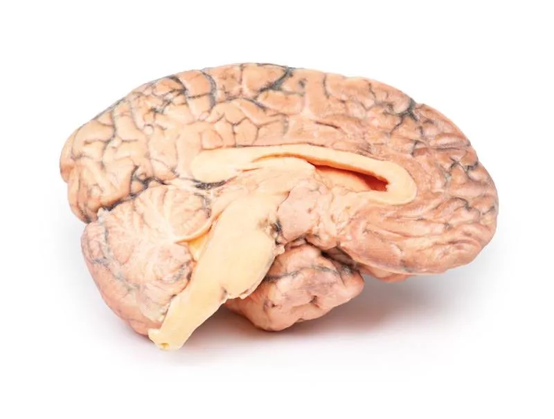
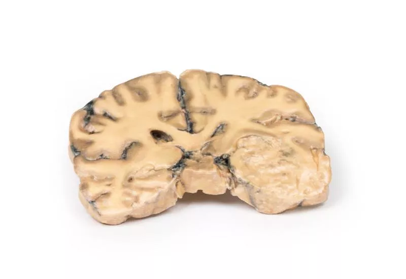
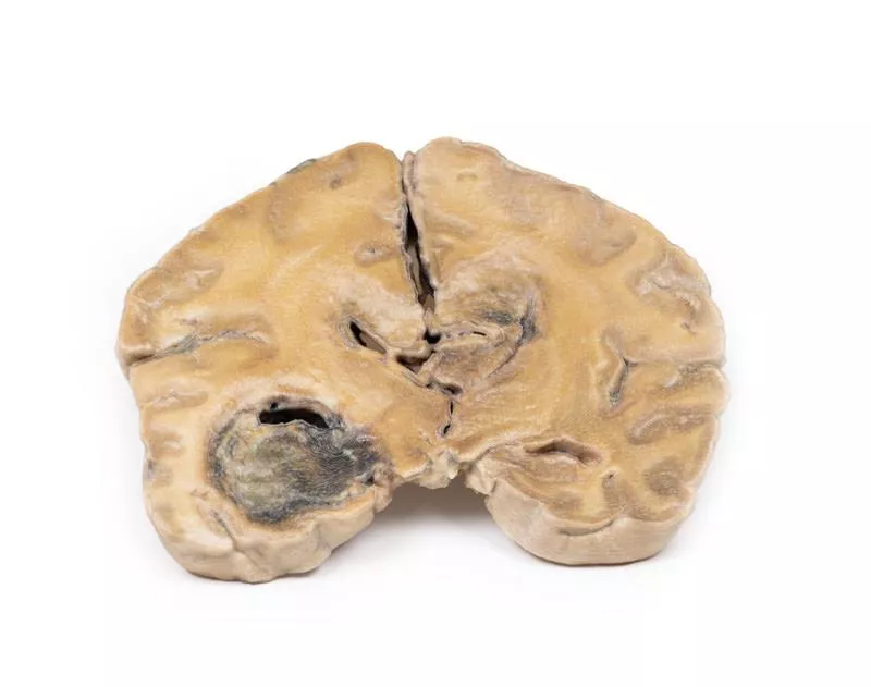
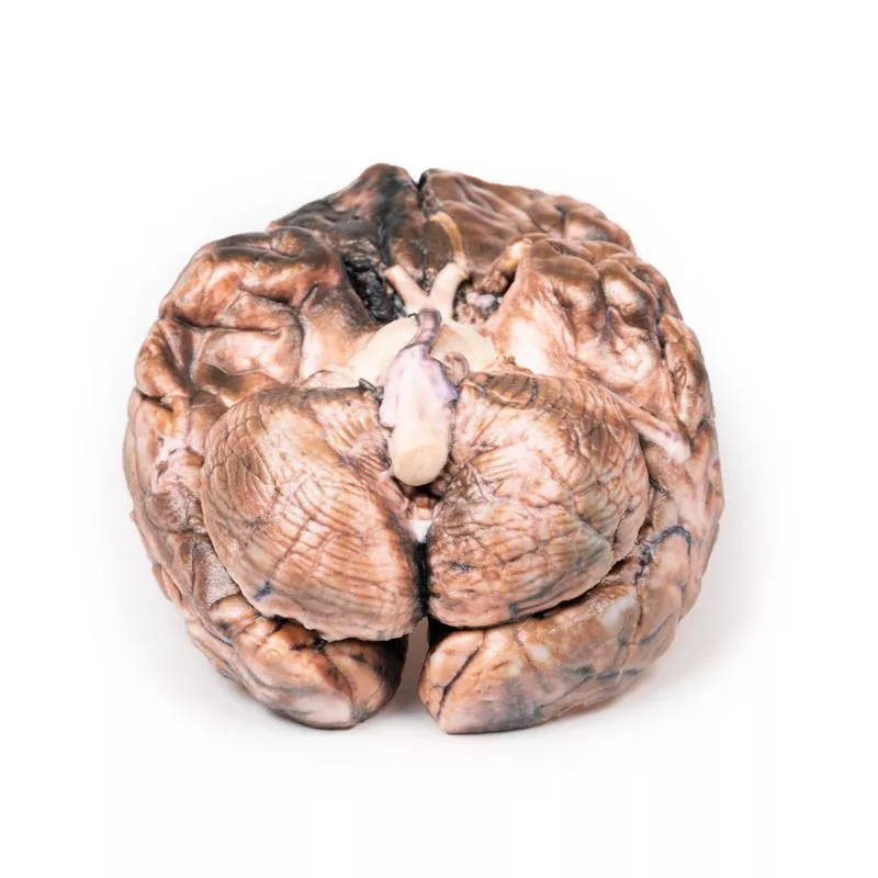
The horizontal brain slice reveals three cystic tumours, predominantly in the right parietal region. The largest measures 5 cm in diameter. Another tumour is seen near its posterior margin, and a third, smaller one is found in the left parietal region. The tumours mainly affect the white matter, with shaggy, friable greyish tissue walls. The largest tumour had ulcerated into the right lateral ventricle, and subfalcine herniation with displacement of the basal ganglia and internal capsule was observed. Histology confirmed metastatic carcinoma. Additional metastases were found in the liver and bone, consistent with a primary breast carcinoma.
Erler-Zimmer
Erler-Zimmer GmbH & Co.KG
Hauptstrasse 27
77886 Lauf
Germany
info@erler-zimmer.de
Achtung! Medizinisches Ausbildungsmaterial, kein Spielzeug. Nicht geeignet für Personen unter 14 Jahren.
Attention! Medical training material, not a toy. Not suitable for persons under 14 years of age.


