Product information "Tetralogy of Fallot"
Clinical History
A 21-month-old boy presented with a 2–3 month history of exhaustion and exertional dyspnoea. He had experienced several short episodes of acute breathing difficulty. On admission, he showed central cyanosis, mild finger clubbing and a harsh systolic murmur at the left sternal edge. Cardiac catheterisation confirmed Tetralogy of Fallot and severe pulmonary oedema. A Willis-Potts surgical anastomosis was performed. Unfortunately, the child died 12 hours post-operatively following acute dyspnoea and left lung consolidation.
Pathology
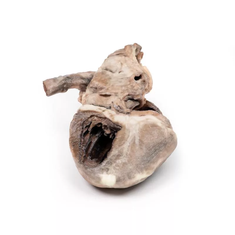
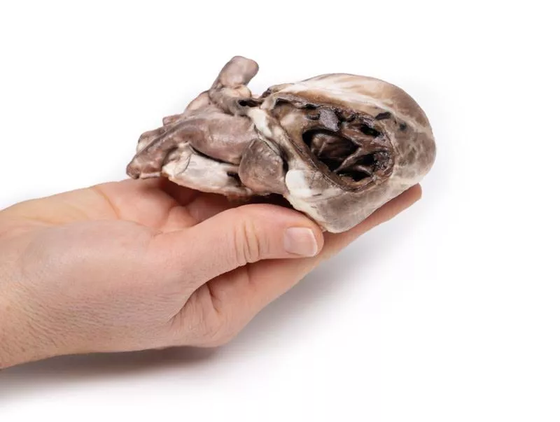
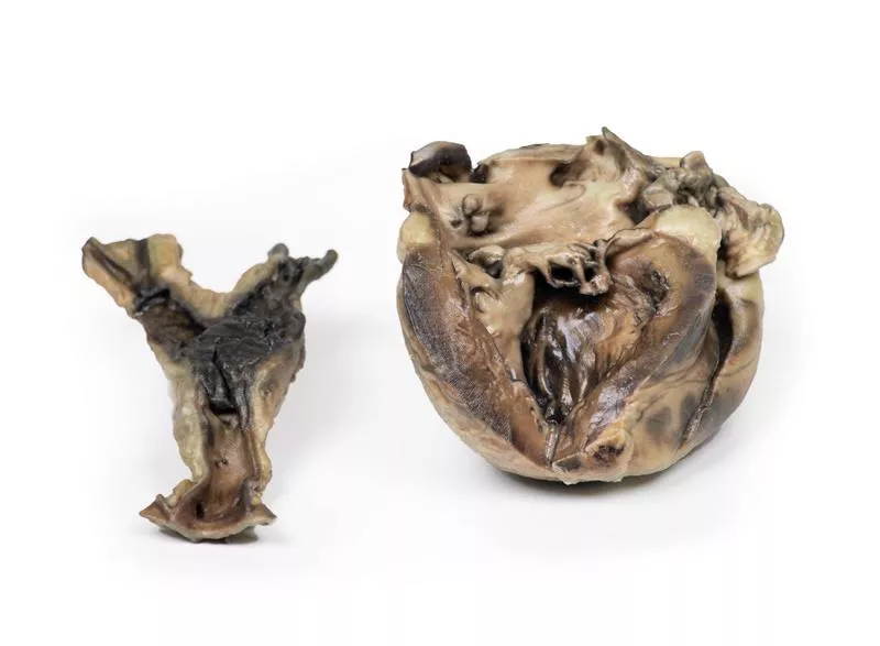
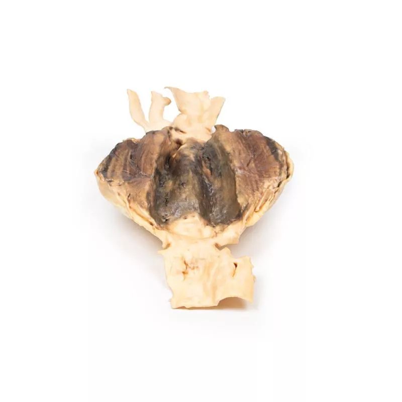
The heart, viewed anteriorly, shows right ventricular hypertrophy and pulmonary outflow tract narrowing. The pulmonary valve is bicuspid and stenosed with visible endocardial fibrosis below. The aorta overrides a large ventricular septal defect (VSD) and communicates with the hypertrophied right ventricle. A probe inserted through the narrowed pulmonary trunk reaches the dilated left pulmonary artery and continues through the surgical anastomosis into the descending aorta. Posteriorly, the opened heart reveals a large atrial septal defect (ASD) and a second smaller ASD. Notably, the left ventricular wall is thinner than the right.
Further Information
Tetralogy of Fallot includes four key defects: 1. Ventricular septal defect, 2. Overriding aorta communicating with both ventricles, 3. Pulmonary stenosis, and 4. Right ventricular hypertrophy. Cyanosis typically appears early and its severity depends on the degree of right ventricular outflow obstruction. Some patients may survive untreated into adulthood, though surgical correction is now standard. Additional defects such as ASD may coexist. While the exact cause is unknown in many cases, genetic links include Down syndrome and DiGeorge syndrome.
Erler-Zimmer
Erler-Zimmer GmbH & Co.KG
Hauptstrasse 27
77886 Lauf
Germany
info@erler-zimmer.de
Achtung! Medizinisches Ausbildungsmaterial, kein Spielzeug. Nicht geeignet für Personen unter 14 Jahren.
Attention! Medical training material, not a toy. Not suitable for persons under 14 years of age.


