Product information "Lobar pneumonia"
Clinical History
There is no clinical history available for this specimen.
Pathology
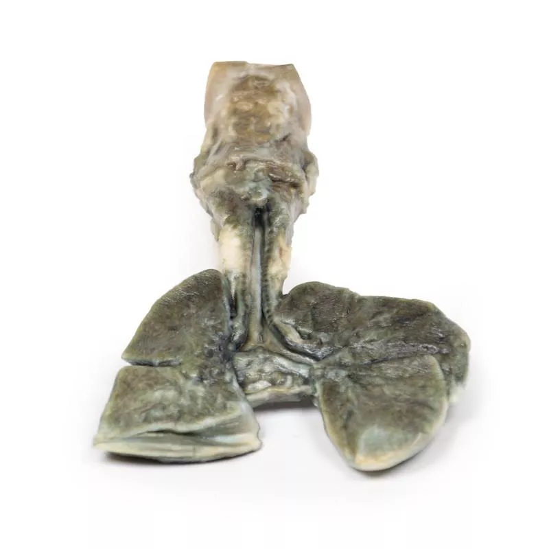
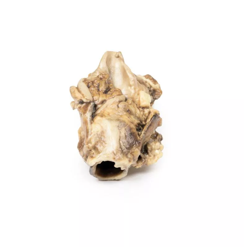
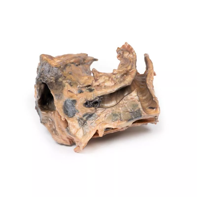
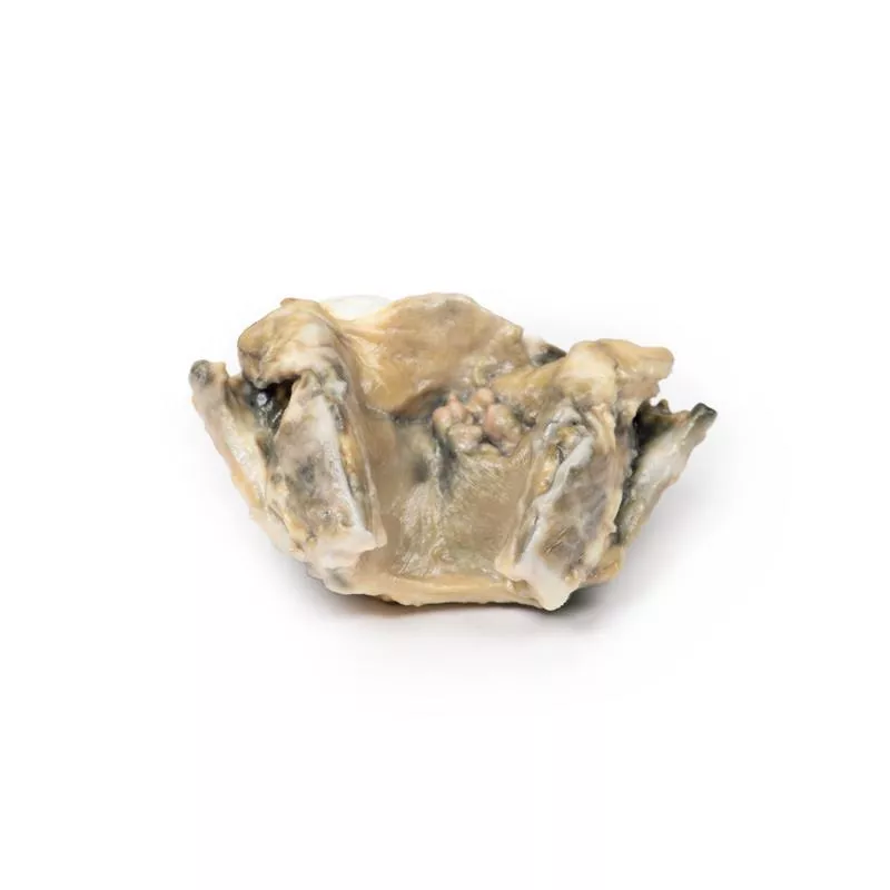
This is a parasagittal section of the right lung, clearly showing the boundaries between the three lobes. The upper and middle lobes are notably congested and hyperaemic, giving them a darker appearance. Smaller areas of similar changes are also visible in the left lung.
Further Information
Lobar pneumonia is characterized by inflammatory exudate filling the alveolar spaces, causing consolidation over a large, continuous area of a lobe. In this case, the affected lobe shows classic red hepatization, resulting from vascular congestion, leakage of red blood cells, and accumulation of neutrophils and fibrin. This leads to a solidified, liver-like appearance of the lung.
Common causative organisms include Streptococcus pneumoniae (pneumococcus), Haemophilus influenzae, Moraxella catarrhalis, and occasionally Mycobacterium tuberculosis or Klebsiella pneumoniae. Legionella pneumophila is another possible cause. Lobar pneumonia can occur as a community-acquired infection, in immunosuppressed patients, or in a hospital setting, though most are community-acquired.
On a chest X-ray, the affected lobe appears radiopaque, with no visible air, clearly indicating lobar pneumonia.
Erler-Zimmer
Erler-Zimmer GmbH & Co.KG
Hauptstrasse 27
77886 Lauf
Germany
info@erler-zimmer.de
Achtung! Medizinisches Ausbildungsmaterial, kein Spielzeug. Nicht geeignet für Personen unter 14 Jahren.
Attention! Medical training material, not a toy. Not suitable for persons under 14 years of age.


