


Product information "Chronic Gastric Ulcer"
Clinical History
An elderly patient with long-standing indigestion collapsed and died from a massive stroke.
Pathology
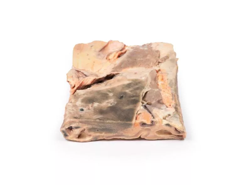
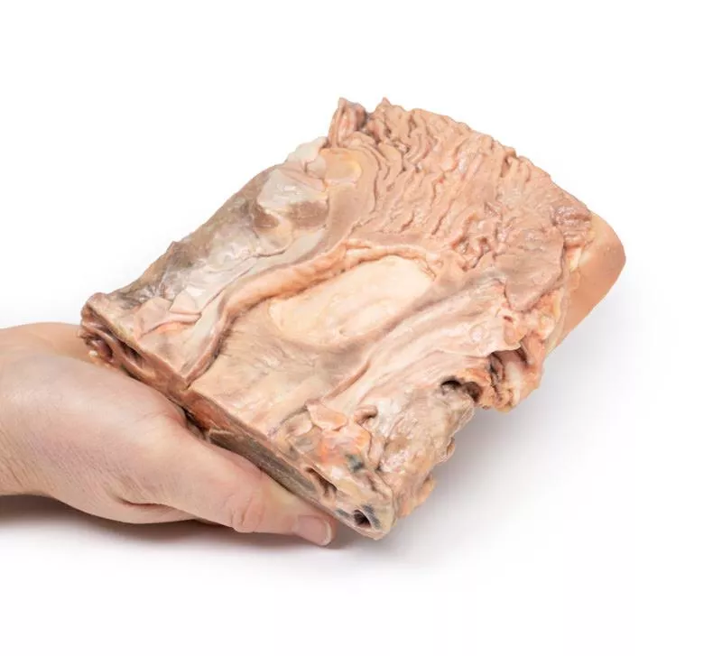
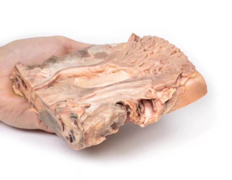
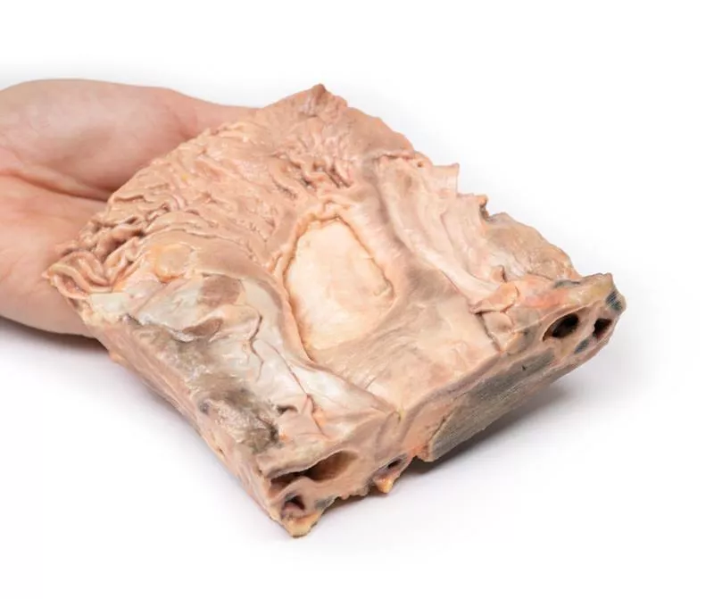
A 2?cm coronal section includes parts of the stomach, diaphragm, liver, and pancreas. A large gastric ulcer (5–6?cm) is located at the lesser curvature near the gastro-oesophageal junction. It has a smooth base and slightly raised edges, with fibrosis pulling mucosal folds inward — a sign of benign ulceration.
Further Information
Gastric ulcers often cause post-meal pain, nausea, and weight loss. Common causes include Helicobacter pylori and NSAIDs. Diagnosis is via endoscopy or H. pylori testing. Treatment involves acid-suppressing drugs (e.g., PPIs), antibiotics for H. pylori, and stopping NSAIDs and smoking. Ulcers affect about 4% of the population.
An elderly patient with long-standing indigestion collapsed and died from a massive stroke.
Pathology
A 2?cm coronal section includes parts of the stomach, diaphragm, liver, and pancreas. A large gastric ulcer (5–6?cm) is located at the lesser curvature near the gastro-oesophageal junction. It has a smooth base and slightly raised edges, with fibrosis pulling mucosal folds inward — a sign of benign ulceration.
Further Information
Gastric ulcers often cause post-meal pain, nausea, and weight loss. Common causes include Helicobacter pylori and NSAIDs. Diagnosis is via endoscopy or H. pylori testing. Treatment involves acid-suppressing drugs (e.g., PPIs), antibiotics for H. pylori, and stopping NSAIDs and smoking. Ulcers affect about 4% of the population.
Login
Erler-Zimmer
Erler-Zimmer Medical GmbH
Hauptstrasse 27
77886 Lauf
Germany
info@erler-zimmer.de
Achtung! Medizinisches Ausbildungsmaterial, kein Spielzeug. Nicht geeignet für Personen unter 14 Jahren.
Attention! Medical training material, not a toy. Not suitable for persons under 14 years of age.
Other customers also bought

Fatty Liver
Clinical HistoryNo clinical details are available.PathologyA liver slice shows a characteristic yellow/grey, greasy appearance on one side. The opposite side has this change limited to the outer margin, while the central area appears darker, likely due to cirrhosis. This is an example of fatty change in the liver.Further InformationFatty change (steatosis) involves triglyceride accumulation in the liver and can be caused by obesity, diabetes, alcohol abuse, starvation, Kwashiorkor, drugs, and toxins. Alcoholism is the most common cause in many populations.

Liver cirrhosis
Clinical HistoryA 63-year-old man was admitted multiple times for bleeding from oesophageal varices. On admission, he was mildly febrile, jaundiced, and showed a severe flapping tremor of his hands. Other findings included ascites, prominent periumbilical veins, and haemorrhoids. He suffered a massive haematemesis and died shortly after.PathologyA liver slice revealed multiple well-demarcated nodules ranging from 1 to 7 mm in diameter. The liver surface was nodular and irregular. This represents cirrhosis of the liver with mixed micro- and macronodular patterns and significant fatty change.Further InformationThe most common cause of cirrhosis and fatty liver change is chronic alcoholism.

Intussusception of small bowel due to metastatic tumour
Clinical HistoryA 66-year-old woman presented with sudden, severe, colicky central abdominal pain, which was somewhat relieved by drawing up her knees. She passed a stool described as containing mucus and blood ("like redcurrant jelly"). On examination, a mass in the left hypochondrium was noted, which hardened during spasms. A laparotomy was performed and the specimen resected.PathologyThe specimen is a 20?cm segment of small bowel with mesentery. A 3?cm polypoid tumour near the proximal margin has invaginated into the bowel lumen, forming a 13?cm long intussusception. The tumour lies at the apex of the invagination. The congested and exudative mucosa suggests early ischaemic necrosis. Although histology is not recorded, the macroscopic features are consistent with a metastatic malignant tumour.Further InformationIntussusception is most common in children, often due to enlarged Peyer’s patches in the distal ileum. In adults, it is rare (1–5?% of bowel obstructions) and typically caused by a polypoid tumour acting as a lead point, dragged forward by peristalsis. Symptoms include intermittent obstruction or severe pain. Abdominal CT often shows the characteristic “target sign”.

Hirschsprung's Disease
Clinical HistoryA 5-year-old boy had a history of constipation since birth. A barium enema revealed a narrow rectum and a dilated sigmoid colon. Surgical resection of the affected bowel segment was attempted, but the patient died during the procedure.PathologyThe specimen shows a massively dilated sigmoid colon with loss of normal mucosal pattern. The adjacent rectum appears normal in size and structure but lacks ganglion cells in the myenteric plexus. This is consistent with Hirschsprung’s disease (congenital aganglionic megacolon).Further InformationHirschsprung’s disease results from the absence of parasympathetic ganglia in a segment of the bowel, most commonly the rectum. This leads to impaired peristalsis and functional obstruction. It is caused by a failure of neural crest cells to migrate during embryogenesis. The proximal colon becomes hypertrophied and dilated, increasing the risk of complications like enterocolitis and perforation.The disease occurs in approximately 1 in 5000 live births, more often in males and those with genetic syndromes such as Down syndrome. Mutations in the RET gene are implicated in many familial and some sporadic cases.Symptoms range from failure to pass meconium in newborns to chronic constipation and abdominal distension in older children. The standard treatment is surgical resection of the aganglionic bowel and connection of the healthy bowel to the rectum.

Ulcerative Colitis
Clinical HistoryA 36-year-old woman was admitted with a 3-week history of bloody diarrhoea and lower abdominal pain. She reported four similar episodes over seven years. Sigmoidoscopy revealed erythematous, ulcerated, and oedematous rectal mucosa. Steroid treatment was started but symptoms persisted, leading to a total colectomy.PathologyThe resected colon showed extensive confluent ulceration with oedematous mucosal islands. Ulcers had necrotic bases and overhanging edges forming pseudo-polyps. Histology revealed acute inflammation, crypt abscesses, necrosis, and ulceration, confirming acute ulcerative colitis (UC).Further InformationUlcerative colitis is a chronic ulcerative inflammatory disease mainly affecting the rectum and extending proximally in a continuous manner. Inflammation is usually limited to the mucosa and superficial submucosa. The cause is unknown. It commonly starts between ages 15–25 and is slightly more common in females. Symptoms include bloody or mucous diarrhoea, urgency, abdominal pain, weight loss, anemia, and fatigue. Complications include toxic megacolon, perforation, and colon cancer. Extraintestinal symptoms may affect eyes, joints, skin, and liver. Treatment includes steroids, disease-modifying drugs, and TNF inhibitors. Colectomy cures intestinal symptoms, but extraintestinal manifestations may continue.

Multiple Polyposis Coli
Clinical HistoryNo clinical details are available for this case.PathologyThe specimens include two segments of sigmoid colon showing numerous sessile and pedunculated polyps up to 1.5 cm, some partially pigmented. There is no visible sign of malignancy.Further InformationMicroscopically, most polyps are tubular adenomas (over 75%), with fewer villous or tubulovillous adenomas. They may show varying degrees of dysplasia, but their histology is similar to sporadic colonic adenomas.Patients with familial adenomatous polyposis (FAP), a hereditary colon cancer syndrome linked to mutations in the APC gene on chromosome 5q21, often undergo prophylactic colectomy. Without treatment, invasive adenocarcinoma almost inevitably develops in one or more polyps, typically about 15 years after adenoma onset. FAP is inherited as an autosomal dominant trait.

Gall Stone Ileus
Clinical HistoryA 54-year-old man was admitted after 12?hours of severe colicky pain, nausea, and vomiting. He also had a 3-year history of intermittent right upper abdominal pain untreated until now. Acute bowel obstruction was diagnosed, and a laparotomy performed.PathologyAn opened segment of small intestine revealed a large, pigmented, rough-surfaced gallstone obstructing the lumen: a classic case of gallstone ileus.Further InformationGallstone ileus is a rare cause (~0.5?%) of bowel obstruction, more often in elderly or female patients. It usually results from a biliary–enteric fistula (sometimes post-sphincterotomy). Stones are typically =2?cm and lodge in the ileum (70?%), or other narrowed sites. Symptoms may be intermittent. Diagnosis is confirmed radiologically (often CT) or during surgery — following Rigler’s triad: small-bowel obstruction, ectopic gallstone, and pneumobilia. Treatment is surgical: remove the stone, close the fistula, and perform cholecystectomy; staged procedures may be required.

Villous adenoma of colon
Clinical HistoryA 70-year-old man was admitted for muscular weakness and passing large amounts of mucus per rectum. He was found to be hypokalaemic. During investigations, a tumour in the sigmoid colon was discovered and later surgically removed.PathologyA 15 cm segment of colon was opened to reveal a large sessile tumour with a velvety surface, measuring 11 x 7 cm, located close to the distal resection margin. The mucosa elsewhere appeared normal, and the serosal surface was unremarkable. Histology confirmed a villous adenoma.Further InformationVillous adenomas can secrete large amounts of mucus and potassium-rich fluid, sometimes causing hypoalbuminaemia or hypokalaemia. They are the least common but most serious type of adenomatous polyps, with invasive carcinoma present in up to 30% of cases at resection.
")
Cholelithiasis (Gallstones)
Clinical HistoryA middle-aged woman experienced recurrent epigastric pain. Endoscopy revealed no ulcers, but a cholangiogram showed a non-functioning gallbladder. She later died from a myocardial infarction.PathologyThe specimen shows a liver section with an opened gallbladder containing six large faceted mixed gallstones – a classic case of cholelithiasis.Further InformationGallstones consist of cholesterol, calcium salts, bilirubin and mucin. Risk factors include age over 50, female sex, pregnancy, diabetes, rapid weight loss, and certain medications. Symptoms range from no complaints to biliary colic, cholecystitis, or pancreatitis. Typical signs are upper abdominal pain, nausea, and vomiting. Diagnosis is mainly done with ultrasound, and in some cases HIDA scan. Treatment begins with analgesia; often followed by laparoscopic cholecystectomy. Though rare, severe complications can be fatal. Importantly, epigastric pain can mimic heart attack symptoms, particularly in women, so cardiac causes must be ruled out with an ECG.

Continuous innovation

Social responsibility

Active customer orientation

Understanding quality

Sustainable actions

ISO 9001 certification