Product information "Lymphoma of the thyroid"
Clinical History
A 68-year-old woman presented with a small, hard thyroid lump. Over six weeks, the mass rapidly enlarged, causing laryngeal stridor and oesophageal obstruction. No lymphadenopathy or splenomegaly was noted.
Pathology
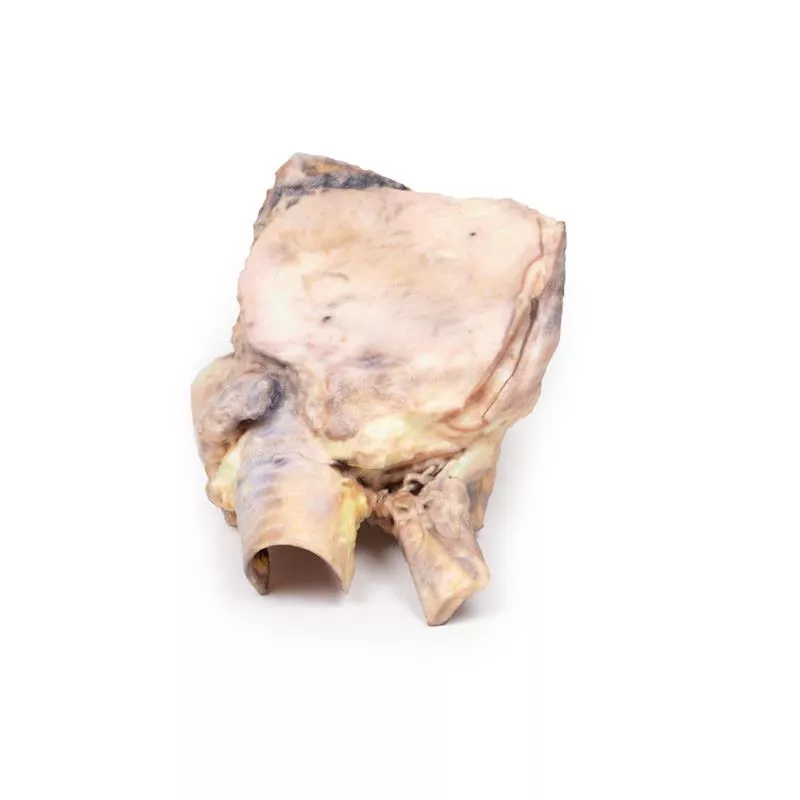
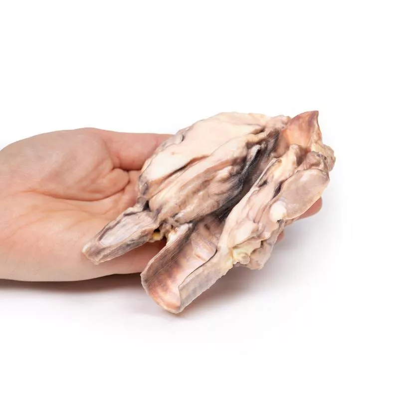
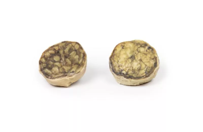
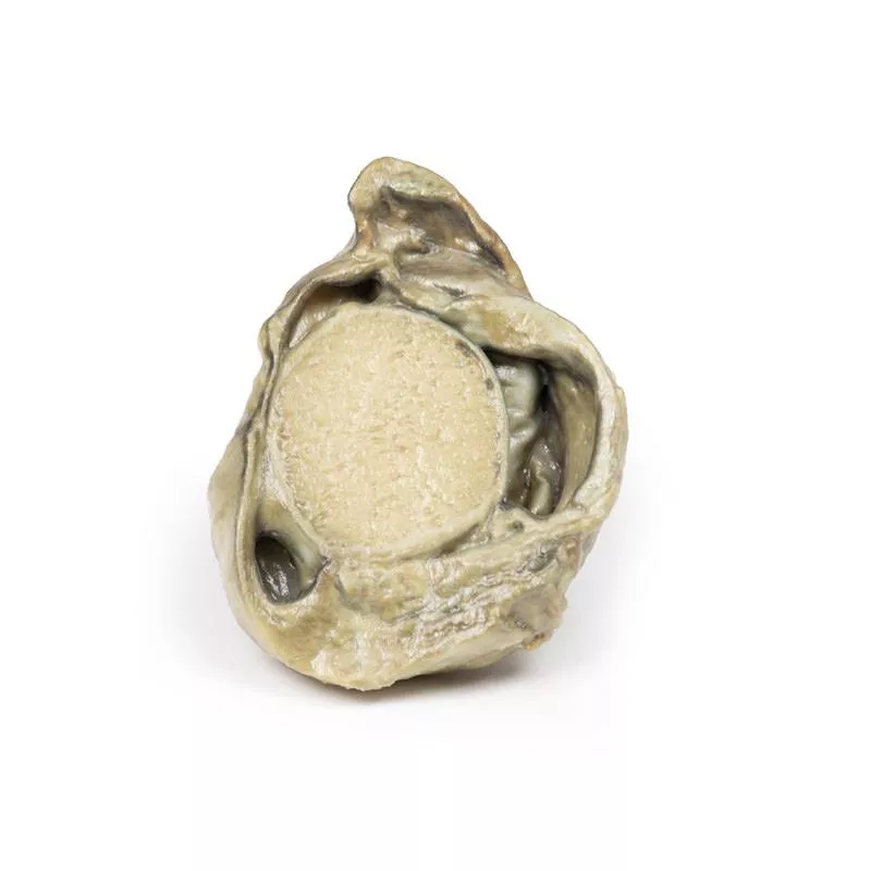
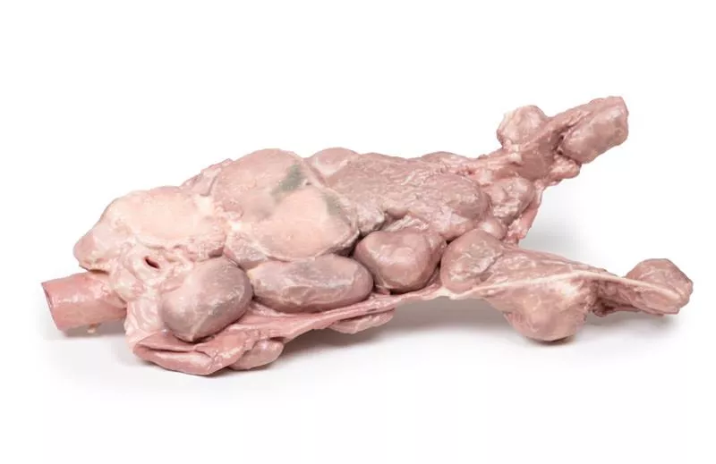
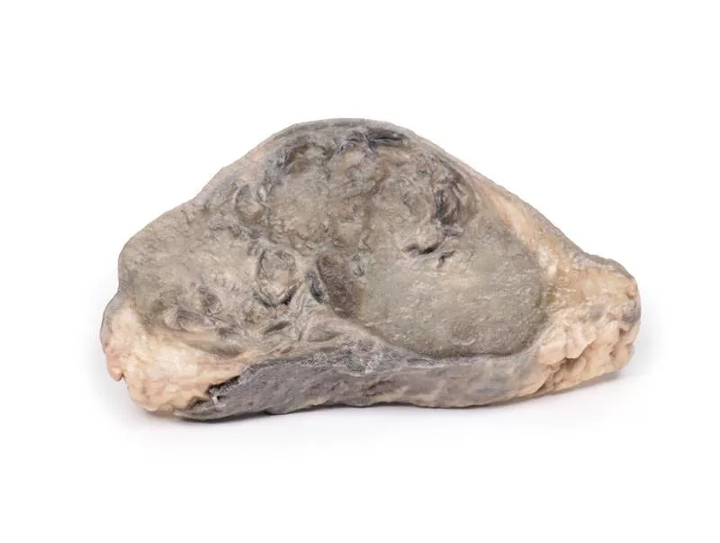
The specimen includes the larynx, thyroid, upper trachea, and oesophagus. The enlarged left thyroid lobe, and to a lesser extent the right, is replaced by homogeneous pale tumour tissue. The tumour compresses the larynx and oesophagus. Histology confirmed lymphoblastic lymphoma of the thyroid. Due to its rarity, anaplastic carcinoma and secondary lymphoma spread must be excluded.
Further Information
Primary thyroid lymphoma is rare but important to consider in thyroid masses. Most are non-Hodgkin lymphomas; lymphoblastic lymphoma is aggressive and usually seen in children. The main known risk factor is chronic autoimmune (Hashimoto’s) thyroiditis, present in about 50% of cases.
Over 90% of patients present with a rapidly enlarging goitre causing compression of trachea, oesophagus, and neck vessels, resulting in symptoms like stridor, hoarseness, dysphagia, and neck pain. Systemic ‘B-symptoms’ may include night sweats, fever, and weight loss.
Diagnosis requires ultrasound with fine needle aspiration or biopsy. Cytology and immunohistochemistry are essential to differentiate lymphoma from Hashimoto’s thyroiditis or carcinoma.
Erler-Zimmer
Erler-Zimmer GmbH & Co.KG
Hauptstrasse 27
77886 Lauf
Germany
info@erler-zimmer.de
Achtung! Medizinisches Ausbildungsmaterial, kein Spielzeug. Nicht geeignet für Personen unter 14 Jahren.
Attention! Medical training material, not a toy. Not suitable for persons under 14 years of age.


