Produktinformationen "Brain (Cerebrum)"
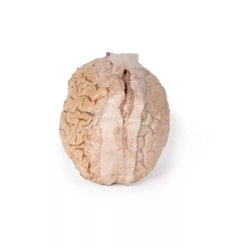
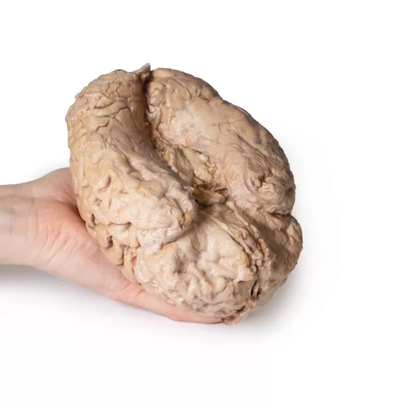
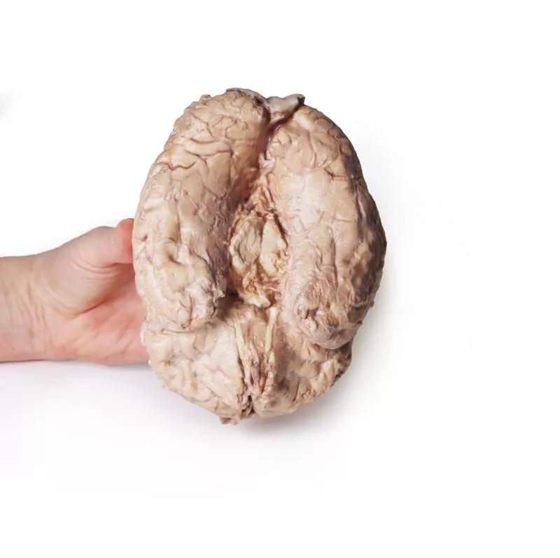
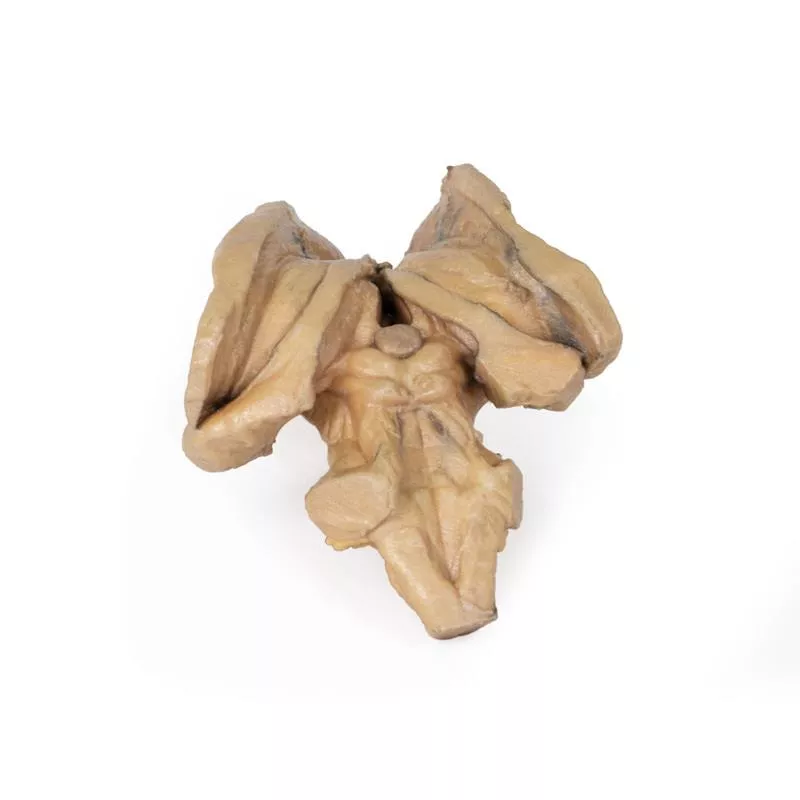
Dieses 3D-Modell bietet eine einzigartige Perspektive auf die Anatomie des Großhirns im Verhältnis zu den Hirnhäuten.
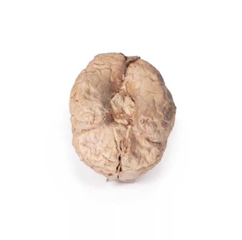
Das Großhirn wurde vom Hirnstamm und Kleinhirn getrennt, wobei nur Teile des Mittelhirns und der Hirnstiele auf der Unterseite sichtbar sind. Angrenzend an den Schnittbereich sind die Riechbahnen und Riechkolben zu sehen, die sich entlang des unteren Randes der Frontallappen des Großhirns erstrecken.
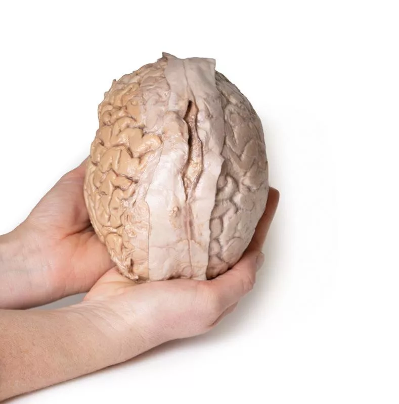
Die unterschiedliche Präparation der linken und rechten Gehirnhälfte ermöglicht einen Einblick in die Organisation des Gehirns und der Hirnhäute, wie sie normalerweise in der Schädelhöhle zu finden sind. In der Mittellinie wurde die Dura mater von anterior (rostral) bis posterior erhalten. Der zentrale Teil der echten (endostalen) Dura wurde geöffnet, um den Sinus sagittalis superior (zwischen der endostalen und meningealen Schicht der Dura mater) freizulegen.
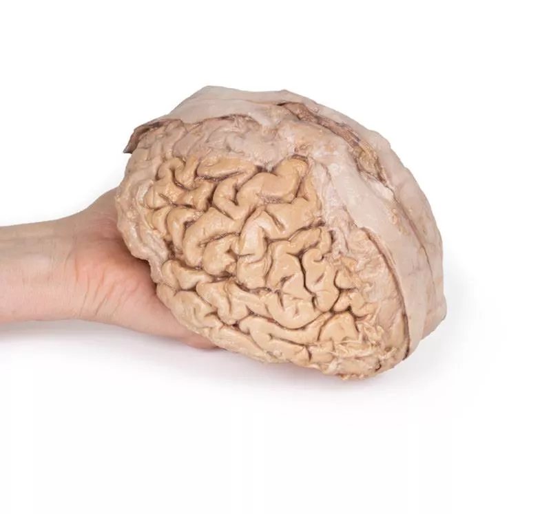
Zahlreiche Arachnoidalgranulationen (Cluster von Arachnoidalzotten) sind innerhalb des geöffneten Sinus sagittalis superior sowie an den Rändern der erhaltenen Dura sichtbar. Auf der rechten Gehirnhälfte wurde die Dura mater vollständig entfernt, um die darunter liegende Arachnoidea freizulegen, die das Erscheinungsbild der darunter liegenden Hirnwindungen und -furchen sowie der Endäste der Hirnarterien verdeckt. Im Gegensatz dazu wurde die Arachnoidea über den größten Teil der Hemisphäre (mit Ausnahme eines Randes als Referenz) präpariert, um die mit Pia mater bedeckten Gyri und Sulci freizulegen. Dies ermöglicht eine klare Sicht auf den Sulcus lateralis und den Sulcus centralis, wobei letzterer die Grenzen des Frontal- und Parietallappens definiert und die primären sensorischen und motorischen Kortexareale auf den Gyri zu beiden Seiten des Sulcus voneinander trennt.
Erler-Zimmer
Erler-Zimmer GmbH & Co.KG
Hauptstrasse 27
77886 Lauf
Germany
info@erler-zimmer.de
Achtung! Medizinisches Ausbildungsmaterial, kein Spielzeug. Nicht geeignet für Personen unter 14 Jahren.
Attention! Medical training material, not a toy. Not suitable for persons under 14 years of age.


