Produktinformationen "Brain Stem, isolated anatomy from midbrain to medulla oblongata"
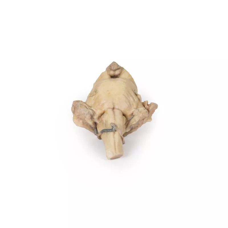
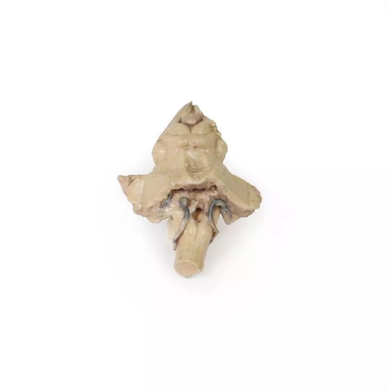
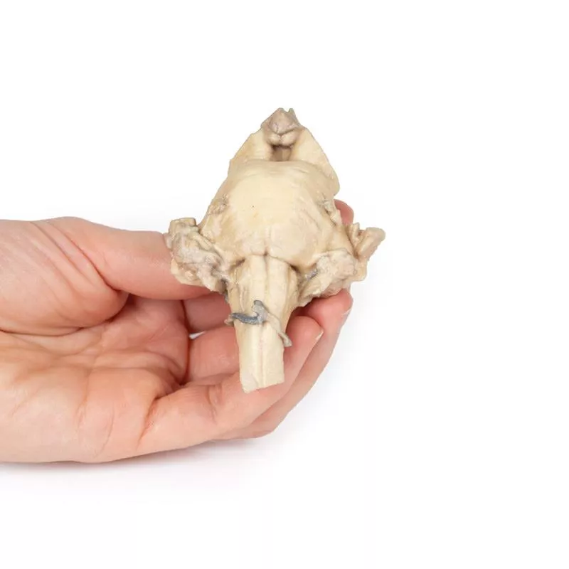
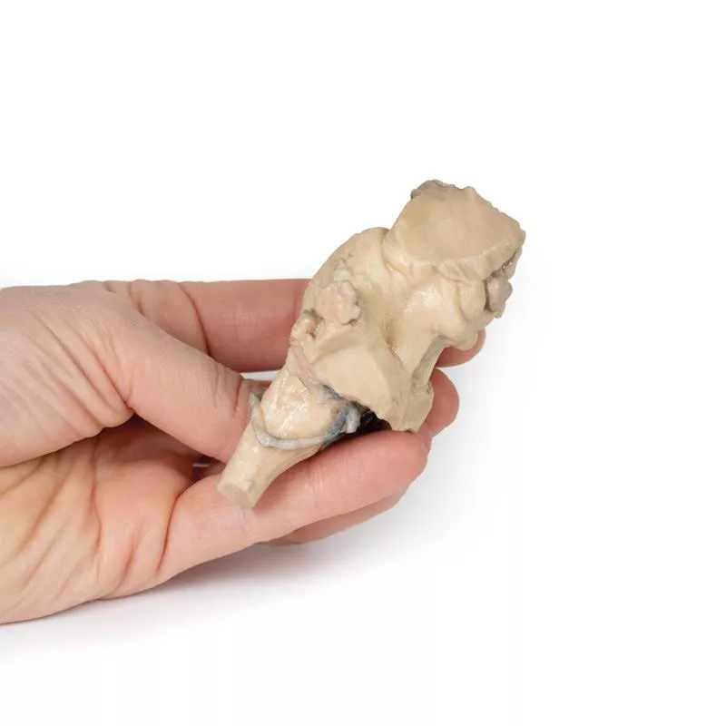
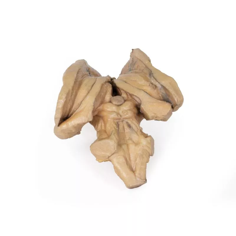
Dieses 3D-Modell bietet einen Blick auf die isolierte Anatomie des Hirnstamms vom Mittelhirn bis zur Medulla oblongata und ergänzt das andere 3D-Modell des Zwischenhirns/Hirnstamms (MP1100) in unserer Serie.
Rostral wurde das 3D-Modell in einem Winkel vom darüber liegenden Zwischenhirn geschnitten, wobei die Mamillarkörper des Hypothalamus zwischen den Hirnstielen (anterior) und der Zirbeldrüse/dem Epithalamus (posterior) erhalten blieben. Posterior sind die Corpora quadrigemina (die oberen und unteren Colliculi) des Mittelhirns neben den oberen Kleinhirnstielen deutlich zu erkennen. Das Kleinhirn selbst wurde entfernt, sodass der Querschnitt der mittleren und unteren Kleinhirnstiele auf jeder Seite sichtbar ist. Unterhalb der geschnittenen Stiele befinden sich der teilweise geöffnete vierte Ventrikel und Reste der hinteren unteren Kleinhirnarterien.
Auf der ventralen Seite des 3D-Modells ist die Pons mit dem Ursprung des Trigeminusnervs (CN V) erhalten geblieben (besonders auf der linken Seite). Unterhalb der Pons auf der Medulla oblongata sind sowohl die Pyramiden als auch die Oliven auf beiden Seiten sichtbar (besonders deutlich auf der rechten Seite).
Erler-Zimmer
Erler-Zimmer GmbH & Co.KG
Hauptstrasse 27
77886 Lauf
Germany
info@erler-zimmer.de
Achtung! Medizinisches Ausbildungsmaterial, kein Spielzeug. Nicht geeignet für Personen unter 14 Jahren.
Attention! Medical training material, not a toy. Not suitable for persons under 14 years of age.


